Debris clearance by microglia: an essential link between degeneration and regeneration
- PMID: 18567623
- PMCID: PMC2640215
- DOI: 10.1093/brain/awn109
Debris clearance by microglia: an essential link between degeneration and regeneration
Abstract
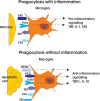
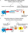
Microglia are cells of myeloid origin that populate the CNS during early development and form the brain's innate immune cell type. They perform homoeostatic activity in the normal CNS, a function associated with high motility of their ramified processes and their constant phagocytic clearance of cell debris. This debris clearance role is amplified in CNS injury, where there is frank loss of tissue and recruitment of microglia to the injured area. Recent evidence suggests that this phagocytic clearance following injury is more than simply tidying up, but instead plays a fundamental role in facilitating the reorganization of neuronal circuits and triggering repair. Insufficient clearance by microglia, prevalent in several neurodegenerative diseases and declining with ageing, is associated with an inadequate regenerative response. Thus, understanding the mechanism and functional significance of microglial-mediated clearance of tissue debris following injury may open up exciting new therapeutic avenues.
Figures


Similar articles
-
Microglial clearance function in health and disease.Neuroscience. 2009 Feb 6;158(3):1030-8. doi: 10.1016/j.neuroscience.2008.06.046. Epub 2008 Jul 1. Neuroscience. 2009. PMID: 18644426 Review.
-
Microglia in central nervous system repair after injury.J Biochem. 2016 May;159(5):491-6. doi: 10.1093/jb/mvw009. Epub 2016 Feb 8. J Biochem. 2016. PMID: 26861995 Review.
-
Microglia biology in health and disease.J Neuroimmune Pharmacol. 2006 Jun;1(2):127-37. doi: 10.1007/s11481-006-9015-5. Epub 2006 Mar 25. J Neuroimmune Pharmacol. 2006. PMID: 18040779 Review.
-
Microglia and neuroprotection: from in vitro studies to therapeutic applications.Prog Neurobiol. 2010 Nov;92(3):293-315. doi: 10.1016/j.pneurobio.2010.06.009. Epub 2010 Jul 4. Prog Neurobiol. 2010. PMID: 20609379 Review.
-
Toll-like receptor 4 deficiency impairs microglial phagocytosis of degenerating axons.Glia. 2014 Dec;62(12):1982-91. doi: 10.1002/glia.22719. Epub 2014 Jul 8. Glia. 2014. PMID: 25042766
Cited by
-
MiR-146b-5p/TRAF6 axis is essential for Ginkgo biloba L. extract GBE to attenuate LPS-induced neuroinflammation.Front Pharmacol. 2022 Aug 24;13:978587. doi: 10.3389/fphar.2022.978587. eCollection 2022. Front Pharmacol. 2022. PMID: 36091773 Free PMC article.
-
Structural plasticity in the dentate gyrus- revisiting a classic injury model.Front Neural Circuits. 2013 Feb 18;7:17. doi: 10.3389/fncir.2013.00017. eCollection 2013. Front Neural Circuits. 2013. PMID: 23423628 Free PMC article. Review.
-
Iron efflux from astrocytes plays a role in remyelination.J Neurosci. 2012 Apr 4;32(14):4841-7. doi: 10.1523/JNEUROSCI.5328-11.2012. J Neurosci. 2012. PMID: 22492039 Free PMC article.
-
Gastrodin Attenuates Lipopolysaccharide-Induced Inflammatory Response and Migration via the Notch-1 Signaling Pathway in Activated Microglia.Neuromolecular Med. 2022 Jun;24(2):139-154. doi: 10.1007/s12017-021-08671-1. Epub 2021 Jun 9. Neuromolecular Med. 2022. PMID: 34109563
-
Aging and sex: Impact on microglia phagocytosis.Aging Cell. 2020 Aug;19(8):e13182. doi: 10.1111/acel.13182. Epub 2020 Jul 29. Aging Cell. 2020. PMID: 32725944 Free PMC article.
References
-
- Ajami B, Bennett JL, Krieger C, Tetzlaff W, Rossi FM. Local self-renewal can sustain CNS microglia maintenance and function throughout adult life. Nat Neurosci. 2007;10:1538–43. - PubMed
-
- Amat JA, Ishiguro H, Nakamura K, Norton WT. Phenotypic diversity and kinetics of proliferating microglia and astrocytes following cortical stab wounds. Glia. 1996;16:368–82. - PubMed
-
- Arnett HA, Mason J, Marino M, Suzuki K, Matsushima GK, Ting JP. TNF alpha promotes proliferation of oligodendrocyte progenitors and remyelination. Nat Neurosci. 2001;4:1116–22. - PubMed
-
- Awasaki T, Ito K. Engulfing action of glial cells is required for programmed axon pruning during Drosophila metamorphosis. Curr Biol. 2004;14:668–77. - PubMed
Publication types
MeSH terms
Substances
Grants and funding
LinkOut - more resources
Full Text Sources
Other Literature Sources
Medical

