Potent anti-inflammatory and neuroprotective effects of TGF-beta1 are mediated through the inhibition of ERK and p47phox-Ser345 phosphorylation and translocation in microglia
- PMID: 18566433
- PMCID: PMC2741684
- DOI: 10.4049/jimmunol.181.1.660
Potent anti-inflammatory and neuroprotective effects of TGF-beta1 are mediated through the inhibition of ERK and p47phox-Ser345 phosphorylation and translocation in microglia
Abstract
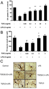
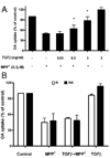
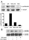
TGF-beta1 is one of the most potent endogenous immune modulators of inflammation. The molecular mechanism of its anti-inflammatory effect on the activation of the transcription factor NF-kappaB has been well-studied; however, the potential effects of TGF-beta1 on other proinflammatory signaling pathways is less clear. In this study, using the well-established LPS and the 1-methyl-4-phenylpyridinium-mediated models of Parkinson's disease, we demonstrate that TGF-beta1 exerts significant neuroprotection in both models via its anti-inflammatory properties. The neuroprotective effects of TGF-beta1 are mainly attributed to its ability to inhibit the production of reactive oxygen species from microglia during their activation or reactivation. Moreover, we demonstrate that TGF-beta1 inhibited LPS-induced NADPH oxidase (PHOX) subunit p47phox translocation from the cytosol to the membrane in microglia within 10 min. Mechanistic studies show that TGF-beta1 fails to protect dopaminergic neurons in cultures from PHOX knockout mice, and significantly reduced LPS-induced translocation of the PHOX cytosolic subunit p47phox to the cell membrane. In addition, LPS-induced ERK phosphorylation and subsequent Ser345 phosphorylation on p47phox were significantly inhibited by TGF-beta1 pretreatment. Taken together, our results show that TGF-beta1 exerted potent anti-inflammatory and neuroprotective properties, either through the prevention of the direct activation of microglia by LPS, or indirectly through the inhibition of reactive microgliosis elicited by 1-methyl-4-phenylpyridinium. The molecular mechanisms of TGF-beta1-mediated anti-inflammatory properties is through the inhibition of PHOX activity by preventing the ERK-dependent phosphorylation of Ser345 on p47phox in microglia to reduce oxidase activities induced by LPS.
Conflict of interest statement
The authors have no financial conflict of interest.
Figures


Similar articles
-
Squamosamide derivative FLZ protects dopaminergic neurons against inflammation-mediated neurodegeneration through the inhibition of NADPH oxidase activity.J Neuroinflammation. 2008 May 28;5:21. doi: 10.1186/1742-2094-5-21. J Neuroinflammation. 2008. PMID: 18507839 Free PMC article.
-
Microglia-mediated neurotoxicity is inhibited by morphine through an opioid receptor-independent reduction of NADPH oxidase activity.J Immunol. 2007 Jul 15;179(2):1198-209. doi: 10.4049/jimmunol.179.2.1198. J Immunol. 2007. PMID: 17617613
-
Clozapine metabolites protect dopaminergic neurons through inhibition of microglial NADPH oxidase.J Neuroinflammation. 2016 May 16;13(1):110. doi: 10.1186/s12974-016-0573-z. J Neuroinflammation. 2016. PMID: 27184631 Free PMC article.
-
Targeting microglia-mediated neurotoxicity: the potential of NOX2 inhibitors.Cell Mol Life Sci. 2012 Jul;69(14):2409-27. doi: 10.1007/s00018-012-1015-4. Epub 2012 May 13. Cell Mol Life Sci. 2012. PMID: 22581365 Free PMC article. Review.
-
Role of microglia in oxidative toxicity associated with encephalomycarditis virus infection in the central nervous system.Int J Mol Sci. 2012;13(6):7365-7374. doi: 10.3390/ijms13067365. Epub 2012 Jun 14. Int J Mol Sci. 2012. PMID: 22837699 Free PMC article. Review.
Cited by
-
Real-time imaging of NADPH oxidase activity in living cells using a novel fluorescent protein reporter.PLoS One. 2013 May 21;8(5):e63989. doi: 10.1371/journal.pone.0063989. Print 2013. PLoS One. 2013. PMID: 23704967 Free PMC article.
-
NADPH oxidases in oxidant production by microglia: activating receptors, pharmacology and association with disease.Br J Pharmacol. 2017 Jun;174(12):1733-1749. doi: 10.1111/bph.13425. Epub 2016 Feb 26. Br J Pharmacol. 2017. PMID: 26750203 Free PMC article. Review.
-
Vibrio vulnificus MO6-24/O lipopolysaccharide stimulates superoxide anion, thromboxane B₂, matrix metalloproteinase-9, cytokine and chemokine release by rat brain microglia in vitro.Mar Drugs. 2014 Mar 26;12(4):1732-56. doi: 10.3390/md12041732. Mar Drugs. 2014. PMID: 24675728 Free PMC article.
-
beta2 Adrenergic receptor activation induces microglial NADPH oxidase activation and dopaminergic neurotoxicity through an ERK-dependent/protein kinase A-independent pathway.Glia. 2009 Nov 15;57(15):1600-9. doi: 10.1002/glia.20873. Glia. 2009. PMID: 19330844 Free PMC article.
-
Identification of a specific α-synuclein peptide (α-Syn 29-40) capable of eliciting microglial superoxide production to damage dopaminergic neurons.J Neuroinflammation. 2016 Jun 21;13(1):158. doi: 10.1186/s12974-016-0606-7. J Neuroinflammation. 2016. PMID: 27329107 Free PMC article.
References
-
- McGeer PL, S I, Boyes BE, McGeer EG. Reactive microglia are positive for HLA-DR in the substantia nigra of Parkinson's and Alzheimer's disease brains. Neurology. 1988;38:1285–1291. - PubMed
-
- Liu B, Hong JS. Role of microglia in inflammation-mediated neurodegenerative diseases: mechanisms and strategies for therapeutic intervention. J Pharmacol Exp Ther. 2003;304:1–7. - PubMed
-
- Aloisi F. The role of microglia and astrocytes in CNS immune surveillance and immunopathology. Adv. Exp. Med. Biol. 1999;468:123–133. - PubMed
-
- McGuire SO, Ling ZD, Lipton JW, Sortwell CE, Collier TJ, Carvey PM. Tumor necrosis factor alpha is toxic to embryonic mesencephalic dopamine neurons. Exp Neurol. 2001;169:219–230. - PubMed
Publication types
MeSH terms
Substances
Grants and funding
LinkOut - more resources
Full Text Sources
Molecular Biology Databases
Miscellaneous

