. 2008 Aug 1;320(1):149-60.
doi: 10.1016/j.ydbio.2008.04.038. Epub 2008 May 10.

Misexpression of ELF5 disrupts lung branching and inhibits epithelial differentiation

Affiliations

Misexpression of ELF5 disrupts lung branching and inhibits epithelial differentiation

David E Metzger et al. Dev Biol. .

Abstract

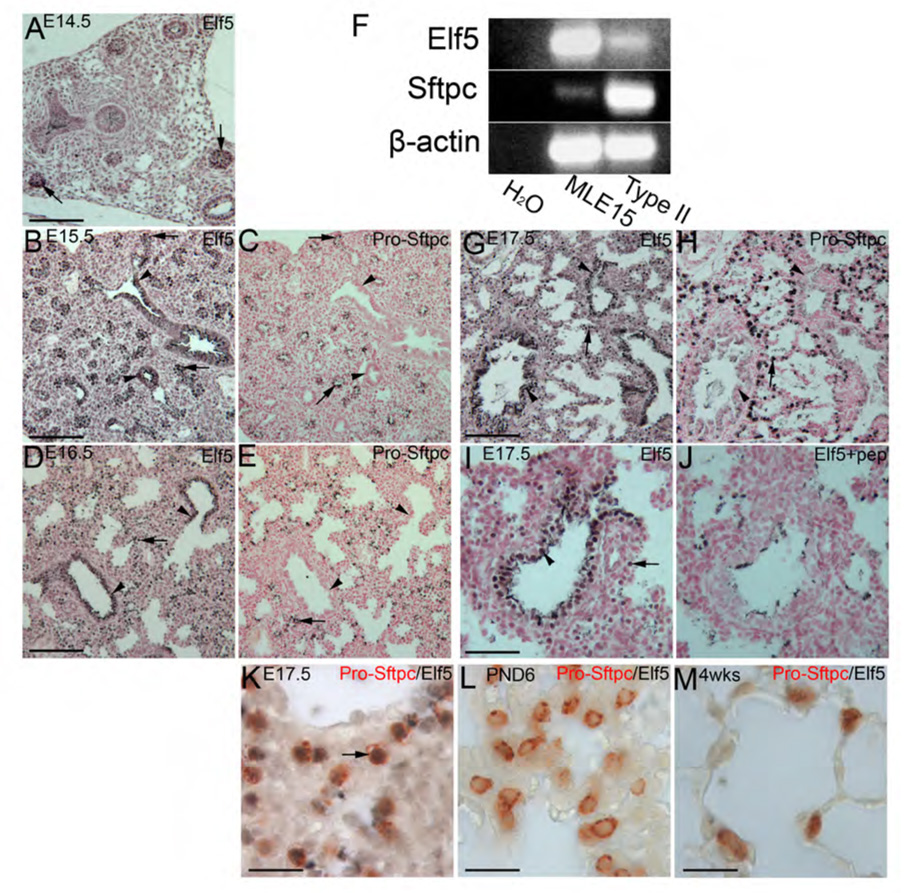
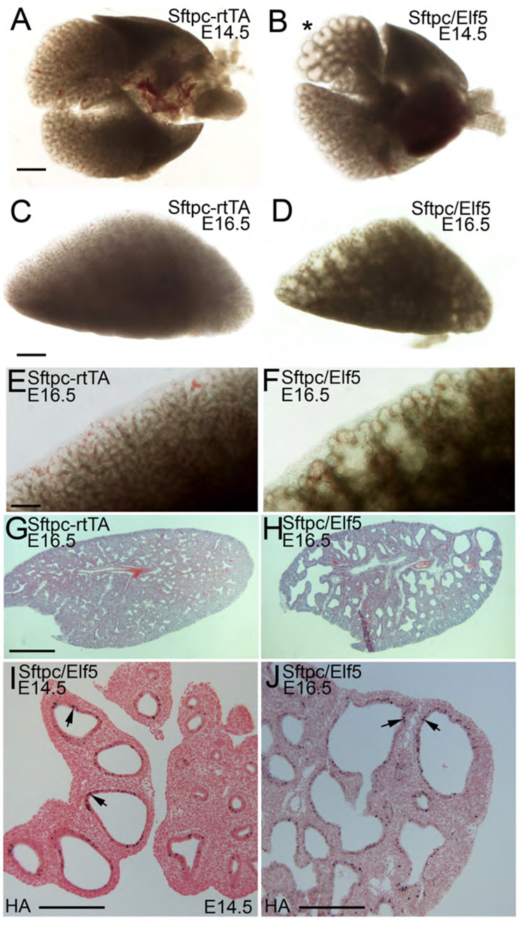
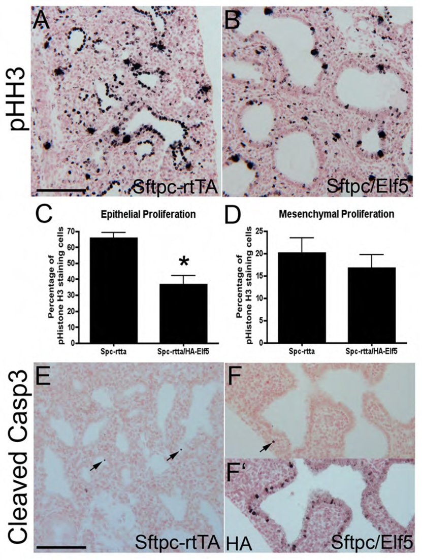
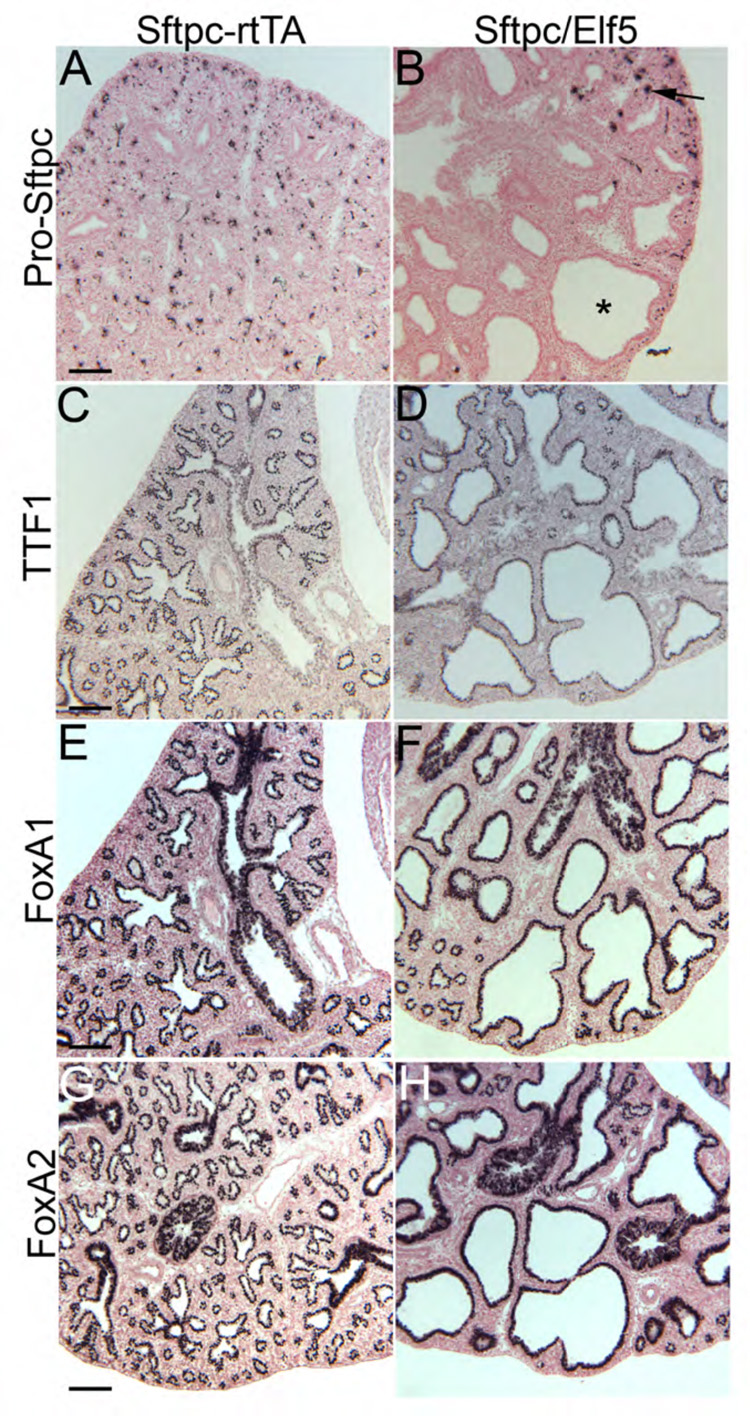
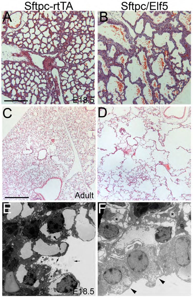
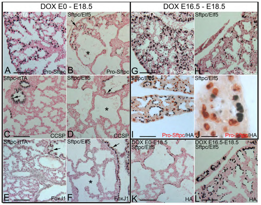
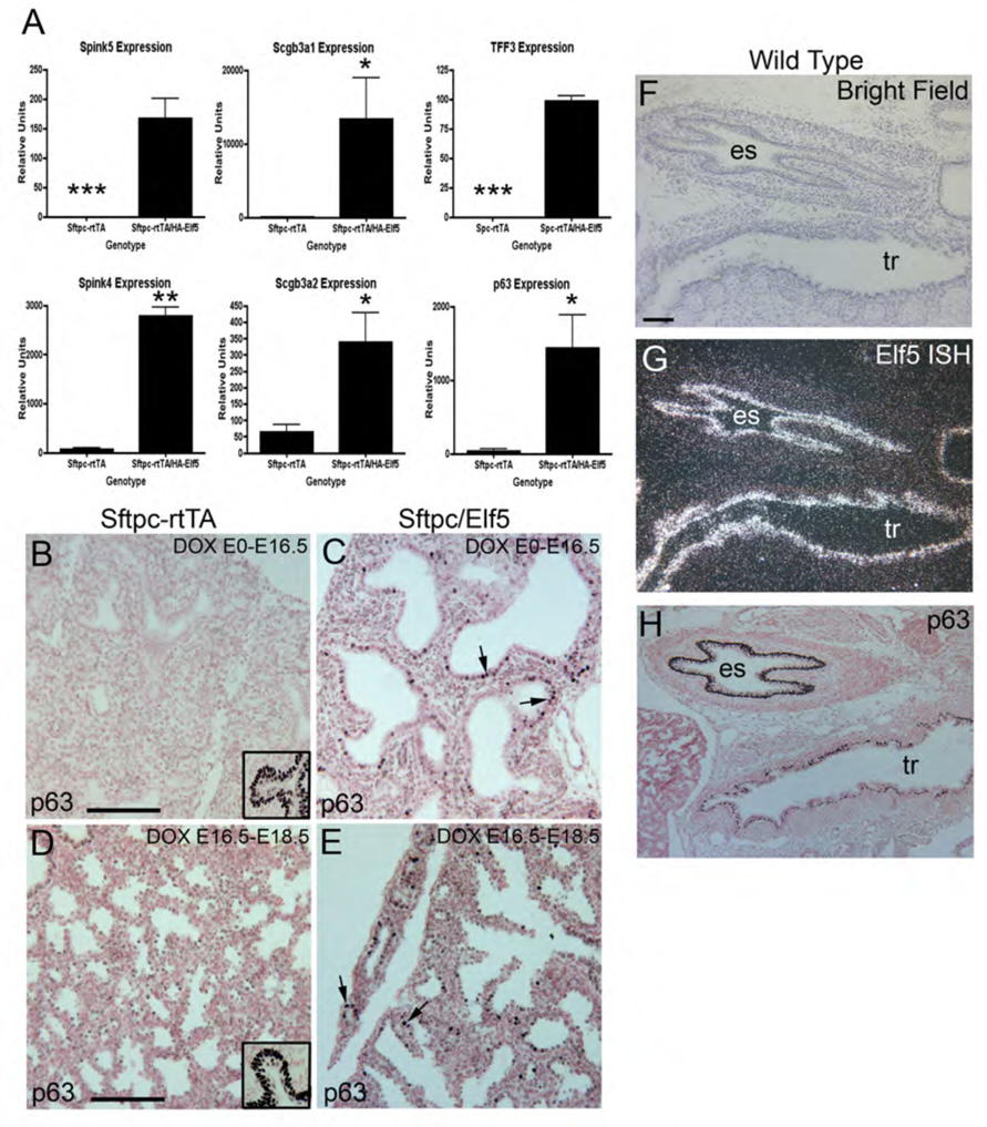
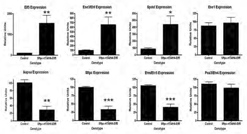
ELF5, an Ets family transcription factor found exclusively in epithelial cells, is expressed in the distal lung epithelium during embryogenesis, then becomes restricted to proximal airways at the end of gestation and postnatally. To test the hypothesis that ELF5 represses distal epithelial differentiation, we generated a transgenic mouse model in which a doxycycline inducible HA-tagged mouse Elf5 transgene was placed under the control of the lung epithelium-specific human SFTPC promoter. We found that expressing high levels of ELF5 during early lung development disrupted branching morphogenesis and produced a dilated epithelium. The effects of ELF5 on morphogenesis were stage-dependent, since inducing the transgene on E16.5 had no effect on branching. ELF5 reduced expression of the distal lung epithelial differentiation markers Erm, Napsa and Sftpc, and type II cell ultrastructural differentiation was immature. ELF5 overexpression did not induce the proximal airway epithelial markers Ccsp and Foxj1, but did induce expression of p63, a marker of basal cells in the trachea and esophagus. High ELF5 levels also induced the expression of genes found in other endodermal epithelia but not normally associated with the lung. These results suggest that precise levels of ELF5 regulate the specification and differentiation of epithelial cells in the lung.


Figures

Fig. 1
Fig. 1
Endogenous expression of ELF5 and Pro-SP-C protein expression during lung development. ELF5 is expressed at the tips of branching lung epithelium (arrows) at E14.5 (A). ELF5 expression continues in branching epithelium (arrows) at E15.5 but is also expressed in the developing airway epithelium (arrowhead) (B). Pro-SP-C is expressed in tips of branching epithelium (arrow) but is not expressed in the proximal airway (arrowheads) (C). ELF5 is present in the airways (arrowheads) and in lung parenchymal cells (arrows) at E16.5 and E17.5 (D,G), while Pro-SP-C is found exclusively in cells in the distal lung parenchyma (arrows) and not the airway (arrowhead) (E,H). ELF5 is seen in many cells in the E17.5 airway (arrowhead) and parenchyma (arrow) (I). Staining on the apical surfaces of some luminal cells is nonspecific as determined by the ELF5 peptide block control (J). Double IHC staining of E17.5 lungs for ELF5 and Pro-SP-C indicates that distal cells expressing ELF5 also stain for Pro-SP-C (K), whereas ELF5 is undetectable in Pro-SP-C positive type II cells in postnatal day 6 (PND6) (L) and 4 week lungs (M). RT-PCR shows an inverse relationship between Elf5 expression and Sftpc expression in MLE15 cells and in isolated type II cells (F). Scale bars: 100 µm in A–E, G, H; 50 µm in I,J and 20 µm in K–M.
Fig. 2
Fig. 2
Misexpression of ELF5 in distal lung epithelial cells disrupts branching morphogenesis at E14.5. Control lungs from an E14.5 SFTPC-rtTA embryo show typical fine branches (A), whereas the lungs from an E14.5 SFTPC/Elf5 embryo display areas of epithelial dilation (B). Note that in this preparation only one lobe appears to be affected (*). A left lung from a control E16.5 SFTPC-rtTA embryo, showing finely branched distal tips (C). The left lung from an E16.5 SFTPC/Elf5 embryo (D) showing widespread dilated epithelium, which are readily apparent at higher magnification (E,F). Histology of E16.5 SFTPC-rtTA (G) and SFTPC/Elf5 (H) lungs demonstrates that the dilated epithelium is widespread, although some areas appear unaffected. Immunostaining for HA in SFTPC/Elf5 lungs on E14.5 (I) and E16.5 (J) shows those cells expressing the HA-Elf5 transgene (arrows). Note that not all of the epithelial cells stain positive. Scale bars: 500 µm in A–D, G, H; 200 µm in E, F, I, J.
Fig. 3
Fig. 3
ELF5 overexpression inhibits epithelial proliferation. IHC for the proliferation marker phospho-Histone H3 (pHH3) in E16.5 SFTPC-rtTA (A) and SFTPC/Elf5 (B) lungs shows decreased epithelial staining in SFTPC/Elf5 lungs. Quantification of the percentage of epithelial cells and mesenchymal cells staining for pHH3 in SFTPC/Elf5 lungs compared to SFTPC-rtTA lungs shows that epithelial proliferation (C) was significantly decreased in SFTPC/Elf5 lungs (p < 0.05), whereas mesenchymal proliferation (D) did not change. IHC for the apoptosis marker cleaved caspase 3 shows that few positive cells are found in either SFTPC-rtTA control lungs (E) or SFTPC/Elf5 lungs (F). A consecutive section to that shown in F stained for HA shows HA-ELF5 is expressed in the area of the epithelium evaluated for cleaved caspase 3 (F’). Scale bar: 100 µm.
Fig. 4
Fig. 4
Effects of ELF5 misexpression on gene expression in E16.5 lungs. Sections of SFTPC-rtTA control lungs (A) and SFTPC/Elf5 lungs (B) stained for Pro-SP-C protein show that it is not detectable in the cystic epithelium of SFTPC/Elf5 lungs (asterisks), although histologically normal regions still express the protein (B; arrow). Control lungs stain uniformly for Pro-SP-C (A). IHC for TTF1 shows strong expression in the epithelium of both control (C) and SFTPC/Elf5 (D) lungs. IHC for FOXA1 (E,F) and FOXA2 (G,H) shows no differences in staining for either protein in SFTPC/Elf5 lungs compared to SFTPC-rtTA controls. Scale bar: 100 µm.
Fig. 5
Fig. 5
Histology of E18.5 SFTPC-rtTA (A) and SFTPC/Elf5 (B) lungs shows that expression of ELF5 transgene disrupts acinar formation, and this persists in adult control (C) and double transgenic (D). Electron micrographs of E18.5 SFTPC-rtTA control (E) and SFTPC/Elf5 (F) lungs reveal ultrastructural differences. SFTPC-rtTA lungs (E) contain normal type II cells that exhibit apical microvilli (*) and cytoplasmic lamellar bodies (arrowhead); surfactant is seen in the alveolar lumen (arrows). In contrast, the cystic epithelium of SFTPC/Elf5 lungs (F) is predominantly composed of immature cuboidal cells lacking apical microvilli (arrowheads) and cytoplasmic lamellar bodies; secreted surfactant could not be found in the cyst lumina. Fibroblasts in the loosely-organized interstitium adjacent to the cystic epithelium contain many lipid droplets (*). Scale bars: 200 µm in A,B; 500 µm in C,D.
Fig. 6
Fig. 6
ELF5 represses SP-C expression in E18.5 lungs. IHC for Pro-SP-C in the lungs of SFTPC/Elf5 embryos that had been exposed to Dox since conception (DOX E0–E18.5) reveals a lack of Pro-SP-C in the epithelial cysts (*) (B), whereas SFTPC-rtTA control lungs show widespread expression Pro-SP-C (A). IHC for CCSP shows endogenous protein expression in the airway epithelium (arrows) of both control (C) and SFTPC/Elf5 lungs (D). Note the lack of CCSP induction in the epithelium of the cysts (*) of double transgenic lungs (D). Similarly, IHC for FOXJ1 in control (E) and SFTPC/Elf5 lungs (F) shows endogenous protein expression in the airway epithelium, but no induction in the cystic epithelium (*) of SFTPC/Elf5 lungs (F). IHC for Pro-SP-C in the lungs of embryos that had been exposed to Dox from E16.5 to E18.5 (DOX E16.5 – E18.5) shows a decided decrease in Pro-SP-C positive cells in SFTPC/Elf5 lungs (H) compared to SFTPC-rtTA controls (G). Double IHC of HA and Pro-SP-C in DOX E16.5 – E18.5 SFTPC/Elf5 lungs (I,J) shows that Pro-SP-C is repressed in cells that stain positive for the HA-Elf5 transgene. IHC for HA in DOX E0–E18.5 SFTPC/Elf5 lungs shows that the transgene is expressed in the cystic epithelium, but is not found in histologically normal areas (K). In contrast, transgene expression is widespread in DOX E16.5 – E18.5 SFTPC/Elf5 lungs (L), but overall histology is normal (compare H and L to A) indicating that the effects of ELF5 misexpression on morphogenesis occur prior to E16.5. Scale bars: 100 µm in A–I,K,L; 20 µm in J.
Fig. 7
Fig. 7
Cystic regions of E16.5 SFTPC/Elf5 lungs were compared by microarray to spatially similar areas of SFTPC-rtTA control lungs to identify upregulated genes. qPCR for expression of 6 of the genes identified-- Spink5, Spink4, Scg3a1, Scgb3a2, Tff3 and p63 -- confirms that all were induced or increased in SFTPC/Elf5 lungs (A; n = 3; * = p < 0.05; ** = p < 0.0001; *** = expression not detected in controls). IHC for p63 in SFTPC-rtTA and SFTPC/Elf5 lungs in E16.5 (DOX E0–E16.5) and E18.5 (DOX E16.5–E18.5) lungs shows that p63 was absent in control lungs (B,D) but induced in double transgenic lungs (C,E). Insets in B and D show control staining for p63 in esophageal epithelium in the same section. Examination of an E17.5 wild-type fetus (F) shows that Elf5 (G) and p63 (H) are endogenously co-expressed in the esophageal (es) and tracheal (tr) epithelium. Scale bars: 100 µm.
Fig. 8
Fig. 8
E16.5 SFTPC/Elf5 lungs were compared to controls by qPCR for expression of other Ets transcription factors (n = 5) Sftpc (n = 5) and Napsa (n = 3). Elf5 was significantly (p < 0.005) increased in SFTPC/Elf5 lungs, as were the Epithelial Specific Ets genes, Ese3 (p < 0.005) and Spdef (p < 0.05). The expression of Sftpc, Napsa, and Erm, however, were significantly (p < 0.0001) reduced in SFTPC/Elf5 lungs. The expression of Pea3 and the Epithelial Specific Ets gene Ese1 did not change significantly.

Similar articles

Cited by

References

    1. Avril-Delplanque A, et al. Aquaporin-3 expression in human fetal airway epithelial progenitor cells. Stem Cells. 2005;23:992–1001. - PubMed
    1. Bellusci S, et al. Fibroblast growth factor 10 (FGF10) and branching morphogenesis in the embryonic mouse lung. Development. 1997;124:4867–4878. - PubMed
    1. Blanpain C, Fuchs E. p63: revving up epithelial stem-cell potential. Nat Cell Biol. 2007;9:731–733. - PubMed
    1. Brasch F, et al. Involvement of napsin A in the C- and N-terminal processing of surfactant protein B in type-II pneumocytes of the human lung. J Biol Chem. 2003;278:49006–49014. - PubMed
    1. Cangemi R, et al. Reduced expression and tumor suppressor function of the ETS transcription factor ESE-3 in prostate cancer. Oncogene. 2007 - PubMed

Publication types

MeSH terms

Substances

LinkOut - more resources