. 2008 Jun;10(6):573-86.
doi: 10.1593/neo.08290.

UNBS5162, a novel naphthalimide that decreases CXCL chemokine expression in experimental prostate cancers

Affiliations

UNBS5162, a novel naphthalimide that decreases CXCL chemokine expression in experimental prostate cancers

Tatjana Mijatovic et al. Neoplasia. 2008 Jun.

Abstract

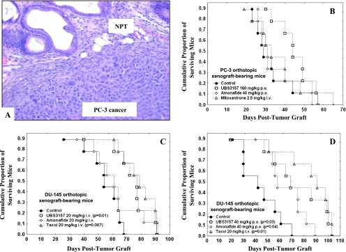
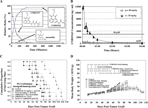
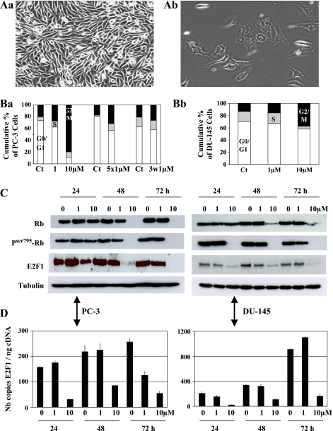
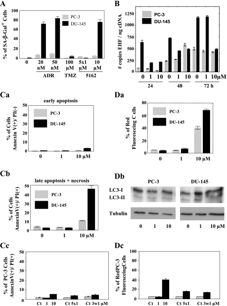
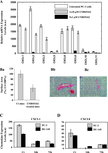
Several naphthalimides have been evaluated clinically as potential anticancer agents. UNBS3157, a naphthalimide that belongs to the same class as amonafide, was designed to avoid the specific activating metabolism that induces amonafide's hematotoxicity. The current study shows that UNBS3157 rapidly and irreversibly hydrolyzes to UNBS5162 without generating amonafide. In vivo UNBS5162 after repeat administration significantly increased survival in orthotopic human prostate cancer models. Results obtained by the National Cancer Institute (NCI) using UNBS3157 and UNBS5162 against the NCI 60 cell line panel did not show a correlation with any other compound present in the NCI database, including amonafide, thereby suggesting a unique mechanism of action for these two novel naphthalimides. Affymetrix genome-wide microarray analysis and enzyme-linked immunosorbent assay revealed that in vitro exposure of PC-3 cells to UNBS5162 (1 microM for 5 successive days) dramatically decreased the expression of the proangiogenic CXCL chemokines. Histopathology additionally revealed antiangiogenic properties in vivo for UNBS5162 in the orthotopic PC-3 model. In conclusion, the present study reveals UNBS5162 to be a pan-antagonist of CXCL chemokine expression, with the compound displaying antitumor effects in experimental models of human refractory prostate cancer when administered alone and found to enhance the activity of taxol when coadministered with the taxoid.


Figures

Figure 1
Figure 1
In vivo UNBS3157-mediated antitumor effects. (A) Typical morphologic illustration of an orthotopic model of refractory prostate cancer using the PC-3 cell line. NPT indicates normal prostate tissue. (B–D) A comparison of the activity of UNBS3157 versus that of amonafide, mitoxantrone, and taxol in two orthotopic models of human hormone refractory PC-3 and DU-145 prostate carcinomas. Drug administration began 7 days after tumor cell graft, which means that the treatment began when the tumors had already begun to grow. All compounds were administered using a schedule of three administrations per week (i.e., week 1: days 7, 9, and 11, etc.) for 4 weeks. The control mice that received the vehicle are represented by black circles. Each treatment group contained nine animals. (B) Compound-induced effects on the survival of PC-3 orthotopic xenograft-bearing mice. UNBS3157 (160 mg/kg) and amonafide (40 mg/kg) were administered orally, whereas mitoxantrone was administered i.v. at a dose of 2.5 mg/kg. (C and D) UNBS3157-induced effects on the survival of DU-145 orthotopic xenograft-bearing mice. UNBS3157 and amonafide were administered at 20 mg/kg i.v. (C) or orally at 40 mg/kg (D) with taxol administered in both experiments i.v. only at 20 mg/kg.
Figure 2
Figure 2
UNBS5162 is the major hydrolysis product of UNBS3157. (A) In vitro rapid and extensive hydrolysis of UNBS3157 to UNBS5162. The kinetics of UNBS3157 degradation in vitro were determined by HPLC-UV (250 nm) analysis, using an Atlantis (Waters) dC18 5 µm, 4.6 x 150 mm analytical column, and a binary gradient system involving: mobile phase A: 0.1% aqueous formic acid and mobile phase B: 0.05% formic acid in acetonitrile; as fully described in the Materials and Methods section. (B) UNBS5162 plasma levels after a single i.v. bolus injection to mice at 20 mg/kg (black triangles) or after a single oral dose of 80 mg/kg (open triangles) of UNBS5162 as a solution in aqueous 0.5% lactic acid. The data are presented as means ± SEM. (C) UNBS5162-mediated in vivo effects on the survival of mice bearing orthotopic xenografts of PC-3 prostate cancer cells. All drug treatments started on the 14th day post-tumor cell implantation into the prostate of immunocompromised mice enabling full tumor development before any treatment. Compounds were administered as follows: 1) UNBS5162 repeat i.v. administration (three times a week for 6 consecutive weeks) at 10 mg/kg (open squares), 2) taxol repeat i.v. administration (once a week for 6 consecutive weeks) at 20 mg/kg (open rhombus), 3) UNBS5162 coadministered at the same time as taxol (open triangles) or 4) after taxol (open circles) at the dose regimens specified previously, and 5) UNBS5162 given three times a week for 6 weeks at 10 mg/kg i.v., followed by 3 additional weeks of 20 mg/kg i.v. taxol at one dose per week (black stars). The control mice which received vehicle alone are represented by black circles. Each treatment group contained nine animals. (D) The effects of UNBS5162 alone or in combination with taxol on mouse body weight as an assessment of compound-associated toxic side effects. The data are presented as means ± SEM.
Figure 3
Figure 3
UNBS5162-induced effects at the level of cell proliferation. (A) Still pictures obtained from 6 days computer-assisted phase-contrast microscopy analysis of untreated (Aa) and 10 µM UNBS5162-treated PC-3 cells (Ab). A double click on panel Aa or Ab will activate the videos (recordings of the 6-day observation period that have been speeded up to approximately 1 minute). To minimize the file size of the generated video clips, they have been compressed using the DivX codec. The codec and a movie player are available at http://divx.com. (B) UNBS5162-induced effects on cell cycle parameters. Flow cytometry analyses of PC-3 (Ba) and DU-145 (Bb) cells left untreated or treated with 1 and 10 µM UNBS5162 for up to 72 hours. PC-3 cells were assessed also on chronic treatment with UNBS5162 (i.e., 1 µM each day for 5 days, “5 x 1” and 1 µM each day for 3 weeks, “3w1”). The open bars represent the proportion of cells in the G0/G1 phase of their cell cycle, whereas the gray and black bars represent the proportion of cells in the S and G2/M phases, respectively. Each experiment was carried out in triplicate. The data are presented as means ± SEM. (C) 1 and 10 µM UNBS5162-mediated effects on Rb protein expression and phosphorylation, and on E2F1 protein expression as evaluated by immunoblot analysis. Tubulin immunoblot was used as the loading control. (D) 1 and 10 µM UNBS5162-mediated effects on E2F1 mRNA accumulation as evaluated by quantitative RT-PCR analysis. The data are presented as the means ± SEM of triplicate samples and expressed as the number of copies per nanogram of column-purified cDNA.
Figure 4
Figure 4
Determination of UNBS5162-mediated effects on senescence induction in human prostate cancer cells. (A) The results of senescence evaluation are presented in terms of the % of β-Gal-positive cells in a given cell population (gray bars for PC-3 cells and black bars for DU-145 cells). Each condition was evaluated in triplicate. The data are presented as the % mean ± SEM of β-Gal-positive cells after the treatments indicated. “5 x 1 µM” means that tumor cells were treated in vitro for 1 day with 1 µM UNBS5162 and then the culture medium was replaced by fresh medium containing 1 µM UNBS5162; this procedure of renewal of cell medium containing 1 µM UNBS5162 was repeated for 5 consecutive days, with determination of senescence being performed 72 hours after the fifth treatment of tumor cells with UNBS5162. All other treatments lasted for 72 hours before senescence assessment. ADR indicates Adriamycin (doxorubicin); TMZ, temozolomide. (B) Determination of UNBS5162-mediated effects on EHF mRNA accumulation as evaluated by quantitative RT-PCR. The data are presented as the means ± SEM of triplicate samples and expressed as the number of copies per nanogram of column-purified cDNA. (C) UNBS5162-induced apoptotic effects as evaluated by flow cytometry with Annexin V-FITC staining of PC-3 (gray bars) or DU-145 (black bars) cells after they have been treated for 72 hours with 0 (Ct, untreated cells), 1, or 10 µM UNBS5162 and evaluated for early apoptotic events [Ca; as revealed by Annexin V (+) and PI (-) staining] and late apoptosis + necrosis [Cb, as revealed by Annexin V (+) and PI (+) staining]. (Cc) Detection of apoptotic events after the chronic treatment of PC-3 cells with 1 µM UNBS5162. (D) UNBS5162-induced proautophagic effects as evaluated by quantification of acidic vesicular organelles (revealed as red fluorescent staining) after acridine orange staining of PC-3 (gray bars) or DU-145 (black bars) cells after they have been treated for 72 hours with 0 (Ct, untreated cells), 1, or 10 µM UNBS5162 (Da). (Db) Cell lysates of PC-3 and DU-145 human prostate cancer cells treated for 72 hours with 0, 1, and 10 µM UNBS5162 were analyzed by Western blot analysis for LC3-I/II expression. Tubulin was used as the loading control. (Dc) Detection of proautophagic effects (revealed as red fluorescent staining) after the chronic treatment of PC-3 cells with 1 µM UNBS5162.
Figure 5
Figure 5
UNBS5162-induced targeting of CXCL chemokines and impact on angiogenesis. (A) Relative expression levels of CXCL chemokine mRNAs in untreated (open bars) and UNBS5162-treated (10 µM for 24 hours, gray bars; 5 x 1 µM, black bars) PC-3 cells, as evaluated by means of full genome microarray analysis. The data are presented as means ± SEM. (B) UNBS5162-induced in vivo anti-angiogenic effects after repeat administration (three times a week for 6 weeks) at 10 mg/kg i.v. to mice implanted with PC-3 orthotopic xenografts. UNBS5162-mediated reduction in angiogenesis in this model was quantified by blood vessel counting of histologic slides prepared from excised tumors subsequently stained with hematoxylin and eosin (Ba). Ten measurements per sample (from nine mice in each experimental group) were performed, and the results are expressed as the percentage of the surface area covered by blood vessels from UNBS5162-treated mice compared with untreated control mice (set at 100%). The data are presented as means ± SEM. Representative slides of untreated (control) excised tumors (Bb) and tumors excised from UNBS5162-treated animals (Bc) after staining with hematoxylin and eosin for blood vessels counting. (C and D) ELISA determination of CXCL1 (C) and CXCL8 (IL-8; D) protein levels in untreated and 5 x 1 µM UNBS5162-treated PC-3 (gray columns) and DU-145 (black columns) cells. In UNBS5162-treated experiments, the supernatants were collected 24 and 72 hours after the last treatment. The concentrations of the two specific chemokines expressed as picograms per milliliters were normalized by the cell number determined in each sample. Each sample was assessed in triplicate. The data are presented as means ± SEM.

Similar articles

Cited by

References

    1. Brana MF, Ramos A. Naphthalimides as anti-cancer agents: synthesis and biological activity. Curr Med Chem Anticancer Agents. 2001;1:237–255. - PubMed
    1. Ratain MJ, Mick R, Berezin F, Janisch L, Schilsky RL, Vogelzang NJ, Lane LB. Phase I study of amonafide dosing based on acetylator phenotype. Cancer Res. 1993;53:2304–2308. - PubMed
    1. Ratain MJ, Rosner G, Allen SL, Costanza M, Van Echo DA, Henderson IC, Schilsky RL. Population pharmacodynamic study of amonafide: a cancer and leukemia group B study. J Clin Oncol. 1995;13:741–747. - PubMed
    1. Brana MF, Cacho M, Garcia MA, de Pascual-Teresa B, Ramos A, Acero N, Llinares F, Munoz-Mingarro D, Abradelo C, Rey-Stolle MF, et al. Synthesis, antitumor activity, molecular modeling, and DNA binding properties of a new series of imidazonaphthalimides. J Med Chem. 2002;45:5813–5816. - PubMed
    1. Bailly C, Carrasco C, Joubert A, Bal C, Wattez N, Hildebrand MP, Lansiaux A, Colson P, Houssier C, Cacho M, et al. Chromophore-modified bisnaphthalimides: DNA recognition, topoisomerase inhibition, and cytotoxic properties of two mono- and bisfuronaphthalimides. Biochemistry. 2003;42:4136–4150. - PubMed

Publication types

LinkOut - more resources