. 2008 Jul;131(Pt 7):1722-35.
doi: 10.1093/brain/awn105. Epub 2008 May 30.

Mitochondrial defects in acute multiple sclerosis lesions

Affiliations

Mitochondrial defects in acute multiple sclerosis lesions

Don Mahad et al. Brain. 2008 Jul.

Abstract

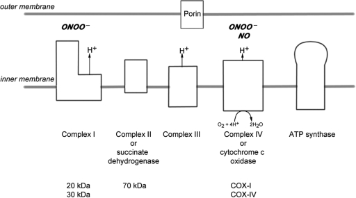
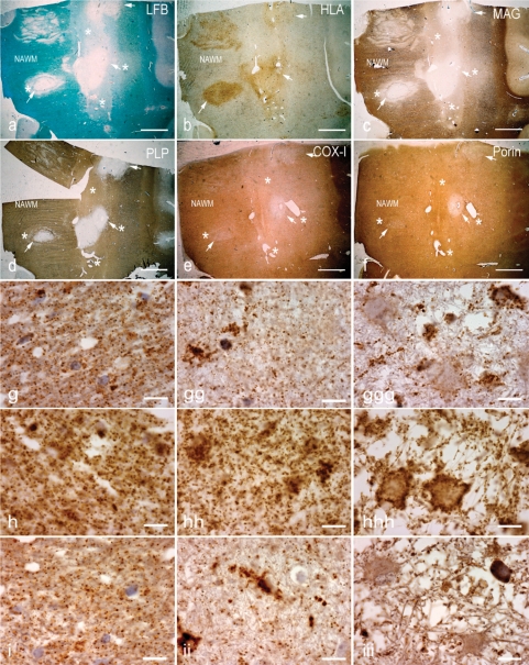
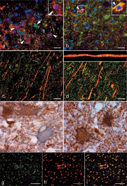
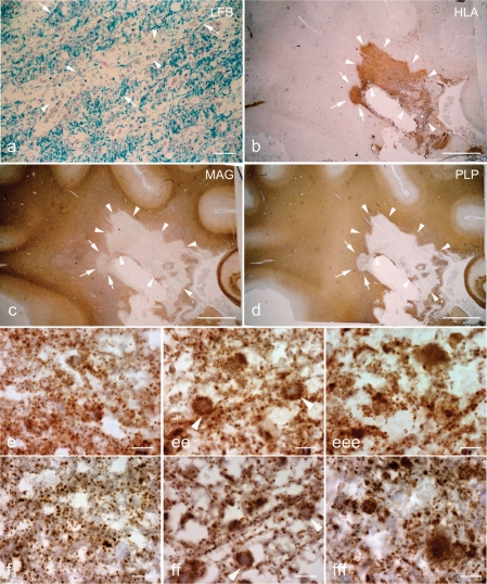
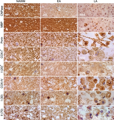
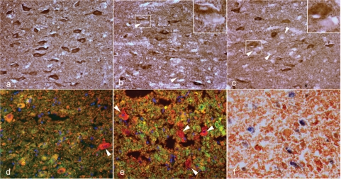
Multiple sclerosis is a chronic inflammatory disease, which leads to focal plaques of demyelination and tissue injury in the CNS. The structural and immunopathological patterns of demyelination suggest that different immune mechanisms may be involved in tissue damage. In a subtype of lesions, which are mainly found in patients with acute fulminant multiple sclerosis with Balo's type concentric sclerosis and in a subset of early relapsing remitting multiple sclerosis, the initial myelin changes closely resemble those seen in white matter stroke (WMS), suggesting a hypoxia-like tissue injury. Since mitochondrial injury may be involved in the pathogenesis of such lesions, we analysed a number of mitochondrial respiratory chain proteins in active lesions from acute multiple sclerosis and from WMS using immunohistochemistry. Functionally important defects of mitochondrial respiratory chain complex IV [cytochrome c oxidase (COX)] including its catalytic component (COX-I) are present in Pattern III but not in Pattern II multiple sclerosis lesions. The lack of immunohistochemically detected COX-I is apparent in oligodendrocytes, hypertrophied astrocytes and axons, but not in microglia. The profile of immunohistochemically detected mitochondrial respiratory chain complex subunits differs between multiple sclerosis and WMS. The findings suggest that hypoxia-like tissue injury in Pattern III multiple sclerosis lesions may be due to mitochondrial impairment.


Figures

Fig. 1
Fig. 1
The mitochondrial respiratory chain. The mitochondrial respiratory chain consists of four complexes (complexes I–IV) and complex V, which is directly involved in ATP synthesis. The complexes are made up of multiple subunits encoded by nuclear DNA and mtDNA, except complex II which is entirely encoded by nuclear DNA. We investigated two subunits of complex I (20 and 30 kDa), one of complex II (70 kDa) and two of complex IV (subunits I and IV of complex IV or COX-I and COX-IV, respectively) in brain tissue. Porin, a voltage gated anion channel located in the outer mitochondrial membrane, was used as a mitochondrial marker. The activity of succinate dehydrogenase (complex II) and COX (complex IV) can be determined at the single cell level using an enzyme histochemical assay (COX/succinate dehydrogenase histochemistry). NO inhibits complex IV activity by competing with oxygen for the oxygen binding sites, of which three out of the four are located in COX-I. Both complexes I and IV are susceptible to peroxynitrite (ONOO) mediated damage.
Fig. 2
Fig. 2
Pattern III multiple sclerosis lesions. (a–d) The brain sections containing Pattern III and Balo's type (with concentric rings of preserved myelin) active multiple sclerosis lesions are stained for LFB (a), HLA (b), MAG (c)and PLP (d). There is reduced density of LFB staining (a, asterisks) in the EA stage, whereas LFB staining is absent (a, arrows) in the LA stage of Pattern III lesions compared with NWM. The neuropathological hallmark of Pattern III lesions is the preferential loss of MAG, as indicated here by the loss of MAG immunoreactivity (c, asterisks) and intact PLP immunoreactivity (d, asterisks) in the EA stage. In contrast, MAG and PLP immunoreactivity is lost in the LA stage of Pattern III lesions (c and d, arrows). e and f: In deeper sections from the same block, there is a decrease in complex IV subunit-I (COX-I) immunoreactivity, which is diffuse in the EA stage (e, asterisks) and most prominent in the LA stage (e, arrows) compared with NWM. The porin immunoreactivity in the EA stage (f, asterisks) of Pattern III lesions is similar to the NWM. However, there is a decrease in porin immunoreactivity in the LA stage (f, arrows). Interestingly, COX-I immunoreactivity is preserved in the concentric rings of the Balo's type multiple sclerosis lesion (e). g–i: Higher magnification images show the punctate COX-I (g), porin (h) and COX-IV (i) immunoreactive elements, typical of mitochondrial staining, in the NWM (left column), EA stage (middle column) and LA stage (right column). There is a global reduction in the number of COX-I immunoreactive elements in the EA (gg) and LA (ggg) stages of Pattern III lesions compared with NWM (g). There are ramified cells consistent with microglia containing dense COX-I immunoreactivity in Pattern III lesions (gg–ggg). The number of porin immunoreactive elements appears reduced in LA (hhh), where there is tissue vacuolation, but not EA (hh) stage compared with NWM (h). The cells with enlarged cytoplasm (consistent with hypertrophied astrocytes) contain a peripheral rim of dense porin (hhh) but not COX-I (ggg) immunoreactivity, and clearly illustrate the disproportionate loss of COX-I compared with porin immunoreactivity in Pattern III lesions. The number of COX-IV immunoreactive elements is also decreased in Pattern III lesions (ii and iii) compared with NWM (i). Scale bars = 8 mm (a–f) and 10 µm (g–i).
Fig. 3
Fig. 3
The oligodendrocytes, axons and hypertrophied astrocytes but not microglia lack COX-I immunoreactivity in Pattern III multiple sclerosis lesions. a–f: The left and right columns of images show COX-I and porin immunoreactivity, respectively. a and b: The double immunofluorescent labelling of COX-I (a, green) and oligodendrocyte marker, CNPase (a, red) identifies oligodendrocytes lacking COX-I immunoreactivity (a, arrowheads and insert) in the EA stage of Pattern III lesions. The cells with COX-I immunoreactivity in the EA stage lack CNPase immunoreactivity (a, arrow). In serial sections, CNPase (red) immunoreactive oligodendrocytes (b, arrowhead and insert) as well as numerous other cells in EA stage contain porin (green) immunoreactive elements. c and d: The double immunofluorescent labelling of COX-I (c, green) and axonal markers [phosphorylated (SMI31) and non-phosphorylated (SMI32) neurofilaments in red] identifies axons with only a small number of COX-I immunoreactive elements (c) in the LA stage of Pattern III lesions. In serial sections, the porin elements (d, green) are more numerous than COX-I elements (c, green) within demyelinated axons in the LA stage of Pattern III lesions. A confocal x–z image (d, insert) confirms the axonal location of the porin immunoreactive elements in the x–y images (d). e and f: The mitochondrial proteins [COX-I (e) and porin (f)] are co-stained with a astrocyte marker, glial fibrillarry acidic protein, using chromogens [DAB (brown) and Vector SG (grey), respectively]. The COX-I immunoreactive elements are absent in a proportion of hypertrophied astrocytes (e) in Pattern III lesions. The hypertrophied astrocytes in serial sections contain abundant porin immunoreactivity (f). g–i: In the LA stage of Pattern III lesions, the activated microglia distributed around the blood vessels and identified by the CD163 (h, red) immunoreactivity contain COX-I immunoreactivity (g and i, green). a–d: Confocal images. Scale bars = 10 µm (a–f) and 50 µm (g–i).
Fig. 4
Fig. 4
Pattern II multiple sclerosis lesions. ad: Sections containing a Pattern II multiple sclerosis lesion are stained for LFB (a), HLA (b), MAG (c) and PLP (d). The EA stage of Pattern II lesions is identified by the perivenous demyelination, which are separated by partly preserved myelin (a, arrows), and abundance of phagocytic macrophages (b) containing myelin debris (a, arrowheads). Both MAG (c) and PLP (d) immunoreactivity are equally lost in the EA and LA stages (c and d, arrows and arrowheads, respectively) of Pattern II lesions. e and f: At higher magnification the Pattern II tissue appears vacuolated with a general reduction in the number of porin immunoreactive elements in EA (ee) and LA (eee) stages compared with NWM (e). f: The reduction in COX-I immunoreactive elements in EA (ff) and LA (fff) stage of Pattern II lesions is relative to the reduction of porin (e) elements. Scale bars = 40 µm (a), 8 mm (b–d) and 10 µm (e and f).
Fig. 5
Fig. 5
WMS lesions. The three columns show immunoreactivity in NWM (left), EA (middle) and LA (right) stages of WMS tissue. a and b: Both CNPase and MBP immunoreactivity are detectable in the NWM, whereas CNPase immunoreactivity is lost while MBP immunoreactivity is intact in the EA stage of WMS lesions. In the LA stage, both CNPase and MBP immunoreactivity are lost except within phagocytic macrophages. c: The porin immunoreactive punctate elements are numerous in the NWM and EA stage. In the LA stage of WMS lesions, there is severe tissue loss and mitochondria are mostly located in phagocytic macrophages. d–h: The number of immunoreactive elements for all mitochondrial respiratory chain complex subunits (COX-I, complex I 20 kDa or ND6, complex I 30 kDa and complex II 70 kDa) except COX-IV appears decreased in EA stage compared with NWM. Furthermore, the majority of phagocytic macrophages in LA stage show a decrease in immunoreactivity for the mitochondrial respiratory chain complex subunits (except for COX-IV) despite the abundance of porin immunoreactive elements (ccc). Asterisk indicates subunits encoded by mtDNA. Scale bars = 10 µm (a–h).
Fig. 6
Fig. 6
The loss of mtDNA encoded respiratory chain subunit immunoreactivity in fixed brain tissue and implications for complex IV activity. (a–e) immunoreactivity in serial sections of fixed hippocampus. (f) activity of complexes II and IV in the snap frozen contralateral hippocampus. There are a number of cells containing complex II 70 kDa, a nuclear encoded subunit, immunoreactivity (a) in the CA2 layer of a fixed hippocampal section from a case with multiple deletions of mtDNA. In contrast, immunoreactivity for mtDNA encoded subunits [complex I 20 kDa or complex IV subunit I (COX-I)] is lacking in a proportion of cells, in serial sections (b and c, arrowheads and inserts). The cells lacking complex I 20 kDa or COX-I immunoreactivity (green) contain mitochondria (red) as shown by double immunofluorecent labelling with porin (d and e, arrowheads). The functional implications of the lack of COX-I immunoreactivity in fixed brain tissue can be clearly seen in a snap frozen section from the contralateral hippocampus where complex IV activity is lacking in a proportion of cells with complex II activity (f, blue).

Similar articles

Cited by

References

    1. Aboul-Enein F, Rauschka H, Kornek B, Stadelmann C, Stefferl A, Bruck W, et al. Preferential loss of myelin-associated glycoprotein reflects hypoxia-like white matter damage in stroke and inflammatory brain diseases. J Neuropathol Exp Neurol. 2003;62:25–33. - PubMed
    1. Almeida A, Almeida J, Bolanos JP, Moncada S. Different responses of astrocytes and neurons to nitric oxide: the role of glycolytically generated ATP in astrocyte protection. Proc Natl Acad Sci USA. 2001;98:15294–9. - PMC - PubMed
    1. Almeida A, Moncada S, Bolanos JP. Nitric oxide switches on glycolysis through the AMP protein kinase and 6-phosphofructo-2-kinase pathway. Nat Cell Biol. 2004;6:45–51. - PubMed
    1. Barnett MH, Prineas JW. Relapsing and remitting multiple sclerosis: pathology of the newly forming lesion. Ann Neurol. 2004;55:458–68. - PubMed
    1. Bolanos JP, Almeida A, Stewart V, Peuchen S, Land JM, Clark JB, et al. Nitric oxide-mediated mitochondrial damage in the brain: mechanisms and implications for neurodegenerative diseases. J Neurochem. 1997;68:2227–40. - PubMed

Publication types

MeSH terms

Substances