. 2008 May 28;28(22):5641-53.
doi: 10.1523/JNEUROSCI.1056-08.2008.

An essential role for Frizzled5 in neuronal survival in the parafascicular nucleus of the thalamus

Affiliations

An essential role for Frizzled5 in neuronal survival in the parafascicular nucleus of the thalamus

Chunqiao Liu et al. J Neurosci. .

Abstract

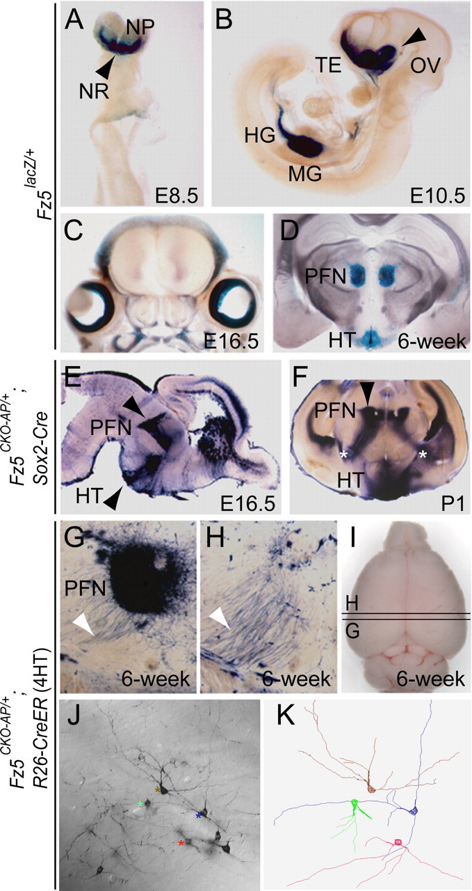
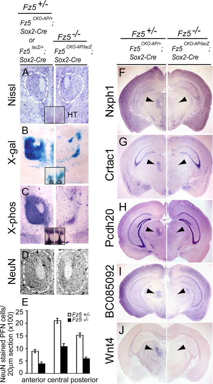
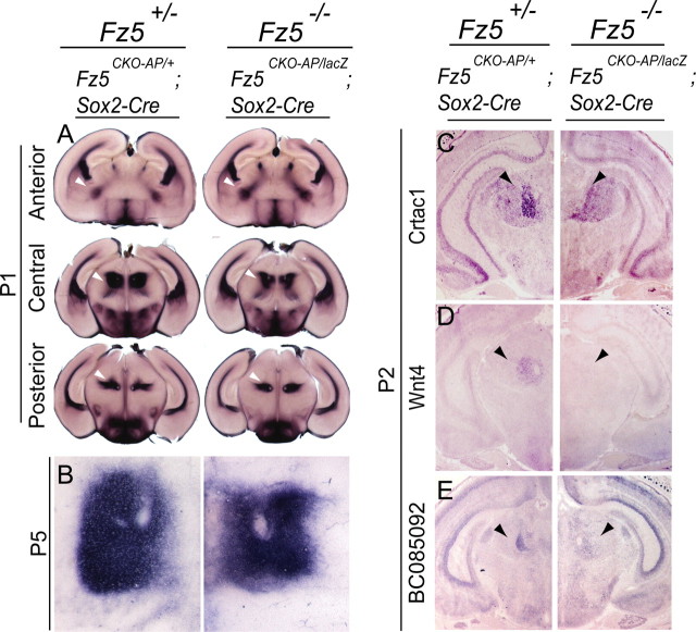
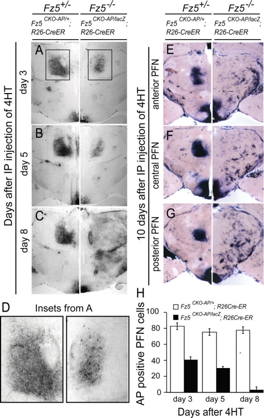
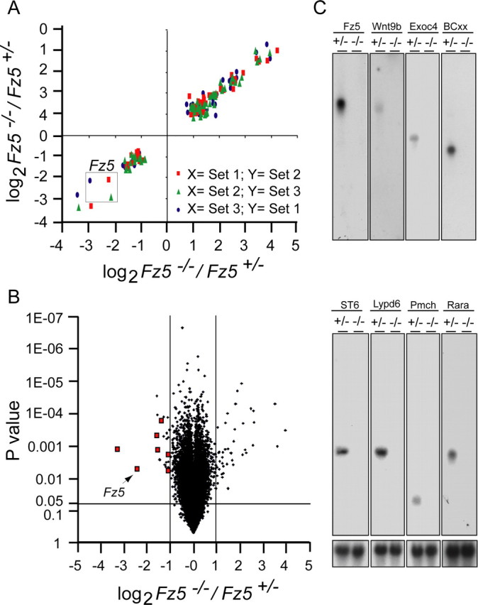
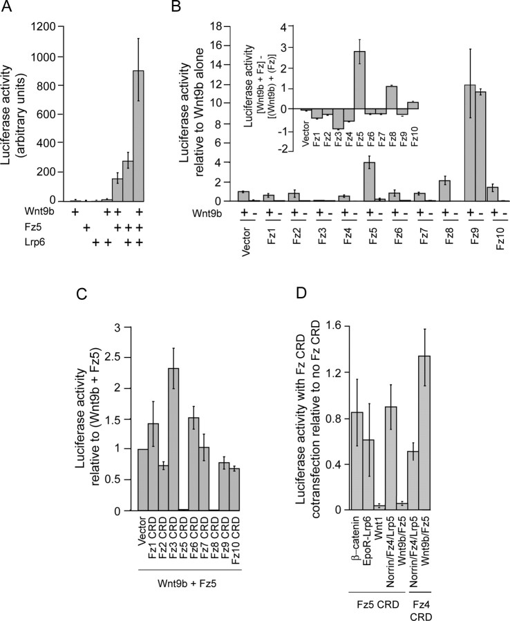
Frizzled5 (Fz5), a putative Wnt receptor, is expressed in the retina, hypothalamus, and the parafascicular nucleus (PFN) of the thalamus. By constructing Fz5 alleles in which beta-galactosidase replaces Fz5 or in which Cre-mediated recombination replaces Fz5 with alkaline phosphatase, we observe that Fz5 is required continuously and in a cell autonomous manner for the survival of adult PFN neurons, but is not required for proliferation, migration, or axonal growth and targeting of developing PFN neurons. A motor phenotype associated with loss of Fz5 establishes a role for the PFN in sensorimotor coordination. Transcripts coding for Wnt9b, the likely Fz5 ligand in vivo, and beta-catenin, a mediator of canonical Wnt signaling, are both downregulated in the Fz5(-/-) PFN, implying a positive feedback mechanism in which Wnt signaling is required to maintain the expression of Wnt signaling components. These data suggest that defects in Wnt-Frizzled signaling could be the cause of neuronal loss in degenerative CNS diseases.


Figures

Figure 1.
Figure 1.
Targeted mutations at the Fz5 locus. A, The WT Fz5 locus consists of a 5′ noncoding exon and a 3′ exon containing the complete Fz5 coding region. A–E, A schematic of the spliced mRNA is shown above the WT or targeted Fz5 gene. 5′ and 3′ Southern blot hybridization probes, derived from regions beyond the edges of the targeting constructs, are indicated. B, Replacement of Fz5 coding sequences with the Escherichia coli lacZ coding region followed by a PGK-Neo selectable marker. C, Fz5 conditional knock-out allele with the following elements (from 5′ to 3′): a Hind III-tagged loxP site in the second exon 5′ of the initiator methionine codon, a second loxP site 3′ of the transcription unit, an AP open reading frame, and a PGK-Neo cassette flanked by frt sites. D, The Fz5 conditional knock-out allele after excision of the PGK-Neo cassette by Flp recombinase. E, Starting with the Fz5 conditional knock-out allele, site-specific excisions by both Flp and Cre results in the replacement of the Fz5 coding region with the AP coding region. F, G, Whole-genome Southern blot hybridization confirms the WT and targeted Fz5 gene structures shown in A–E. The locations of the Hind III and BglII restriction fragments visualized by whole-genome Southern blotting with the 5′ and 3′ probes, respectively, are shown by horizontal lines above A–E.
Figure 2.
Figure 2.
Highly restricted expression of the Fz5 gene during development and in adulthood. A, B, X-gal-stained whole mounts of Fz5 lacZ/+ embryos show Fz5 expression in the neural ridge (NR) at the anterior of the neural plate (NP) at E8.5 (A) and in the telencephalon (TE), optic vesicle (OV), hindgut (HG), and midgut (MG) at E10.5 (B). C, D, X-gal-stained coronal vibratome sections of Fz5 lacZ/+ heads show Fz5 expression in the retinas at E16.5 (C) and in the PFN and hypothalamus (HT) at 6 weeks of age (D). E, F, NBT/BCIP staining of vibratome sections of Fz5 CKO-AP/+;Sox2-Cre (Fz5 +/−) brains. A sagittal section at E16.5 (E) and a coronal section at P1 (F) show Fz5 expression in the PFN and hypothalamus. A fiber tract, marked by an asterisk in F, originates in each PFN and courses inferiorly, laterally, and anteriorly to the ipsilateral striatum. G–I, Coronal sections through the PFN (G) and just anterior to the PFN (H) from a 6-week-old Fz5 CKO-AP/+;R26-creER mouse injected intraperitoneally 14 d earlier with 40 μg/g 4HT and stained with NBT/BCIP. The locations of the two sections are shown in I. White arrowheads, AP-labeled axons coursing from the PFN to the striatum. J, K, Morphologies of Fz5AP-expressing PFN neurons after sparse Cre-mediated recombination in an NBT/BCIP-stained 200 μm vibratome section from an Fz5 CKO-AP/+;R26-creER mouse injected at 6 weeks of age with 40 μg/g 4HT. J, DIC image at a single focal plane with the cell bodies of four neurons indicated by colored asterisks. K, Neurolucida tracing of these four neurons from a stack of 200 1 μm Z-series sections.
Figure 3.
Figure 3.
Progressive loss of neurons in the postnatal Fz5 −/− PFN. A–E, Nissl, X-gal, NBT/BCIP, and anti-NeuN staining of coronal sections of ∼8-week-old Fz5 CKO-AP/+;Sox2-Cre or Fz5 lacZ/+;Sox2-Cre (Fz5 +/−) and Fz5CKO-AP/lacZ;Sox2-Cre (Fz5 −/−) brains showing the PFN region of the thalamus. A–C, Insets, The hypothalamic region from the same sections, which has equivalent cell densities and levels of Fz5 expression in Fz5 +/− and Fz5 −/− brains. A, D, The PFN is outlined by white dashes. A, C, D, Age-matched Fz5 CKO-AP/+;Sox2-Cre (Fz5 +/−) and Fz5CKO-AP/lacZ;Sox2-Cre (Fz5 −/−) mice. B, Age-matched Fz5 lacZ/+;Sox2-cre (Fz5 +/−) and Fz5CKO-AP/lacZ;Sox2-Cre (Fz5 −/−) mice. E, Mean and SD of the number of NeuN-expressing cells per 20 μm coronal section in the anterior, central, and posterior regions of the PFN, as delimited in D (3 sections/sample). F–J, In situ hybridization with the indicated probes to coronal sections through the PFN region of the thalamus from age-matched adult Fz5 CKO-AP/+;Sox2-Cre (Fz5 +/−) and Fz5CKO-AP/lacZ;Sox2-Cre (Fz5 −/−) brains. Each probe hybridizes to the Fz5 +/− but not the Fz5 −/− PFN (arrowheads). The abbreviated probe names and National Center for Biotechnology Information accession numbers are Nxph1 (BC079608), Crtac1 (BC024472), Pcdh20 (BC024927), and BC085092, unknown protein (BC085092).
Figure 4.
Figure 4.
PFN neurons develop normally in Fz5 −/− mice, but multiple genes are downregulated in the early postnatal PFN. A, NBT/BCIP staining of coronal vibratome sections from P1 Fz5 CKO-AP/+;Sox2-Cre (Fz5 +/−) and Fz5CKO-AP/lacZ;Sox2-Cre (Fz5 −/−) brains showing the PFN and its projection to the striatum (arrowheads). B, NBT/BCIP staining of the PFN at P5 in Fz5 CKO-AP/+;Sox2-Cre (Fz5 +/−) and Fz5CKO-AP/lacZ;Sox2-Cre (Fz5 −/−) brains. C–E, In situ hybridization to coronal sections through the PFN (arrowheads) from P2 Fz5 CKO-AP/+;Sox2-Cre (Fz5 +/−) and Fz5CKO-AP/lacZ;Sox2-Cre (Fz5 −/−) brains using probes derived from Crtac1, Wnt4, and BC085092 cDNAs.
Figure 5.
Figure 5.
Rapid loss of adult PFN neurons after timed deletion of the Fz5 coding region. A–D, A single intraperitoneal injection of 40 μg/g 4HT in adult Fz5 CKO-AP/+;R26-creER mice induces Cre-mediated recombination, which generates a large number of AP-expressing Fz5 +/− PFN neurons with little or no loss of cell viability. The same 4HT treatment in Fz5CKO-AP/lacZ;R26-creER mice is followed by a rapid loss of AP-expressing Fz5 −/− neurons. The boxed regions in A, which are enlarged in D, show the loss of AP-expressing neurons within 3 d of 4HT injection. E–G, Ten days after a single intraperitoneal injection of 40 μg/g 4HT in adulthood, AP-expressing Fz5 −/− PFN neurons are eliminated throughout the anterior–posterior extent of the Fz5CKO-AP/lacZ;R26-creER PFN, but AP-expressing Fz5 +/− neurons persist throughout the Fz5 CKO-AP/+;R26-creER PFN. As seen at the bottom of A–C and E–G, 4HT exposure in adulthood leads to equivalently efficient induction of AP-expressing cells in the hypothalamus in Fz5 CKO-AP/+;R26-creER and Fz5CKO-AP/lacZ;R26-creER mice, with no apparent effect on the survival of Fz5 +/− or Fz5 −/− hypothalamic cells. H, Mean ± SD of the number of AP-expressing cell bodies in three 20 μm sections of the PFN after 4HT injection; data were obtained from the experiment shown in A–C.
Figure 6.
Figure 6.
Weight gain and motor coordination deficits in Fz5 −/− mice. A, Fz5CKO-AP/lacZ;Sox2-Cre (Fz5 −/−) mice weigh, on average, ∼15–20% less than their Fz5 CKO-AP/+;Sox2-Cre (Fz5 +/−) littermates (n > 5). B, Fz5CKO-AP/lacZ;Sox2-Cre (Fz5 −/−) and Fz5 CKO-AP/+;Sox2-Cre (Fz5 +/−) mice show similar levels of free wheel running per 24 h. Each symbol represents one mouse. C, Fz5CKO-AP/lacZ;Sox2-Cre (Fz5 −/−) mice have impaired performance in a rotorod test. Results are shown for three different rotation speeds. Each mouse was tested three times for each rotation speed. The average time to falling from the rotorod is shown.
Figure 7.
Figure 7.
Microarray hybridization of adult Fz5 +/− versus Fz5 −/− PFN. A, PFN RNA was prepared from three independent dissections of unfixed vibratome-sectioned brains from 6- to 8-week-old Fz5 CKO-AP/+;Sox2-Cre (Fz5 +/−) and Fz5CKO-AP/lacZ;Sox2-Cre (Fz5 −/−) mice. For each RNA sample, tissue was pooled from three or more brains and hybridized to Affymetrix 430 2.0 chips, representing 39,000 mouse transcripts. The figure shows pairwise comparisons among the three independent experiments for those transcripts that showed a more than twofold change in Fz5 −/−:Fz5 +/− abundance ratios. For genes with more than one entry in the Affymetrix spreadsheet, the scatter plot shows only the entry with the greatest change in transcript abundance. B, The data from the three independent microarray hybridization experiments are shown as a scatter plot with the average Fz5 −/−:Fz5 +/− transcript abundance ratio on the horizontal axis and the p value for each ratio on the vertical axis. For genes with more than one entry in the Affymetrix spreadsheet and transcripts abundance changes of greater than twofold, the scatter plot shows only the entry with the greatest change in transcript abundance. Red squares, Probes used for RNA blot hybridization in C. C, Blot hybridization to PFN RNA dissected from unfixed vibratome-sectioned age-matched adult brains. Probes are from transcripts that were found to be downregulated in the Fz5CKO-AP/lacZ;Sox2-Cre (Fz5 −/−) PFN relative to the Fz5 lacZ/+;Sox2-cre (Fz5 +/−) PFN as determined by in situ hybridization (Figs. 3, 4) or microarray hybridization. Top, The same four blots that were used in the bottom images, stripped and reprobed. Bottom, The GAPDH transcript serves as a control. The abbreviated probe names and National Center for Biotechnology Information accession numbers are as follows: BCxx, a gene of unknown function (BC126943); Exoc4, exocyst component 4 (BC034644); Lypd6, LY6/PLAUR domain protein (BC070462); Pmch, premelanin concentrating hormone (BC048543); Rara, retinoic acid receptor α (BC010216); ST6, (α-N-acetyl-neuraminyl-2,3-β-galactosyl-1,3)-N-acetylgalactosaminide α-2,6-sialyltransferase (BC058387).
Figure 8.
Figure 8.
Downregulation of Wnt9b and β-catenin transcripts in the Fz5 −/− PFN. A–F, In situ hybridization to coronal sections through the PFN from P2 (left) and 8-week-old (right) Fz5 CKO-AP/+;Sox2-Cre (Fz5 +/−) and Fz5CKO-AP/lacZ;Sox2-Cre (Fz5 −/−) brains, using probes derived from Fz5, Wnt9b, and β-catenin cDNAs. Each probe hybridizes to the PFN in the Fz5 +/−, but not in the Fz5 −/− brain (arrowheads).
Figure 9.
Figure 9.
Evidence that Wnt9b and Fz5 can synergistically activate the canonical Wnt signaling pathway. A, Luciferase activity caused by canonical Wnt signaling after transient cotransfection of the 293/STF luciferase reporter cell line with the indicated plasmids. B, Relative specificity of Wnt9b for Fz5 compared with other Frizzleds in cotransfected 293/STF cells. Note that Fz9 has strong constitutive activity in the absence of Wnt9b. Inset, The excess luciferase activity attributable to Wnt9b-Fz interactions calculated by subtracting the sum of the luciferase activity of cells individually transfected with Wnt9b and with the indicated Frizzled from the luciferase activity of cells cotransfected with Wnt9b and the same Frizzled. C, Inhibition of Wnt9b-Fz5 signaling in 293/STF cells by coexpression of plasma-membrane localized, myc-tagged, and GPI-anchored fusion proteins with the CRD from each of the 10 mouse Frizzleds. The Fz5 and Fz8 CRDs produce nearly complete inhibition of canonical Wnt signaling; the remaining eight Frizzled CRDs produce little or no inhibition. D, Specificity of inhibition of Frizzled signaling by Fz5 CRD myc-GPI. The histogram shows the ratio of 293/STF luciferase activity in the presence of Fz5 CRD-myc-GPI or Fz4 CRD-myc-GPI relative to the 293/STF luciferase activity in the absence of a CRD-myc-GPI inhibitor. All histograms show the mean ± SD of triplicate transfections.

Similar articles

Cited by

References

    1. Aymerich MS, Barroso-Chinea P, Perez-Manso M, Munoz-Patino AM, Moreno-Igoa M, Gonzalez-Hernandez T, Lanciego JL. Consequences of unilateral nigro-striatal denervation on the thalamostriatal pathway in rats. Eur J Neurosci. 2006;23:2099–2108. - PubMed
    1. Backman CM, Zhang Y, Hoffer BJ, Tomac AC. Tetracycline-inducible expression systems for the generation of transgenic animals: a comparison of various inducible systems carried in a single vector. J Neurosci Methods. 2004;139:257–262. - PubMed
    1. Badea TC, Wang Y, Nathans J. A noninvasive genetic/pharmacologic strategy for visualizing cell morphology and clonal relationships in the mouse. J Neurosci. 2003;23:2314–2322. - PMC - PubMed
    1. Carmichael J, Sugars KL, Bao YP, Rubinsztein DC. Glycogen synthase kinase-3β inhibitors prevent cellular polyglutamine toxicity caused by the Huntington's disease mutation. J Biol Chem. 2002;277:33791–33798. - PubMed
    1. Carr KD, Bak TH. Medial thalamic injection of opioid agonists: mu-agonist increases while kappa-agonist decreases stimulus thresholds for pain and reward. Brain Res. 1988;441:173–184. - PubMed

MeSH terms