. 2008 Aug;28(15):4896-914.
doi: 10.1128/MCB.01775-07. Epub 2008 May 27.

Pharmacoproteomics of a metalloproteinase hydroxamate inhibitor in breast cancer cells: dynamics of membrane type 1 matrix metalloproteinase-mediated membrane protein shedding

Affiliations

Pharmacoproteomics of a metalloproteinase hydroxamate inhibitor in breast cancer cells: dynamics of membrane type 1 matrix metalloproteinase-mediated membrane protein shedding

Georgina S Butler et al. Mol Cell Biol. 2008 Aug.

Abstract

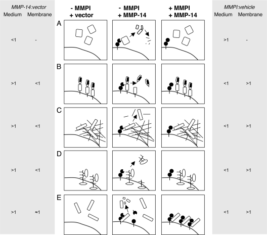
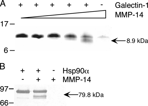
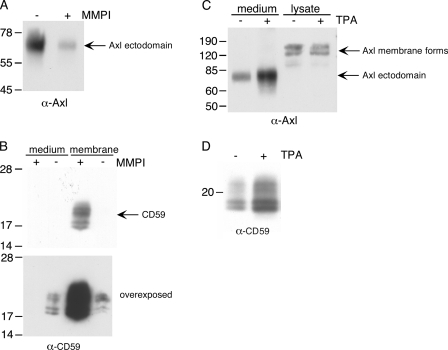
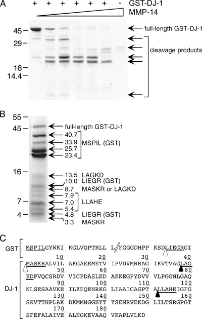
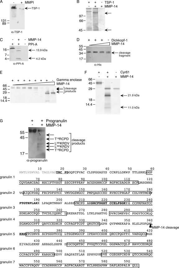
Broad-spectrum matrix metalloproteinase (MMP) inhibitors (MMPI) were unsuccessful in cancer clinical trials, partly due to side effects resulting from limited knowledge of the full repertoire of MMP substrates, termed the substrate degradome, and hence the in vivo functions of MMPs. To gain further insight into the degradome of MMP-14 (membrane type 1 MMP) an MMPI, prinomastat (drug code AG3340), was used to reduce proteolytic processing and ectodomain shedding in human MDA-MB-231 breast cancer cells transfected with MMP-14. We report a quantitative proteomic evaluation of the targets and effects of the inhibitor in this cell-based system. Proteins in cell-conditioned medium (the secretome) and membrane fractions with levels that were modulated by the MMPI were identified by isotope-coded affinity tag (ICAT) labeling and tandem mass spectrometry. Comparisons of the expression of MMP-14 with that of a vector control resulted in increased MMP-14/vector ICAT ratios for many proteins in conditioned medium, indicating MMP-14-mediated ectodomain shedding. Following MMPI treatment, the MMPI/vehicle ICAT ratio was reversed, suggesting that MMP-14-mediated shedding of these proteins was blocked by the inhibitor. The reduction in shedding or the release of substrates from pericellular sites in the presence of the MMPI was frequently accompanied by the accumulation of the protein in the plasma membrane, as indicated by high MMPI/vehicle ICAT ratios. Considered together, this is a strong predictor of biologically relevant substrates cleaved in the cellular context that led to the identification of many undescribed MMP-14 substrates, 20 of which we validated biochemically, including DJ-1, galectin-1, Hsp90alpha, pentraxin 3, progranulin, Cyr61, peptidyl-prolyl cis-trans isomerase A, and dickkopf-1. Other proteins with altered levels, such as Kunitz-type protease inhibitor 1 and beta-2-microglobulin, were not substrates in biochemical assays, suggesting an indirect affect of the MMPI, which might be important in drug development as biomarkers or, in preclinical phases, to predict systemic drug actions and adverse side effects. Hence, this approach describes the dynamic pattern of cell membrane ectodomain shedding and its perturbation upon metalloproteinase drug treatment.


Figures

FIG. 1.
FIG. 1.
Hypothesis: the MMPI attenuates shedding and release of cleaved proteins into the conditioned medium. Without MMP-14 (left panels, −MMPI + vector), no MMP-14-mediated processing occurs. With MMP-14 but in the absence of the MMPI (center panels, −MMPI +MMP-14), active MMP-14 on the cell membrane (A) processes secreted proteins, which may result in further cleavages and clearance by MMPs or other proteases; (B) sheds membrane-associated or integral membrane proteins or their binding partners from the cell surface; (C) processes or releases proteins from extracellular and pericellular matrix; or (D) sheds directly or indirectly mobilizes secreted proteins from cell binding sites, e.g., by processing proteoglycans or integrins. These events will be blocked by a broad-spectrum MMPI (right panels, +MMPI +MMP-14). In the presence of an MMPI, soluble substrates increase in the conditioned medium (A). Whether the ratio changes or not will depend upon the rate of clearance of any fragments which will still be quantified as labeled tryptic peptides. Previously shed cell- or matrix-associated proteins decrease in the conditioned medium (B, C, and D), which coincides with their increase in the membrane or matrix. A similar response might be caused by MMPI-induced dominant-negative effects (E). Autodegradation of MMP-14 (center panel) is prevented by the MMPI, leading to an accumulation of mature MMP-14 at the cell surface (right panel). These inhibited MMP-14 molecules could act as “substrate traps,” binding substrates (and other interacting molecules) at exosites without cleavage and release. Hence, shed and soluble proteins would be titrated from the conditioned medium and sequestered at the cell surface. The predicted ICAT ratios for cells transfected with MMP-14 compared with empty vector (MMP-14/vector) and cells transfected with MMP-14 treated with inhibitor drug or vehicle (MMPI/vehicle) are shown adjacent to each panel for proteins in the conditioned medium (Medium) or cell membrane fractions (Membrane).
FIG. 2.
FIG. 2.
Confirmation of MMP-14 processing of galectin-1 and Hsp90α in vitro. (A) MMP-14 cleavage of galectin-1 with increasing enzyme/substrate molar ratio (1:500 to 1:10) was analyzed on 15% Tris-Tricine SDS-PAGE. (B) Analysis of MMP-14 proteolysis of Hsp90α on 10% SDS-PAGE. Arrows indicate cleaved protein fragments, and apparent molecular masses are shown. Positions of molecular mass markers (103 Da) are indicated.
FIG. 3.
FIG. 3.
Biochemical validation of novel MMP-14 substrates. (A) Conditioned medium proteins (20 μg) from MDA-MB-231 cells transfected with MMP-14 or empty vector (left panel, MMP-14/vector) and MMP-14-transfected cells treated with prinomastat (10 μM) or vehicle (right panel, MMPI/vehicle) were subjected to ICAT multidimensional LC-MS-MS analysis (ratios shown) or were separated by 11% SDS-PAGE. CRIM-1 was detected by Western blotting using an anti-CRIM-1 goat polyclonal antibody raised against the extracellular domain. (B) MMP-14 cleavage of recombinant follistatin-related protein 3 with a C-terminal His tag incubated with increasing enzyme/substrate molar ratios (1:100, 1:50, 1:10, and 1:5) was analyzed with 12.5% Tris-Tricine SDS-PAGE and silver stained or Western blotted (1:100, 1:50, and 1:10) with an anti-polyhistidine antibody. Comparison of these two analyses reveals near comigration of MMP-14 autolytic degradation products just above the 30.9-kDa band in the silver-stained gel. (C) Recombinant pentraxin 3 (Ptx3) with a C-terminal His tag was electrophoresed on 12.5% Tris-Tricine SDS-PAGE and subjected to Western blotting with an anti-polyhistidine antibody. (D) Recombinant human Niemann-Pick type C2 (NPC2) incubated with or without MMP-14 was electrophoresed on 12.5% Tris-Tricine SDS-polyacrylamide gels and silver stained. (E) Iduronate-2-sulfatase (IDS) samples, after incubation with MMP-14, were electrophoresed on 15% Tris-Tricine SDS-polyacrylamide gels and silver stained. All samples were incubated for 18 h at 37°C. Western blotting was carried out using appropriate Alexa-Fluor 680-conjugated immunoglobulin G secondary antibodies (Molecular Probes) and detected with an Odyssey infrared scanner (LiCor). MMP-14 did not cross-react with these antibodies. Arrows indicate cleaved protein fragments, and apparent molecular masses are shown. Positions of molecular mass markers as 103 Da are indicated.
FIG. 4.
FIG. 4.
Regulation of ectodomain shedding of Axl receptor tyrosine kinase and CD59 by MMPI and 12-O-tetradecanoylphorbol-13-acetate (TPA). Conditioned medium or membrane fractions (5 μg total protein) from MMP-14-transfected MDA-MB-231 cells treated with 10 μM prinomastat (+MMPI) or vehicle (−MMPI) were analyzed by SDS-PAGE and Western blotting. (A) Axl receptor tyrosine kinase (Axl) in conditioned medium was detected on 10% polyacrylamide gels, using an antibody specific for the Axl receptor tyrosine kinase ectodomain. (B) CD59 was detected on 13% polyacrylamide gels, using the monoclonal antibody BRIC 229. Panels show results from a single Western blot, but the lower panel is overexposed to show CD59 in the medium sample. Blots were developed with appropriate horseradish peroxidase-conjugated secondary antibodies and enhanced chemiluminescence. Positions of molecular mass markers as 103 Da are shown. Conditioned medium or cell lysates (5 μg total protein) from MDA-MB-231 cells (transfected with empty vector) treated with 0.2 ng/ml TPA (+) or untreated (−) under serum-free conditions for 72 h were subjected to reducing SDS-PAGE and Western blotting. Axl receptor tyrosine kinase (C) and CD59 (D) were detected as described above.
FIG. 5.
FIG. 5.
DJ-1 is a substrate of MMP-14. (A) GST-DJ-1 (435 pmol) was incubated for 18 h at 37°C with increasing concentrations of MMP-14 (GST-DJ-1/MMP-14 molar ratios of 1:0, 1,000:1, 500:1, 250:1, 100:1, and 50:1), or MMP-14 was incubated alone (in amounts equivalent to a 100:1 ratio). (B) GST-DJ-1, digested with 500:1 MMP-14, was blotted onto polyvinylidene difluoride membrane. Arrows indicate bands subjected to Edman sequencing. Apparent molecular weights and sequences obtained for the first five residues are shown. Samples were electrophoresed on 12.5% (A) and 15% Tris-Tricine (B) SDS-polyacrylamide gels and stained with Coomassie brilliant blue R250. Molecular mass markers as 103 Da are shown. (C) Schematic diagram of GST-DJ-1. The start and end of the pGEX-5X-1 vector fusion protein sequence are shown, followed by the entire DJ-1 sequence. N-terminal sequences identified by Edman degradation are underlined. Cleavage sites within DJ-1 are indicated by arrowheads and those in the GST fusion protein by open arrowheads.
FIG. 6.
FIG. 6.
Validation of candidate MMP substrates identified in conditioned medium. (A) Conditioned medium (10 μg total protein) from MMP-14-transfected MDA-MB-231 cells treated with vehicle (−MMPI) or 10 μM prinomastat (+MMPI) (nonreduced) was separated by 12% SDS-PAGE, and TSP-1 (TSP-1) was detected by Western blotting and enhanced chemiluminescence using the mouse monoclonal antibody MAII, which recognizes the heparin binding domain of TSP-1. (B) Recombinant TSP-1 was incubated with or without MMP-14, and cleavage products were analyzed by SDS-PAGE on 9% gels by silver staining. Fragments are indicated by arrows. (C) Samples of peptidyl-prolyl cis-trans isomerase A (PPI-A) incubated with or without MMP-14 were analyzed on 15% Tris-Tricine SDS-polyacrylamide gels, Western blotted with a rabbit anti-PPI-A polyclonal antibody. (D) Recombinant dickkopf-1 with a C-terminal His tag incubated with increasing concentrations of MMP-14 (1:50, 1:10, and 1:5 molar ratio enzyme/substrate) was analyzed on 12.5% Tris-Tricine SDS-polyacrylamide gels. Western blotting was carried out with an anti-polyhistidine antibody. (E) Gamma enolase was incubated with increasing concentrations of MMP-14 (1:1,000, 1:500, 1:250, 1:100, 1:50, and 1:10 enzyme/substrate molar ratio), or MMP-14 was incubated alone (equivalent to a 1:10 ratio). Products were analyzed on 12.5% Tris-Tricine SDS-polyacrylamide gels by silver staining. (F) Cyr61 cleavage fragments produced upon incubation with MMP-14 were visualized on 15% Tris-Tricine SDS-polyacrylamide gels by silver staining. (G) Progranulin processing by MMP-14 was visualized on 12.5% Tris-Tricine SDS-polyacrylamide gels, Western blotted with a rabbit polyclonal antibody raised against progranulin. Fragments are shown by arrows, with N-terminal sequences obtained by Edman degradation. The sequence of progranulin is shown in the bottom panel: residues 1 to 17 constitute the signal sequence which is removed; residue T18 is the mature N terminus of progranulin; constituent granulins are boxed; N-terminal sequences of fragments are underlined; peptides identified by ICAT are in bold; the MMP-14 cleavage site is indicated by an arrow. All recombinant proteins were incubated for 18 h at 37°C with MMP-14. Western blots were detected using species-appropriate Alexa-Fluor 680-conjugated secondary antibodies (Molecular Probes) on an Odyssey infrared scanner (LiCor), unless otherwise stated. Arrows indicate cleaved protein fragments, and apparent molecular weights and masses are shown. Positions of molecular mass markers as 103 Da are indicated.

Similar articles

Cited by

References

    1. Abdul, M., and N. Hoosein. 2000. Changes in beta-2 microglobulin expression in prostate cancer. Urol. Oncol. 5168-172. - PubMed
    1. Allain, F., C. Vanpouille, M. Carpentier, M. C. Slomianny, S. Durieux, and G. Spik. 2002. Interaction with glycosaminoglycans is required for cyclophilin B to trigger integrin-mediated adhesion of peripheral blood T lymphocytes to extracellular matrix. Proc. Natl. Acad. Sci. USA 992714-2719. - PMC - PubMed
    1. Allegrini, G., F. A. Goulette, J. W. Darnowski, and P. Calabresi. 2004. Thrombospondin-1 plus irinotecan: a novel antiangiogenic-chemotherapeutic combination that inhibits the growth of advanced human colon tumor xenografts in mice. Cancer Chemother. Pharmacol. 53261-266. - PubMed
    1. Andolfo, A., W. R. English, M. Resnati, G. Murphy, F. Blasi, and N. Sidenius. 2002. Metalloproteases cleave the urokinase-type plasminogen activator receptor in the D1-D2 linker region and expose epitopes not present in the intact soluble receptor. Thromb. Haemostasis 88298-306. - PubMed
    1. Ashworth, J. L., G. Murphy, M. J. Rock, M. J. Sherratt, S. D. Shapiro, C. A. Shuttleworth, and C. M. Kielty. 1999. Fibrillin degradation by matrix metalloproteinases: implications for connective tissue remodelling. Biochem. J. 340171-181. - PMC - PubMed

Publication types

MeSH terms