

Review
doi: 10.1007/978-0-387-69080-3_4.
Cdk1, Plks, Auroras, and Neks: the mitotic bodyguards
Affiliations
- PMID: 18497029
- PMCID: PMC2533106
- DOI: 10.1007/978-0-387-69080-3_4
Item in Clipboard
Review
Cdk1, Plks, Auroras, and Neks: the mitotic bodyguards
Adv Exp Med Biol.
2008.
No abstract available
Figures
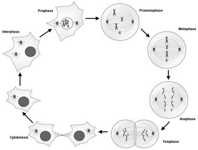
During the interphase the cell’s nucleus is well defined, with two pairs of centrioles adjacent to the nucleus. At the end of the interphase, the genome has been duplicated but the chromosomes are not distinguishable. When prophase starts, the nucleoli disappear and the chromatin starts to coil and fold into observable chromosomes, the spindle forms and the centrosomes move apart. During prometaphase, the nuclear membrane breaks down and some of spindle microtubules attach to sister chromatids at the kinetochores. The microtubules start to deplace the chromatid pairs to form a metaphase plate. At the metaphase the chromosomes have moved to the center of the dividing cell along the metaphase plate. Identical chromatids are attached to kinetochore fibers radiating from opposite ends of the parent cell. The sister chromatids begin to separate at the anaphase when the spindle microtubules pull separating chromosomes to opposite poles. During telophase, daughter nuclei begin to assemble with nuclear envelopes appearing around chromosomes. Nucleoli reappear and chromosomes decondense. The last step of mitosis is the cytokinesis step. It occurs when a contractile ring of actin and myosin filaments constricts the plasma membrane at the equator, triggering the physical division of the two daughter cells.
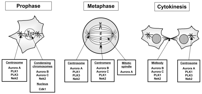
One of the clue to succeed in mitosis for mitotic kinases, is to be “at the right place at the right moment”. The short summary of where the kinases have been found gives an idea of the complexity of the controls insured by mitotic protein kinases.
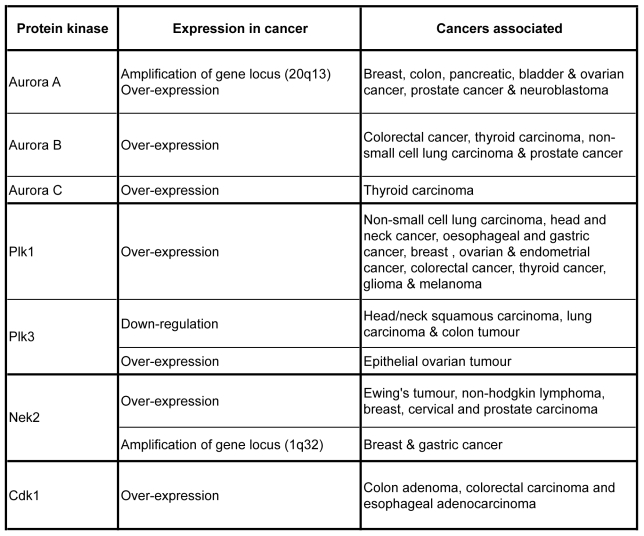
As mitosis regulators, the expression levels of the major mitotic kinases are crucial for cell division. In many cancers, up and down regulation of their expression have been observed, underlying the importance to follow the expression level of mitotic kinases for developing new targeted therapy.
Similar articles
-
[Regulation of mitosis by mitotic kinases].Tanpakushitsu Kakusan Koso. 2009 Mar;54(4 Suppl):441-6. Tanpakushitsu Kakusan Koso. 2009. PMID: 21089489 Review. Japanese. No abstract available.
-
Protein kinases controlling the onset of mitosis.Cell Mol Life Sci. 2006 Apr;63(7-8):781-95. doi: 10.1007/s00018-005-5515-3. Cell Mol Life Sci. 2006. PMID: 16465440 Free PMC article. Review.
-
Never say never. The NIMA-related protein kinases in mitotic control.Trends Cell Biol. 2003 May;13(5):221-8. doi: 10.1016/s0962-8924(03)00056-4. Trends Cell Biol. 2003. PMID: 12742165 Review.
-
Phosphatases: providing safe passage through mitotic exit.Nat Rev Mol Cell Biol. 2011 Jul 13;12(8):469-82. doi: 10.1038/nrm3149. Nat Rev Mol Cell Biol. 2011. PMID: 21750572 Review.
-
Mitotic kinases: the key to duplication, segregation, and cytokinesis errors, chromosomal instability, and oncogenesis.Pharmacol Ther. 2006 Sep;111(3):974-84. doi: 10.1016/j.pharmthera.2006.02.006. Epub 2006 Apr 17. Pharmacol Ther. 2006. PMID: 16603252 Review.
Cited by
-
Potential functional variants in SMC2 and TP53 in the AURORA pathway genes and risk of pancreatic cancer.Carcinogenesis. 2019 Jun 10;40(4):521-528. doi: 10.1093/carcin/bgz029. Carcinogenesis. 2019. PMID: 30794721 Free PMC article.
-
Identification of Candidate Cyclin-dependent kinase 1 (Cdk1) Substrates in Mitosis by Quantitative Phosphoproteomics.Mol Cell Proteomics. 2016 Jul;15(7):2448-61. doi: 10.1074/mcp.M116.059394. Epub 2016 May 1. Mol Cell Proteomics. 2016. PMID: 27134283 Free PMC article.
-
Integrative network analysis identifies key genes and pathways in the progression of hepatitis C virus induced hepatocellular carcinoma.BMC Med Genomics. 2011 Aug 8;4:62. doi: 10.1186/1755-8794-4-62. BMC Med Genomics. 2011. PMID: 21824427 Free PMC article.
-
Coordination of Protein Kinase and Phosphoprotein Phosphatase Activities in Mitosis.Front Cell Dev Biol. 2018 Mar 22;6:30. doi: 10.3389/fcell.2018.00030. eCollection 2018. Front Cell Dev Biol. 2018. PMID: 29623276 Free PMC article. Review.
-
Identification of Candidate Casein Kinase 2 Substrates in Mitosis by Quantitative Phosphoproteomics.Front Cell Dev Biol. 2017 Nov 22;5:97. doi: 10.3389/fcell.2017.00097. eCollection 2017. Front Cell Dev Biol. 2017. PMID: 29214152 Free PMC article.
References
-
- Draetta G, Beach D. Activation of cdc2 protein kinase during mitosis in human cells: cell cycle-dependent phosphorylation and subunit rearrangement. Cell. 1988;54(1):17–26. - PubMed
-
- Draetta G, Luca F, Westendorf J, Brizuela L, Ruderman J, Beach D. Cdc2 protein kinase is complexed with both cyclin A and B: evidence for proteolytic inactivation of MPF. Cell. 1989;56(5):829–38. - PubMed
-
- Karaiskou A, Perez LH, Ferby I, Ozon R, Jessus C, Nebreda AR. Differential regulation of Cdc2 and Cdk2 by RINGO and cyclins. J Biol Chem. 2001;276(38):36028–34. - PubMed
Publication types
MeSH terms
Substances
LinkOut - more resources
Full Text Sources
Other Literature Sources
Miscellaneous
