. 2008 Aug;82(15):7264-75.
doi: 10.1128/JVI.00737-08. Epub 2008 May 21.

Severe acute respiratory syndrome coronavirus infection causes neuronal death in the absence of encephalitis in mice transgenic for human ACE2

Affiliations

Severe acute respiratory syndrome coronavirus infection causes neuronal death in the absence of encephalitis in mice transgenic for human ACE2

Jason Netland et al. J Virol. 2008 Aug.

Abstract

Infection of humans with the severe acute respiratory syndrome coronavirus (SARS-CoV) results in substantial morbidity and mortality, with death resulting primarily from respiratory failure. While the lungs are the major site of infection, the brain is also infected in some patients. Brain infection may result in long-term neurological sequelae, but little is known about the pathogenesis of SARS-CoV in this organ. We previously showed that the brain was a major target organ for infection in mice that are transgenic for the SARS-CoV receptor (human angiotensin-converting enzyme 2). Herein, we use these mice to show that virus enters the brain primarily via the olfactory bulb, and infection results in rapid, transneuronal spread to connected areas of the brain. This extensive neuronal infection is the main cause of death because intracranial inoculation with low doses of virus results in a uniformly lethal disease even though little infection is detected in the lungs. Death of the animal likely results from dysfunction and/or death of infected neurons, especially those located in cardiorespiratory centers in the medulla. Remarkably, the virus induces minimal cellular infiltration in the brain. Our results show that neurons are a highly susceptible target for SARS-CoV and that only the absence of the host cell receptor prevents severe murine brain disease.


Figures

FIG. 1.
FIG. 1.
SARS-CoV antigen staining in the lungs of K18-hACE2 Tg and wild-type mice. K18-hACE2 Tg (A, C, E, and F) and non-Tg (B and D) mice were infected intranasally with 2.4 × 10⁴ PFU of SARS-CoV (Urbani strain). Three mice from each group were sacrificed at days 1 to 6 p.i. Lungs were harvested, fixed, sectioned, and stained with anti-SARS-CoV N protein MAb. Viral antigen was localized to the conducting airway of Tg and non-Tg mice at day 1 (A and B) and day 2 p.i. (C and D). Antigen was detected in the alveoli at day 4 p.i (E and F). (G and H) Scoring of antigen staining in the airway and in alveoli. Airway antigen was scored by determining the percentage of infected airways; alveolar staining was scored based on the percentage of lung that was infected. In both cases, the scoring is as follows: 0, 0%; 1, 10 to 20%; 2, 30 to 40%; 3, 50 to 60%; 4, 70 to 80%; 5, 90 to 100%. *, P < 0.05. Original magnifications: ×20 (A to E) and ×2 (F). uninf, uninfected.
FIG. 2.
FIG. 2.
Lung pathology in SARS-CoV-infected K18-hACE2 Tg and non-Tg mice. Lungs were harvested from K18-hACE2 Tg (A, B, and D to H) and non-Tg (C) mice daily from days 1 to 6 p.i., fixed, sectioned, and stained with hematoxylin and eosin. (A) Epithelial cell sloughing from the airway of a Tg animal at day 1 p.i. (B and C) Peribronchial inflammation in both Tg (B) and non-Tg (C) mice at day 2 p.i. (D) Interstitial infiltrates in Tg mouse at day 3 p.i. (E and F) Lungs from Tg mice exhibited intense neutrophilic infiltrates at day 4 p.i. (E) with foreign debris present in many lesions (F, arrows), consistent with aspiration pneumonia. (G and H) Staining of adjacent sections shows that inflammation (G) and SARS-CoV antigen (H) overlap in some regions of the lung (arrows). (I to K) Lungs from three Tg and three non-Tg mice at each day p.i. were scored for cell sloughing (I), airway inflammation (J), and cellular proliferation (K). Cell sloughing scores were based on the number of sloughed/necrotic cells per airway as follows: 0, none detected; 1, rare; 2, 1 to 3; 3, 4 to 6; 4, 7 to 9; and 5, 10 or more. Inflammation scores were assigned as follows: 1, uncommon individual cells; 2, detectable peribronchiolar/perivascular aggregates; 3, moderate cellular aggregates; 4, cellular aggregates with vascular margination; 5, cellular aggregates with vascular margination and edema or alveolar cellular infiltration. Proliferation was scored based on the number of mitotic figures per lung section as follows: 0, no mitotic figures; 1, rare; 2, 2 to 3; 3, 4 to 6; 4, 7 to 9; 5, 10 or more. *, P < 0.05. Original magnifications: ×40 (A, C, D, and F), ×20 (B and E), and ×4 (G and H).
FIG. 3.
FIG. 3.
Intracranial infection of K18-hACE2 mice with different dosages of SARS-CoV. Tg mice were intracranially inoculated with 3.2 × 10⁴ (high dose) (A-C) or 320 (low dose) (D to F) PFU of SARS-CoV, and lungs and brains were harvested at day 4 p.i. Tissue was fixed, sectioned, and stained for SARS-CoV antigen and with hematoxylin. (A) Viral antigen was detected throughout the brain after infection with 3.2 × 10⁴ PFU of SARS-CoV. (B and C) Aspiration pneumonia was detected in the lungs of animals infected with 3.2 × 10⁴ PFU in the absence of viral antigen (n = 7). (D) The distribution of viral antigen in the brains of animals infected with 320 PFU was limited. (E and F) Lungs of mice inoculated with 320 PFU of SARS-CoV showed no evidence of aspiration pneumonia or viral antigen (n = 4). (G) Viral titers in the lungs of Tg-positive mice after intracranial or intranasal (2.4 × 10⁴ PFU) virus inoculation at day 4 p.i. (n = 3 for all groups) (H to J) Viral antigen is detected in the dorsal vagal complex in mice inoculated at low dose intracranially (H), high dose intracranially (I) and intranasally (J). nT, nucleus tractus solitari; AP, area postrema; nX, dorsal motor nucleus of the vagus; nXII, motor hypoglossal nucleus; 4v, fourth ventricle; Cer, cerebellum. Original magnifications: ×1 (A, B, and E), ×40 (C and F), ×2 (D), and ×5 (H to J).
FIG. 4.
FIG. 4.
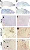
Viral antigen distribution in the brain following intranasal inoculation. Brains were harvested from mice infected intranasally with 2.4 × 10⁴ PFU and stained for viral antigen. (A to C) Antigen distribution in the brain at 60 h (A) and 3 (B), 4 (C), and 5 (D) days p.i. (E to J) The brain stem (E, G, and I) and hypothalamus (F, H, and J) were examined for viral antigen and inflammation. (E and F) Naïve controls. (G and H) SARS-CoV-infected brains exhibited antigen exclusively in neurons with no obvious inflammation at day 6 p.i. (compare to naïve controls). (I and J) JHMV-infected brains showed less extensive neuronal infection but severe perivascular inflammation (closed arrows) and meningitis (open arrows) at day 6 p.i. Original magnifications: ×1 (A to D) and ×5 (E to J).
FIG. 5.
FIG. 5.
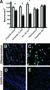
Infection results in neuronal death but does not induce apoptosis. (A) Brain sections from age-matched uninfected (n = 4) (white bars) mice and mice at day 4 p.i. (n = 4) (black bars) were stained with cresyl violet, and the number of neurons in the indicated regions were quantified by light microscopy. For each region, three adjacent fields were counted and averaged. Infected mice showed a significant reduction in neurons in the cingulate cortex, infralimbic cortex, and anterior olfactory nucleus (Ant Olf Nuc) compared to uninfected controls. No difference was detected in the hippocampus and cerebellar nucleus (Nuc). *, P < 0.005. (B to E) Brain sections from infected (D and E) and DNase-treated uninfected mice (B and C) were assayed for apoptosis by TUNEL assay. TUNEL-positive cells (green) were detected in DNase 1-treated brains (B and C) but were not present in SARS-CoV-infected brains at either day 4 (D) or day 6 (E). Nuclei were labeled with TOPRO-3 (blue). Original magnification, ×40 for all images.
FIG. 6.
FIG. 6.
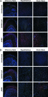
SARS-CoV infection induces increased numbers of activated microglia but not astrocytes. Brain sections from uninfected and SARS-CoV- and JHMV-infected mice at day 6 p.i. were stained for viral antigen (green) and GFAP (astrocytes) or Iba1 (microglia/macrophage) (red). Nuclei were labeled with TOPRO-3 (blue). (A) Limited GFAP labeling was detected in uninfected and SARS-CoV-infected brains, while numerous GFAP cells were found in JHMV-infected brains. (B) No Iba1 immunoreactivity was found in uninfected brains while Iba1-positive cells were present in both SARS-CoV- and JHMV-infected samples. Original magnification, ×10 for all images.
FIG. 7.
FIG. 7.
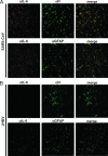
IL-6 is expressed by SARS-CoV-infected but not JHMV-infected neurons. Brain sections were stained with anti-IL-6 antibody and either anti-SARS-CoV N, anti-JHMV N, or anti-GFAP antibody and analyzed by confocal microscopy. (A) In SARS-CoV-infected brains at 4 days p.i., IL-6 is expressed mostly by infected neurons but also by GFAP-positive astrocytes. (B) In JHMV-infected brains, most infected neurons do not express IL-6, and the majority of IL-6-positive cells are astrocytes. Images of SARS-CoV-infected brain depict the cortex. Images of JHMV-infected brain depict the midbrain (top row) and medulla (bottom row). Original magnification, ×40 for all images.

Similar articles

Cited by

References

    1. Akashi, H., Y. Inaba, Y. Miura, K. Sato, S. Tokuhisa, M. Asagi, and Y. Hayashi. 1981. Propagation of the Kakegawa strain of bovine coronavirus in suckling mice, rats and hamsters. Arch. Virol. 67367-370. - PMC - PubMed
    1. Allan, S. M., and N. J. Rothwell. 2001. Cytokines and acute neurodegeneration. Nat. Rev. Neurosci. 2734-744. - PubMed
    1. Aloisi, F. 2001. Immune function of microglia. Glia 36165-179. - PubMed
    1. Arbour, N., R. Day, J. Newcombe, and P. J. Talbot. 2000. Neuroinvasion by human respiratory coronaviruses. J. Virol. 748913-8921. - PMC - PubMed
    1. Astic, L., D. Saucier, P. Coulon, F. Lafay, and A. Flamand. 1993. The CVS strain of rabies virus as transneuronal tracer in the olfactory system of mice. Brain Res. 619146-156. - PubMed

Publication types

MeSH terms

LinkOut - more resources