. 2008 Jul 25;283(30):20848-56.
doi: 10.1074/jbc.M710186200. Epub 2008 May 20.

Modification of Drosophila p53 by SUMO modulates its transactivation and pro-apoptotic functions

Affiliations

Modification of Drosophila p53 by SUMO modulates its transactivation and pro-apoptotic functions

Federico Mauri et al. J Biol Chem. .

Abstract

Conjugation to SUMO is a reversible post-translational modification that regulates several transcription factors involved in cell proliferation, differentiation, and disease. The p53 tumor suppressor can be modified by SUMO-1 in mammalian cells, but the functional consequences of this modification are unclear. Here, we demonstrate that the Drosophila homolog of human p53 can be efficiently sumoylated in insect cells. We identify two lysine residues involved in SUMO attachment, one at the C terminus, between the DNA binding and oligomerization domains, and one at the N terminus of the protein. We find that sumoylation helps recruit Drosophila p53 to nuclear dot-like structures that can be marked by human PML and the Drosophila homologue of Daxx. We demonstrate that mutation of both sumoylation sites dramatically reduces the transcriptional activity of p53 and its ability to induce apoptosis in transgenic flies, providing in vivo evidence that sumoylation is critical for Drosophila p53 function.


Figures

FIGURE 1.
FIGURE 1.
Identification of two sumoylation sites in Drosophila p53. A, schematic structure of human and Drosophila p53, with respective sumoylation sites. The transactivation (TA), DNA binding (DBD), and oligomerization (OD) domains are indicated. B, sumoylation of p53. Wild-type HT-Dmp53 and the indicated mutants were transfected in S2 cells with or without plasmids expressing GFP-dSUMO or its non-conjugatable version GFP-dSUMOΔC. Lysates were separated by SDS-PAGE. HT-Dmp53 and GFP-dSUMO were detected by immunoblotting. C, various sumoylated forms migrate differently. The indicated p53 mutants were transfected in S2 cells and analyzed by immunoblotting in the same gel. D, p53 is conjugated to endogenous SUMO. Wild-type HT-Dmp53 and the double-lysine KRKR mutant were transfected in S2 cells. Lysates were immunoprecipitated with a monoclonal antibody to the RGS-His tag, and revealed with an antibody to Drosophila SUMO (bottom right). Expression of HT-Dmp53 proteins was also analyzed in the immunoprecipitate (bottom left) and in total lysates (input). The antibody to Drosophila SUMO has a weak cross-reactivity to p53 (asterisk). Arrows indicate p53 modified with one or two SUMO molecules.
FIGURE 2.
FIGURE 2.
Nuclear localization of exogenous p53 in tissue culture and developing eye imaginal discs. A, wild-type and non sumoylatable p53 form nuclear dots in cultured cells. S2 cells were transfected with the indicated constructs, plated on concanavalin A-coated coverslips before fixation, and analyzed by confocal immunofluorescence using a monoclonal anti-Dmp53 antibody. Nuclei were visualized by Hoechst staining (scale bar, 5 mm). B, p53 sumoylation mutants display differential localization with respect to GFP-dSUMO and human PML IV. Wild-type p53 and the indicated mutants were co-transfected with GFP-dSUMO or human PML IV in S2 cells. Cells were treated as above. Localization of p53 proteins (red) and GFP-dSUMO or PML IV (green) was analyzed by confocal microscopy. Only merged images are shown, where yellow indicates co-localization. The complete set of single images for all the mutants are available as supplemental Figs. S1 and S2. C, Drosophila p53 (green), SUMO (red), and DAPI (blue) expression in the developing eye imaginal disc. High levels of p53 expression in the posterior of the developing eye imaginal disc were obtained using GMR-Gal4 to drive expression of GUSp53 transgenes. Overexpressed wild-type p53 accumulates endogenous SUMO in subnuclear domains. Overexpressed p53KRKR also forms nuclear dots, but recruits much less SUMO (scale bar, 5 μm). D, Drosophila p53 (green) and DAPI (blue) expression in irradiated developing eye imaginal discs. Overview (A) and high magnification (B and C) of moderately expressed wild-type p53, forming nuclear dots in an untreated eye disc. Overview (D) and high magnification (E and F) of wild-type p53 4 h after X-irradiation. Overview (G) and high magnification (H and I) of p53KRKR forming dots in an untreated eye disc. Overview (J) and high magnification (K and L) of p53KRKR 4 h after X-irradiation.
FIGURE 3.
FIGURE 3.
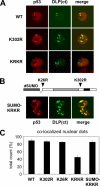
Sumoylation affects localization of p53 to nuclear dots marked by DLP. A, mutation of the lysines affects p53 co-localization with DLP. Confocal analysis of the nuclear localization of wild-type p53 and lysine mutants with respect to GFP-DLP(ct) in transfected S2 cells. B, fusion to SUMO induces full co-localization of p53 KRKR with DLP. Confocal analysis of the nuclear localization of the SUMO-KRKR chimera with respect to GFP-DLP(ct). The structure of the SUMO-KRKR chimera is schematically drawn in the same panel: Drosophila SUMO (amino acids 1–85) is fused to residue 18 of p53 KRKR. Images refer to a single Z section. C, quantification of p53 nuclear dots co-localized with GFP-DLP(ct) dots, assayed with the indicated constructs. More than 460 nuclear p53 dots were counted per mutant, in three independent experiments.
FIGURE 4.
FIGURE 4.
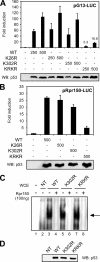
Mutation of both sumoylation sites affects transcriptional activity of p53 but not its DNA binding. A, transactivation of a human p53-responsive promoter. The pG13-LUC reporter plasmid was transfected in S2 cells together with increasing amounts of vector expressing wild-type p53 or sumoylation mutants. A plasmid constitutively expressing β-galactosidase was included as a control for transfection efficiency. p53 transcriptional activity was measured by luciferase assay, while the levels of expressed proteins were analyzed by immunoblotting of the same lysates (lower panel). Fold induction values of the p53 KRKR mutant are indicated. Error bars indicate S.E. (n = 4). B, transactivation of a Drosophila p53-responsive promoter. The pRpr150-LUC reporter carrying the p53 binding site from the Reaper DNA-damage responsive enhancer was transfected and assayed as described above. Error bars indicate S.E. (n = 3). C, EMSA. Wild-type p53 and lysine mutants were tested for sequence-specific DNA binding by gel shift, using a double-stranded oligonucleotide containing the p53-responsive element form the Reaper enhancer (Rpr150). Specificity of the binding was confirmed by competition with cold Rpr150 oligonucleotide. Lane 1, free probe. Lanes 2–9, whole cell lysates from S2 cells untransfected (NT) or transfected with the indicated p53 constructs. D, protein levels of transfected p53 mutants were assayed by immunoblotting of the lysates used for EMSA.
FIGURE 5.
FIGURE 5.
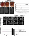
p53KRKR does not induce apoptosis as efficiently as wild-type p53, and is unable to fully rescue DNA damage-induced apoptosis. A–F, high levels of p53 expression in the posterior of the developing eye imaginal disc were obtained using GMR-Gal4 to drive expression of GUSp53 transgenes. A, wild-type adult eye. B, adult eye overexpressing wild-type p53. C, adult eye overexpressing p53KRKR. D–F, TUNEL staining for apoptotic cells in eye imaginal discs. D, wild-type eye imaginal disc. E, eye disc overexpressing wild-type p53. F, eye disc overexpressing p53KRKR. Scale bar: 20 μm. G, distribution profiles of the distance of TUNEL-positive cells from the furrow in p53+-expressing cells versus p53KRKR-expressing cells. All samples were normalized to calculate the mean percentage of apoptotic cells at a given distance from the furrow out of the total number of apoptotic cells in the disc. Distribution profiles were generated to plot percent of apoptotic cells at each distance from the furrow (n = 5). H–O, cleaved caspase-3 staining of eye imaginal discs mock-treated, or 4 h after X-irradiation. In the absence of a Gal4 driver, the Glass/multimer promoter of GUSp53 transgenes expresses levels of p53 that can rescue DNA damage-induced apoptosis in a p53 mutant tissue, but are too low to induce apoptosis without an external stress. The transgene expression domain in the posterior of each eye disc is indicated with brackets. H–K, untreated eye discs. L–O, eye discs stained for cleaved capase-3 4 h after X-irradiation. P, quantification of relative volumes of cleaved caspase-3 staining in the regions marked by brackets. See “Experimental Procedures” for details of caspase quantification. Bars indicate S.E. (n = 5). A two-tailed Student's t test was used to determine the significance of the observed changes.

Similar articles

Cited by

References

    1. Vousden, K. H., and Lane, D. P. (2007) Nat. Rev. Mol. Cell. Biol. 8275 -283 - PubMed
    1. Harris, S. L., and Levine, A. J. (2005) Oncogene 242899 -2908 - PubMed
    1. Watson, I. R., and Irwin, M. S. (2006) Neoplasia 8655 -666 - PMC - PubMed
    1. Bode, A. M., and Dong, Z. (2004) Nat. Rev. Cancer 4793 -805 - PubMed
    1. Toledo, F., and Wahl, G. M. (2006) Nat. Rev. Cancer 6909 -923 - PubMed

Publication types

MeSH terms

Substances