. 2008 Jun;172(6):1717-28.
doi: 10.2353/ajpath.2008.071054. Epub 2008 May 15.

Stat3 promotes metastatic progression of prostate cancer

Affiliations

Stat3 promotes metastatic progression of prostate cancer

Junaid Abdulghani et al. Am J Pathol. 2008 Jun.

Abstract

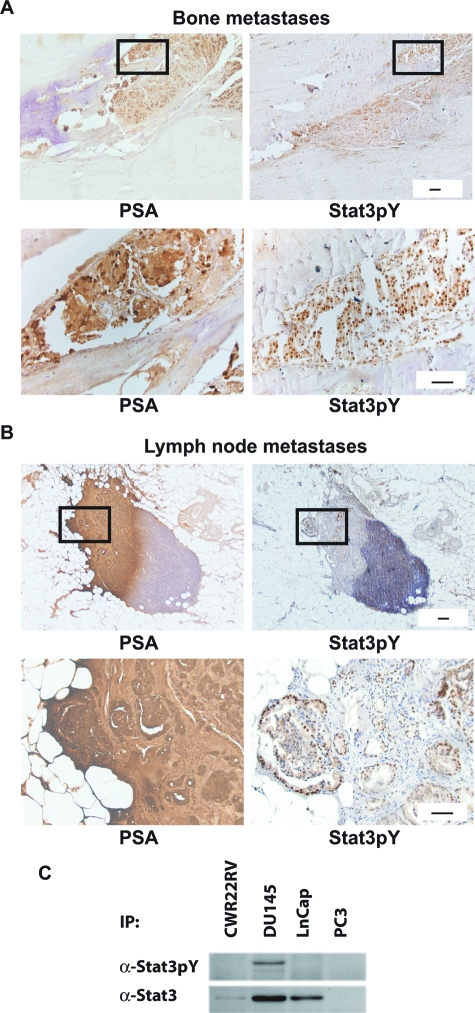
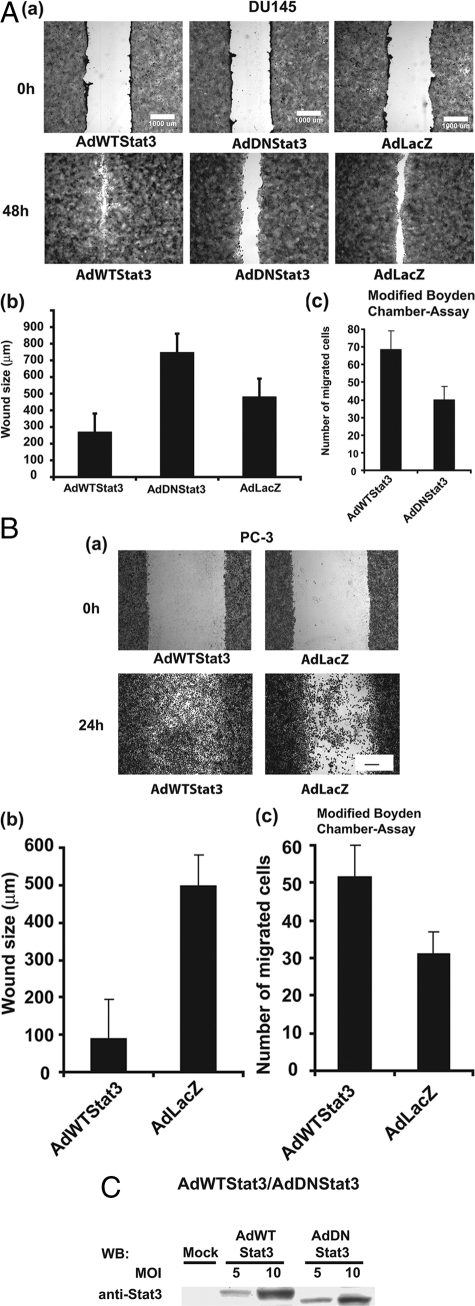
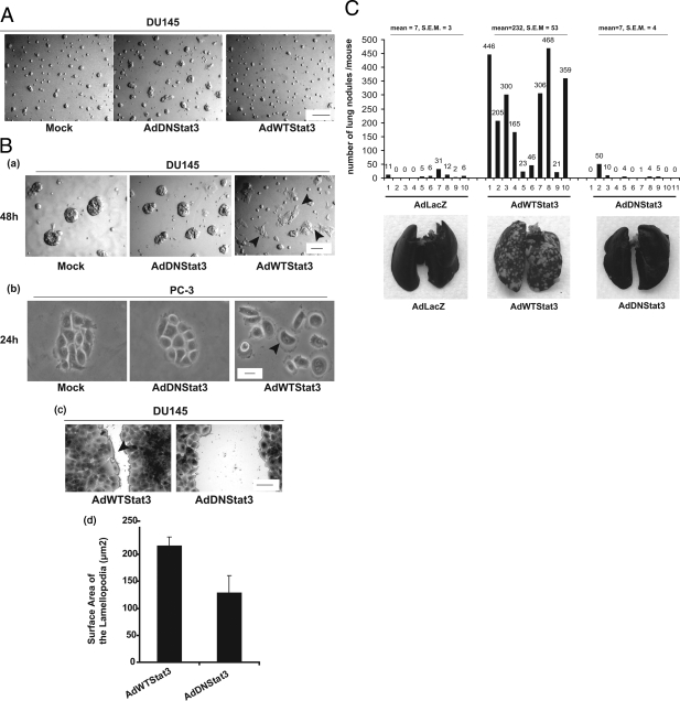
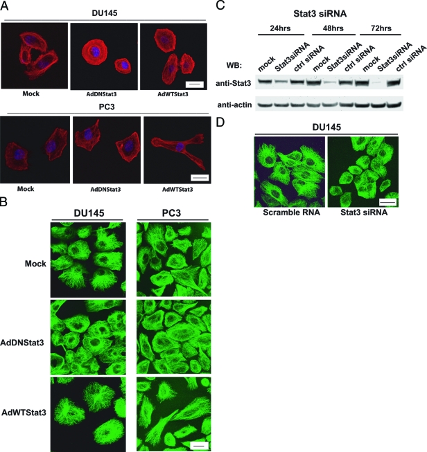
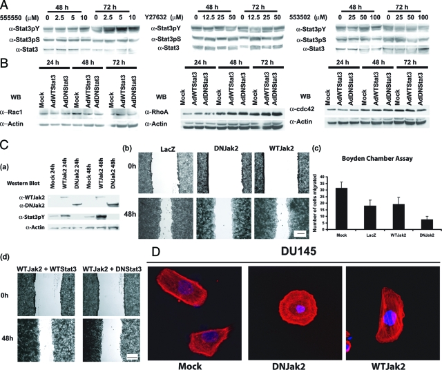
There are currently no effective therapies for metastatic prostate cancer because the molecular mechanisms that underlie the metastatic spread of primary prostate cancer are unclear. Transcription factor Stat3 is constitutively active in malignant prostate epithelium, and its activation is associated with high histological grade and advanced cancer stage. In this work, we hypothesized that Stat3 stimulates metastatic progression of prostate cancer. We show that Stat3 is active in 77% of lymph node and 67% of bone metastases of clinical human prostate cancers. Importantly, adenoviral gene delivery of wild-type Stat3 (AdWTStat3) to DU145 human prostate cancer cells increased the number of lung metastases by 33-fold in an experimental metastasis assay compared with controls. Using various methods to inhibit Stat3, we demonstrated that Stat3 promotes human prostate cancer cell migration. Stat3 induced the formation of lamellipodia in both DU145 and PC-3 cells, further supporting the concept that Stat3 promotes a migratory phenotype of human prostate cancer cells. Moreover, Stat3 caused the rearrangement of cytoplasmic actin stress fibers and microtubules in both DU145 and PC-3 cells. Finally, inhibition of the Jak2 tyrosine kinase decreased both activation of Stat3 and prostate cancer cell motility. Collectively, these data indicate that transcription factor Stat3 is involved in metastatic behavior of human prostate cancer cells and may provide a therapeutic target to prevent metastatic spread of primary prostate cancer.


Figures

Figure 1
Figure 1
Activation of Stat3 in human prostate cancer metastases to lymph nodes and to bone. Activation of Stat3 in prostate cancer metastases was analyzed by immunohistochemical staining using a polyclonal anti-Stat3pY antibody in paraffin-embedded tissue sections. 3,3′-Diaminobenzidine (3,3′-DAB) was used as a chromogen and Mayer hematoxylin as a counterstain. Biotin-streptavidin-amplified peroxidase-antiperoxidase immunodetection shows intense positive reactions for active Stat3 in the nuclei of prostate cancer cells metastasized to bone (A) and lymph nodes (B) (right). Parallel sections were immunostained for prostate-specific antigen (PSA) to verify the location of prostate cancer cells in the metastases tissues (left). C: Stat3 protein is expressed in CWR22Rv1, DU145, and LNCaP human prostate cancer cells, whereas PC-3 cells are negative for Stat3. Stat3 is constitutively active only in DU145 cells. Immunoprecipitated Stat3 was blotted with anti-phosphoTyrStat3 (anti-Stat3pY) pAb and parallel samples were immunoblotted with anti-Stat3 mAb as indicated. Scale bars = 50 μm.
Figure 2
Figure 2
Stat3 promotes motility of prostate cancer cells. DU145 (A) and PC-3 cells (B) were infected with wild-type Stat3 adenovirus (AdWTStat3), dominant-negative Stat3 adenovirus (AdDNStat3), or adenovirus expressing β-galactosidase (AdLacZ) at m.o.i. of 10, as indicated. Identical scratches were made in parallel wells (n = 8) using a 1000-μl pipette tip 24 hours after the adenoviral infection. The cells were fixed with 0.5% crystal violet at 0 hours, 24 hours, 48 hours, and 72 hours. The wells were photographed (Nikon stereoscope), (a) and wound size measured (b). The peak differences between the treatment groups were observed at 48 hours and at 24 hours for DU145 (A) and PC-3 (B) cells, respectively, after the scratches were made. The mean values of four independent experiments each with two parallel wells per treatment (total, n = 8 per group) are presented and SD values are indicated. For modified Boyden chamber assays, DU145 and PC-3 cells were exposed to infection with adenovirus carrying either DNStat3 or WTStat3 at a m.o.i. of 10 for 90 minutes. Motility of DU145 (A) and PC-3 (B) cells through uncoated filters into the lower chamber of the microchemotaxis chambers filled with media containing 5% FBS was measured after 24 hours (c). Migrated cells were fixed, stained, and counted using phase contrast microscope in triplicate filters in three individual experiments (mean ± SD). C: Dose-dependent expression of WTStat3 or DNStat3 in PC-3 cells was detected by immunoblotting of the whole-cell lysates with anti-Stat3 pAb 24 hours after adenoviral gene transfer (m.o.i., 5 and 10). Scale bars = 1000 μm.
Figure 3
Figure 3
Stat3 disrupts homotypic cluster formation, enhances lamellipodia formation of human prostate cancer cells, and increases frequency of experimental prostate cancer metastases to lungs of nude mice. A: DU145 cells were either mock-infected or infected with adenovirus carrying WTStat3 or DN-Stat3 at a m.o.i. of 10 for 90 minutes. Twenty-four hours after the adenoviral infection, the cells were seeded on Matrigel. After 96 hours, morphological alterations of DU145 cells were visualized and photographed under phase contrast microscopy (Nikon stereoscope). Representative images from three independent experiments are shown. B: DU145 cells (a) and PC-3 cells (b) were infected with a m.o.i. of 10 of AdWTStat3 or AdDNStat3 or the cells were mock-infected and seeded on Matrigel spread on glass coverslips. Lamellipodia formation was photographed at 48 hours (DU145) and 24 hours (PC-3) by phase contrast stereomicroscope. The areas of lamellipodia along the wound margins in the wound filling assays of DU145 cells were measured using the Metamorph imaging system (c) and the surface areas of lamellipodia were calculated per cell (d) ±SD. C: Athymic nude mice were injected with DU145 cells infected with adenovirus expressing WTStat3, DNStat3, or LacZ at a m.o.i. of 10 (1 × 10⁶ cells per mouse) through the tail vein. After 8 weeks, the lungs were harvested and stained with India ink, bleached with Fekete’s solution, and scored for surface lung metastases. Note that adenoviral gene delivery of WTStat3 expression in DU145 cells results in a significant increase in metastases (33-fold increase) (mean, 232; SEM = 53) compared to AdLacZ (mean, 7; SEM = 3) or AdDNStat3 (mean, 7; SEM = 4) infected cells (top). Representative photographs of India ink-stained lungs derived from athymic nude mice injected with LacZ, WTStat3, or DNStat3 expressing DU145 cells after 8 weeks (bottom). Scale bars: 300 μm (A); 150 μm (Ba); 10 μm (Bb); 50 μm (Bc).
Figure 4
Figure 4
Stat3 induces polarization of actin cytoskeleton and microtubules of human prostate cancer cells. DU145 and PC-3 cells grown on cover glasses were mock-infected or infected with m.o.i. 10 of adenovirus expressing WTStat3 or DNStat3. After 48 hours, the cells were fixed, permeabilized, and incubated with rhodamine-conjugated phalloidin (A) or α-tubulin (B) using FITC-conjugated secondary antibodies. Cells were visualized and photographed under fluorescence microscopy (Zeiss LSM 510 imaging software). Representative images from one of three independent experiments are shown. A: Note the actin bundles in the cell bodies extending to filopodia in prostate cancer cells infected with adenovirus expressing WTStat3 compared to the controls. The actin cytoskeleton in AdDNStat3-infected DU145 cells is predominantly arranged circularly around the nuclei. B: In DU145 and PC-3 cells expressing WTStat3, the microtubules are radiating from the centrosome with their ends extending toward the plasma membrane, whereas in cells infected with AdDNStat3 the microtubule network is not polarized. C: SiRNA inhibition of Stat3 expression in DU145 cells. DU145 cells transfected with siRNA targeted to Stat3 (Stat3 siRNA) (100 pmol/well), with scrambled siRNA (ctrl siRNA) or transfection reagent alone (mock) as controls were harvested and immunoblotted with anti-Stat3 pAb 24 hours after the transfection. Stripped filters were reblotted with anti-actin pAb to demonstrate equal loading. D: DU145 cells grown on glass coverslips were transfected with Stat3 siRNA (100 pmol/well) with scrambled siRNA as control. After 30 hours, the cells were fixed and immunostained for α-tubulin as described above. Note the disorganization of the microtubule network in Stat3 siRNA-transfected cells. Scale bars: 10 μm (A, top; B); 4 μm (A, bottom); 15 μm (D).
Figure 5
Figure 5
Jak2, but not RhoGTPases, contributes to the Stat3-induced migration of prostate cancer cells. A: DU145 cells were treated with pharmacological inhibitors of RhoA (555550 and Y27632) and Rac1 (553502) for 24, 48, and 72 hours at indicated concentrations. The levels of active forms of Stat3 (Stat3pY and Stat3pS) were analyzed by Western blotting from the whole cell lysates. Parallel samples were immunoblotted with anti-Stat3 mAb as indicated. B: Stat3 does not regulate Rac1, RhoA, or Cdc42 expression in DU145 cells. DU145 cells were infected with adenovirus expressing either WTStat3 or DNStat3 at a m.o.i. of 10. At 24, 48, and 72 hours after the adenoviral gene delivery, the levels of Rac1, RhoA, and Cdc42 protein expression were determined by Western blot analysis. The filters were stripped and reblotted with anti-actin pAb. C: Suppression of Jak2 inhibits phosphorylation of Stat3 and migration of DU145 cells. DU145 cells were infected with adenovirus expressing wild-type Jak2 (AdWTJak2) or dominant-negative Jak2 (AdDNJak2) at a m.o.i. of 10. a: The whole cell lysates were analyzed for Jak2 expression, tyrosine phosphorylation of Stat3, and for actin expression by Western blotting 24 and 48 hours after adenoviral gene delivery. To test the effect of Jak2 on prostate cancer cell migration, identical scratches were made in parallel wells of DU145 cells 24 hours after infection with AdWTJak2, AdDNJak2, or AdLacZ (m.o.i., 10) for 90 minutes. b: Photographs of the fixed cells (at 0, 24, 48, and 72 hours) after the adenoviral gene delivery were captured by phase contrast microscope (Nikon Eclipse TS100) equipped with a digital imaging system (Digital Sight DS-L1). Promotion of cell migration by WTJak2 was confirmed by modified Boyden chamber assays. DU145 cells were exposed to infection with adenovirus carrying either WTJak2, DNJak2, or LacZ at a m.o.i. of 10 for 90 minutes. c: Motility of DU145 cells through uncoated filters into the lower chamber of the microchemotaxis chambers filled with media containing 5% FBS was measured after 24 hours. Migrating cells were fixed, stained, and counted using phase contrast microscope in triplicate filters in three individual experiments (mean ± SD). To test whether Stat3 suppression will inhibit the cell migration induced by WTJak2, DU145 cells were infected with the combinations of adenoviral expressing WTJak2 and DNStat3 or WTJak2 and WTStat3 each at a m.o.i. of 5 for 90 minutes. Identical scratches were made in parallel wells and photographs of the fixed cells were captured by phase contrast microscope at 48 hours after the adenoviral gene delivery. D: Jak2 induces polarization of actin cytoskeleton in DU145 and PC-3 prostate cancer cells. DU145 and PC-3 cells grown on cover glasses were mock-infected or infected after 16 hours with a m.o.i. of 10 of AdWTJak2 or AdDNJak2 adenovirus. After 48 hours, the cells were fixed, permeabilized, and incubated with rhodamine-conjugated phalloidin using FITC-conjugated secondary antibodies. Cells were visualized and photographed under fluorescence microscopy (Zeiss LSM 510 imaging software). Representative images from one of three independent experiments are shown. Scale bars: 500 μm (Cb); 1000 μm (Cd).

Comment in

Similar articles

Cited by

References

    1. Arya M, Bott SR, Shergill IS, Ahmed HU, Williamson M, Patel HR. The metastatic cascade in prostate cancer. Surg Oncol. 2006;15:117–128. - PubMed
    1. Ihle JN. The Stat family in cytokine signaling. Curr Opin Cell Biol. 2001;13:211–217. - PubMed
    1. Levy DE, Darnell JE., Jr Stats: transcriptional control and biological impact. Nat Rev Mol Cell Biol. 2002;3:651–662. - PubMed
    1. Wen Z, Zhong Z, Darnell JE., Jr Maximal activation of transcription by Stat1 and Stat3 requires both tyrosine and serine phosphorylation. Cell. 1995;82:241–250. - PubMed
    1. Mora LB, Buettner R, Seigne J, Diaz J, Ahmad N, Garcia R, Bowman T, Falcone R, Fairclough R, Cantor A, Muro-Cacho C, Livingston S, Karras J, Pow-Sang J, Jove R. Constitutive activation of Stat3 in human prostate tumors and cell lines: direct inhibition of Stat3 signaling induces apoptosis of prostate cancer cells. Cancer Res. 2002;62:6659–6666. - PubMed

Publication types

MeSH terms