

Dopaminergic activation of estrogen receptors induces fos expression within restricted regions of the neonatal female rat brain
- PMID: 18478050
- PMCID: PMC2359852
- DOI: 10.1371/journal.pone.0002177
Dopaminergic activation of estrogen receptors induces fos expression within restricted regions of the neonatal female rat brain
Abstract
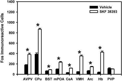
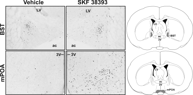
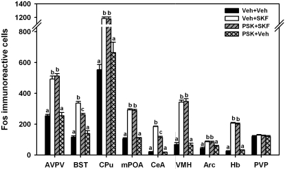
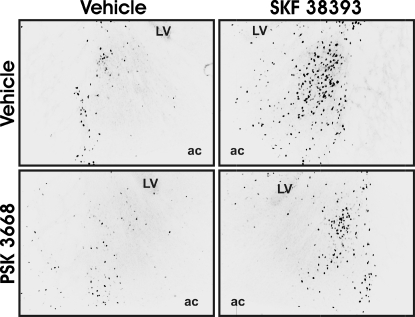
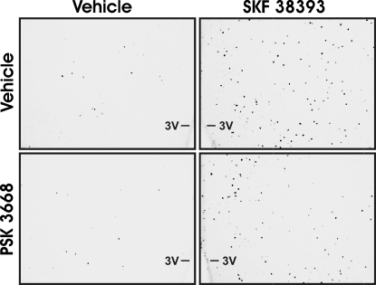
Steroid receptor activation in the developing brain influences a variety of cellular processes that endure into adulthood, altering both behavior and physiology. Recent data suggests that dopamine can regulate expression of progestin receptors within restricted regions of the developing rat brain by activating estrogen receptors in a ligand-independent manner. It is unclear whether changes in neuronal activity induced by dopaminergic activation of estrogen receptors are also region specific. To investigate this question, we examined where the dopamine D1-like receptor agonist, SKF 38393, altered Fos expression via estrogen receptor activation. We report that dopamine D1-like receptor agonist treatment increased Fos protein expression within many regions of the developing female rat brain. More importantly, prior treatment with an estrogen receptor antagonist partially reduced D1-like receptor agonist-induced Fos expression only within the bed nucleus of the stria terminalis and the central amygdala. These data suggest that dopaminergic activation of estrogen receptors alters neuronal activity within restricted regions of the developing rat brain. This implies that ligand-independent activation of estrogen receptors by dopamine might organize a unique set of behaviors during brain development in contrast to the more wide spread ligand activation of estrogen receptors by estrogen.
Conflict of interest statement
Figures
Similar articles
-
Dopaminergic activation of estrogen receptors in neonatal brain alters progestin receptor expression and juvenile social play behavior.Endocrinology. 2005 Sep;146(9):3705-12. doi: 10.1210/en.2005-0498. Epub 2005 May 26. Endocrinology. 2005. PMID: 15919740
-
D1 dopamine receptor agonist (SKF-38393) induction of Fos immunoreactivity in progestin receptor-containing areas of female rat brain.J Neuroendocrinol. 1997 May;9(5):385-94. doi: 10.1046/j.1365-2826.1997.00594.x. J Neuroendocrinol. 1997. PMID: 9181492
-
Definition of the developmental transition from dopaminergic to photic regulation of c-fos gene expression in the rat suprachiasmatic nucleus.Brain Res Mol Brain Res. 1995 Oct;33(1):136-48. doi: 10.1016/0169-328x(95)00117-b. Brain Res Mol Brain Res. 1995. PMID: 8774955
-
The atypical dopamine D1 receptor agonist SKF 83959 induces striatal Fos expression in rats.Eur J Pharmacol. 2005 Dec 28;528(1-3):88-94. doi: 10.1016/j.ejphar.2005.11.003. Epub 2005 Dec 1. Eur J Pharmacol. 2005. PMID: 16324697
-
Widespread expression of functional D1-dopamine receptors in fetal rat brain.Brain Res Dev Brain Res. 1997 Aug 18;102(1):105-15. doi: 10.1016/s0165-3806(97)00091-6. Brain Res Dev Brain Res. 1997. PMID: 9298239
Cited by
-
Auditory learning in an operant task with social reinforcement is dependent on neuroestrogen synthesis in the male songbird auditory cortex.Horm Behav. 2020 May;121:104713. doi: 10.1016/j.yhbeh.2020.104713. Epub 2020 Feb 19. Horm Behav. 2020. PMID: 32057821 Free PMC article.
-
Estrogen synthesis and signaling pathways during aging: from periphery to brain.Trends Mol Med. 2013 Mar;19(3):197-209. doi: 10.1016/j.molmed.2012.12.007. Epub 2013 Jan 22. Trends Mol Med. 2013. PMID: 23348042 Free PMC article. Review.
-
Methamphetamine-enhanced female sexual motivation is dependent on dopamine and progesterone signaling in the medial amygdala.Horm Behav. 2015 Jan;67:1-11. doi: 10.1016/j.yhbeh.2014.10.004. Epub 2014 Nov 11. Horm Behav. 2015. PMID: 25448531 Free PMC article.
-
Minireview: Tipping the balance: ligand-independent activation of steroid receptors.Mol Endocrinol. 2015 Mar;29(3):349-63. doi: 10.1210/me.2014-1315. Epub 2015 Jan 27. Mol Endocrinol. 2015. PMID: 25625619 Free PMC article. Review.
-
Winning territorial disputes selectively enhances androgen sensitivity in neural pathways related to motivation and social aggression.Proc Natl Acad Sci U S A. 2010 Jul 6;107(27):12393-8. doi: 10.1073/pnas.1001394107. Epub 2010 Jul 6. Proc Natl Acad Sci U S A. 2010. PMID: 20616093 Free PMC article.
References
-
- Weisz J, Ward IL. Plasma testosterone and progesterone titers of pregnant rats, their male and female fetuses, and neonatal offspring. Endocrinology. 1980;106:306–316. - PubMed
-
- Amateau SK, Alt JJ, Stamps CL, McCarthy MM. Brain estradiol content in newborn rats: sex differences, regional heterogeneity, and possible de novo synthesis by the female telencephalon. Endocrinology. 2004;145:2906–2917. - PubMed
-
- Simerly RB. Wired for reproduction: organization and development of sexually dimorphic circuits in the mammalian forebrain. Annu Rev Neurosci. 2002;25:507–36. 507-536. - PubMed
-
- Cooke B, Hegstrom CD, Villeneuve LS, Breedlove SM. Sexual differentiation of the vertebrate brain: principles and mechanisms. Front Neuroendocrinol. 1998;19:323–362. - PubMed
-
- Mong JA, McCarthy MM. Steroid-induced developmental plasticity in hypothalamic astrocytes: implications for synaptic patterning. J Neurobiol. 1999;40:602–619. - PubMed
Publication types
MeSH terms
Substances
Grants and funding
LinkOut - more resources
Full Text Sources
