. 2008 Jun;36(11):3655-66.
doi: 10.1093/nar/gkn249. Epub 2008 May 12.

Mechanism of high-mobility group protein B enhancement of progesterone receptor sequence-specific DNA binding

Affiliations

Mechanism of high-mobility group protein B enhancement of progesterone receptor sequence-specific DNA binding

Sarah C Roemer et al. Nucleic Acids Res. 2008 Jun.

Abstract

The DNA-binding domain (DBD) of progesterone receptor (PR) is bipartite containing a zinc module core that interacts with progesterone response elements (PRE), and a short flexible carboxyl terminal extension (CTE) that interacts with the minor groove flanking the PRE. The chromosomal high-mobility group B proteins (HMGB), defined as DNA architectural proteins capable of bending DNA, also function as auxiliary factors that increase the DNA-binding affinity of PR and other steroid receptors by mechanisms that are not well defined. Here we show that the CTE of PR contains a specific binding site for HMGB that is required for stimulation of PR-PRE binding, whereas the DNA architectural properties of HMGB are dispensable. Specific PRE DNA inhibited HMGB binding to the CTE, indicating that DNA and HMGB-CTE interactions are mutually exclusive. Exogenous CTE peptide increased PR-binding affinity for PRE as did deletion of the CTE. In a PR-binding site selection assay, A/T sequences flanking the PRE were enriched by HMGB, indicating that PR DNA-binding specificity is also altered by HMGB. We conclude that a transient HMGB-CTE interaction alters a repressive conformation of the flexible CTE enabling it to bind to preferred sequences flanking the PRE.


Figures
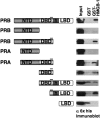
Figure 1.
Figure 1.
Regions of PR and HMGB-1 required for protein interaction. PR constructs expressed in Sf9 cells with N-terminal 6× histidine tags were used in GST pull-down assay to detect HMGB-1 interaction. The position of the CTE in different PR constructs is indicated by a black line. Free GST or GST-HMGB-1 were immobilized on glutathione–Sepharose resins and incubated with Sf9 cell extracts containing his-tagged PR constructs. Bound PR was eluted from the resins and detected by western immunoblotting with a monoclonal mouse antibody (clone 1162/F6) to 6× his tag. Equal amounts of each PR protein were added as determined in advance by western immunoblot. Western blot lanes are: 10% input PR, free GST and GST-HMGB-1.
Figure 2.
Figure 2.
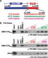
The role of the CTE in HMGB-1 interaction. (A) Schematic of PR core DBD and CTE plus sequence of synthetic peptides corresponding to aa 636 to 654 in the CTE, scrambled sCTE peptide and a control unrelated peptide (URP). (B) CTE peptide specifically competed for binding of HMGB-1 to PR. Free GST and GST-HMGB-1 were immobilized to glutathione–Sepharose resins and incubated with purified PR DBD670 (0.5 µM/1.5 µg) or full-length PR-B (0.05 µM/1.5 µg) in the absence and presence of varying amounts of peptides. Peptides were added in molar excess over receptor; 50- to 250-fold for PR DBD670 and 500- to 5000-fold for PR-B. Resins were washed, eluted and bound receptor was detected by western blotting with antibody for PR DBD.
Figure 3.
Figure 3.
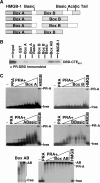
Regions HMGB-1 required for protein interaction. (A) Schematic of HMGB-1 constructs used in GST pull downs and EMSA. (B) Binding of HMGB-1 domains to PR DBD-CTE651. Whole-cell extracts prepared from bacterial cultures expressing GST, GST-box A, GST-box A with the basic linker (ABasic), GST-box B, GST-box B with basic linker (BBasic), GST-box A and box B (Box AB) or GST-HMGB-1 were immobilized on glutathione–Sepharose resin and incubated with 0.4 µM PR DBD–CTE651. Resins were washed, eluted and bound protein was detected by western blotting with antibody to PR DBD. (C) Enhancement of PR-DNA binding requires full-length HMGB-1 as shown by EMSA. Increasing amounts (0.5–7.0 μM) of HMG boxes were incubated with 32P labeled PRE in the presence of (2.0 nM) PR-A and compared to PR-A DNA binding alone and in the presence of (350 nM) full-length HMGB-2. Increasing concentrations of di-box (Box AB 0.5–2.0 μM) were incubated with 32P labeled PRE in the absence of PR-A to demonstrate Box AB's high-affinity nonspecific DNA binding. DNA binding was detected by EMSA as described in Materials and methods section.
Figure 4.
Figure 4.
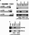
HMGB-1 protein interaction with CTE of PR is disrupted in the presence of PRE DNA. (A) Constructs of PR DBD with different lengths of CTE (aa 670, aa 651 and aa 641) were used in GST pull-down assays to detect HMGB-1 interaction. (B) Free GST or HMGB-1-GST fusion proteins were immobilized to glutathione–Sepharose resins as in Figure 1 and were incubated with PR DBD–CTE670, PR DBD–CTE651 or PR DBD–CTE641 in the absence (–DNA) or presence of PRE oligonucleotide (0.2 µM) or a nonspecific (N.S.) oligonucleotide DNA. Resins were washed, eluted and the bound protein was detected by western blotting with antibody for PR DBD. (C) GST pull-down assays with full-length PR were conducted as above in B and bound protein was detected by western blotting with a monoclonal antibody for PR.
Figure 5.
Figure 5.
The role of the CTE in PR-DNA binding. (A and B) CTE peptide enhances PR–DNA binding in a manner dependent on the presence of CTE in the receptor. Increasing amounts (2–32 nM) of (A) PR DBD-CTE670 or (B) PR DBD–CTE641 were incubated with 32P labeled PRE in the presence or absence of CTE, sCTE and URP peptide (0.75 µM), and DNA binding was detected by EMSA as described in Materials and methods section. Binding was graphed as fraction PRE bound and represent average values ± standard error of the mean (SEM) from a minimum of three independent experiments.
Figure 6.
Figure 6.
HMGB-1 influences PRE flanking DNA sequence recognized by PR. Shown in the Sequence logo format (Weblogo) are the sequences selected by PR DBD–CTE648 alone or in the presence of HMGB-1 (+HMGB-1) as aligned by CONSENSUS. The sequence positions of the inverted repeat PREs are shown on the x-axis and the information content of the alignment for each position is shown on the y-axis.
Figure 7.
Figure 7.
Functional effect of mutations in the DNA intercalating residues of HMGB-1. (A) Schematic of HMGB-1 domains. Mut-HMGB-1 contains alanine substitutions for the residues that intercalate in the DNA (Phe37, Phe102 and Ile121) as described previously (45,62). (B) Binding of wild-type (Wt) and mutant (Mut) HMGB-1 with PRE DNA as detected by EMSA. Increasing concentrations of WT HMGB-1 or Mut HMGB-1 proteins (0–5 µM) were incubated with a single concentration of [32P]-labeled PRE (0.6 nM) under conditions in the absence of competitor DNA that permit the detection of nonsequence-specific DNA binding. The distinct mobilities of Wt-HMGB-1 and Mut HGMB-1 complexes are indicated by the arrows. Free and shifted DNA bands were quantitated and graphed as a fraction of bound DNA. (C) DNA-bending properties of HMGB-1 Wt and Mut proteins as analyzed by circularization assay. HMGD, HMGB-1 Wt, HMGB-1 Mut (2 μM) or no protein was incubated with 10-mer linear duplex DNA in the presence of DNA ligase. Reactions were digested with exocnuclease III to eliminate linear DNA fragments. HMGD as a positive control was used to generate a ladder of known DNA circle sizes. Formation of DNA circles was detected by EMSA and cyber green staining (Invitrogen) as described in Materials and methods section.
Figure 8.
Figure 8.
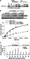
Mutations in DNA intercalating residues of HMGB-1 did not influence enhancement of PR-DNA binding in vitro but reduced the effect of HMGB-1 on PR transcriptional activity in cells. (A) Protein interaction of HMGB-1 with PR DBD was not affected by mutations in the intercalating residues of HMGB-1. Free GST, GST-Wt HMGB-1 or GST-Mut HMGB-1 were immobilized to glutathione–Sepharose resins and incubated with increasing concentrations of purified PR DBD–CTE670 (0.25 µM to 1.0 µM). Resins were washed, eluted and bound protein was detected by western blotting with antibody for PR DBD (input is 10%). (B) Effect of Wt HMGB-1 and Mut HMGB-1 on binding of PR to PRE DNA as detected by EMSA. Increasing concentrations of purified PR DBD–CTE648 (0–30 nM) were incubated with a single concentration of [32P]-labeled PRE (0.6 nM). The free and protein bound PRE complexes were separated by native gel electrophoresis, quantitated, and graphed as fraction of bound PRE–DNA. (C) Mut HMGB-1 had reduced ability to enhance transcriptional activity of PR in cells. Cos-1 cells were cotransfected with PRE2-tk-LUC reporter, PR-B, Wt HMGB-1 or Mut HMGB-1. Cells were treated with 10 nM R5020 (synthetic progesterone analog) or an equal volume of EtOH for 20 h. Luciferase activity was determined and normalized to internal control β-galactosidase activity. Normalized luciferase activity with vehicle-treated cells was set to 1.0 and all other treated groups were calculated as fold >1.0. Error bars indicate standard mean of the error (n = 5) for doses 0–40 and n = 3 for dose 80.
Figure 9.
Figure 9.
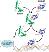
Proposed mechanism of action of HMGB. Prior to binding DNA, HMGB (green) interacts with the CTE (red) of PR (core DBD in blue) and disrupts a repressive intra-molecular interaction between the CTE and core DBD (only single domain models are shown for simplicity). HMGB interaction also promotes CTE binding to the DNA minor groove flanking the PRE. HMGB binding to PR is transient and dissociates upon stable binding of the CTE to DNA. Dissociated HMGB interacts with DNA/chromatin in the vicinity of the promoter to affect RNA pol II and general transcription factors (GTFs) in down stream steps in the process of steroid receptor-mediated gene transcription.

Similar articles

Cited by

References

    1. Beato M, Klug J. Steroid hormone receptors: an update. Hum. Reprod. Update. 2000;6:225–236. - PubMed
    1. Evans RM. The steroid and thyroid hormone receptor superfamily. Science. 1988;240:889–895. - PMC - PubMed
    1. Khorasanizadeh S, Rastinejad F. Nuclear-receptor interactions on DNA-response elements. Trends Biochem. Sci. 2001;26:384–390. - PubMed
    1. Mangelsdorf DJ, Thummel C, Beato M, Herrlich P, Schutz G, Umesono K, Blumberg B, Kastner P, Mark M, Chambon P, et al. The nuclear receptor superfamily: the second decade. Cell. 1995;83:835–839. - PMC - PubMed
    1. Gearhart MD, Holmbeck SM, Evans RM, Dyson HJ, Wright PE. Monomeric complex of human orphan estrogen related receptor-2 with DNA: a pseudo-dimer interface mediates extended half-site recognition. J. Mol. Biol. 2003;327:819–832. - PubMed

Publication types