

Cell water dynamics on multiple time scales
- PMID: 18436650
- PMCID: PMC2359779
- DOI: 10.1073/pnas.0709585105
Cell water dynamics on multiple time scales
Abstract
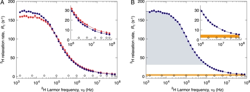
Water-biomolecule interactions have been extensively studied in dilute solutions, crystals, and rehydrated powders, but none of these model systems may capture the behavior of water in the highly organized intracellular milieu. Because of the experimental difficulty of selectively probing the structure and dynamics of water in intact cells, radically different views about the properties of cell water have proliferated. To resolve this long-standing controversy, we have measured the (2)H spin relaxation rate in living bacteria cultured in D(2)O. The relaxation data, acquired in a wide magnetic field range (0.2 mT-12 T) and analyzed in a model-independent way, reveal water dynamics on a wide range of time scales. Contradicting the view that a substantial fraction of cell water is strongly perturbed, we find that approximately 85% of cell water in Escherichia coli and in the extreme halophile Haloarcula marismortui has bulk-like dynamics. The remaining approximately 15% of cell water interacts directly with biomolecular surfaces and is motionally retarded by a factor 15 +/- 3 on average, corresponding to a rotational correlation time of 27 ps. This dynamic perturbation is three times larger than for small monomeric proteins in solution, a difference we attribute to secluded surface hydration sites in supramolecular assemblies. The relaxation data also show that a small fraction ( approximately 0.1%) of cell water exchanges from buried hydration sites on the microsecond time scale, consistent with the current understanding of protein hydration in solutions and crystals.
Conflict of interest statement
The authors declare no conflict of interest.
Figures

Similar articles
-
Specific cellular water dynamics observed in vivo by neutron scattering and NMR.Phys Chem Chem Phys. 2010 Sep 21;12(35):10154-60. doi: 10.1039/c0cp01048k. Epub 2010 Aug 16. Phys Chem Chem Phys. 2010. PMID: 20714607
-
Time scales of water dynamics at biological interfaces: peptides, proteins and cells.Faraday Discuss. 2009;141:131-44; discussion 175-207. doi: 10.1039/b806194g. Faraday Discuss. 2009. PMID: 19227355
-
Neutron scattering reveals extremely slow cell water in a Dead Sea organism.Proc Natl Acad Sci U S A. 2007 Jan 16;104(3):766-71. doi: 10.1073/pnas.0601639104. Epub 2007 Jan 10. Proc Natl Acad Sci U S A. 2007. PMID: 17215355 Free PMC article.
-
Dynamics at the protein-water interface from 17O spin relaxation in deeply supercooled solutions.Biophys J. 2008 Sep 15;95(6):2951-63. doi: 10.1529/biophysj.108.135194. Epub 2008 Jun 27. Biophys J. 2008. PMID: 18586840 Free PMC article.
-
Protein hydration dynamics in solution: a critical survey.Philos Trans R Soc Lond B Biol Sci. 2004 Aug 29;359(1448):1207-23; discussion 1223-4, 1323-8. doi: 10.1098/rstb.2004.1499. Philos Trans R Soc Lond B Biol Sci. 2004. PMID: 15306377 Free PMC article. Review.
Cited by
-
Is buffer a good proxy for a crowded cell-like environment? A comparative NMR study of calmodulin side-chain dynamics in buffer and E. coli lysate.PLoS One. 2012;7(10):e48226. doi: 10.1371/journal.pone.0048226. Epub 2012 Oct 30. PLoS One. 2012. PMID: 23118958 Free PMC article.
-
NMR Provides Unique Insight into the Functional Dynamics and Interactions of Intrinsically Disordered Proteins.Chem Rev. 2022 May 25;122(10):9331-9356. doi: 10.1021/acs.chemrev.1c01023. Epub 2022 Apr 21. Chem Rev. 2022. PMID: 35446534 Free PMC article. Review.
-
Effects of External Perturbations on Protein Systems: A Microscopic View.ACS Omega. 2022 Nov 30;7(49):44556-44572. doi: 10.1021/acsomega.2c06199. eCollection 2022 Dec 13. ACS Omega. 2022. PMID: 36530249 Free PMC article. Review.
-
Site-specific hydration dynamics in the nonpolar core of a molten globule by dynamic nuclear polarization of water.J Am Chem Soc. 2011 Apr 20;133(15):5987-95. doi: 10.1021/ja111515s. Epub 2011 Mar 28. J Am Chem Soc. 2011. PMID: 21443207 Free PMC article.
-
Profiling of dynamics in protein-lipid-water systems: a time-resolved fluorescence study of a model membrane protein with the label BADAN at specific membrane depths.Eur Biophys J. 2010 Mar;39(4):647-56. doi: 10.1007/s00249-009-0538-6. Epub 2009 Sep 16. Eur Biophys J. 2010. PMID: 19760185 Free PMC article.
References
-
- Makarov V, Pettitt BM, Feig M. Solvation and hydration of proteins and nucleic acids: A theoretical view of simulation and experiment. Acc Chem Res. 2002;35:376–384. - PubMed
-
- Raschke TM. Water structure and interactions with protein surfaces. Curr Opin Struct Biol. 2006;16:152–159. - PubMed
-
- Levy Y, Onuchic JN. Water mediation in protein folding and molecular recognition. Annu Rev Biophys Biomol Struct. 2006;35:389–415. - PubMed
-
- Ball P. Water as an active constituent in cell biology. Chem Rev. 2008;108:74–108. - PubMed
-
- Luby-Phelps K. Cytoarchitecture and physical properties of cytoplasm: Volume, viscosity, diffusion, intracellular surface area. Int Rev Cytol. 2000;192:189–221. - PubMed
Publication types
MeSH terms
Substances
LinkOut - more resources
Full Text Sources
Other Literature Sources
