. 2008 Apr 21;181(2):241-54.
doi: 10.1083/jcb.200710019.

Aurora B kinase and protein phosphatase 1 have opposing roles in modulating kinetochore assembly

Affiliations

Aurora B kinase and protein phosphatase 1 have opposing roles in modulating kinetochore assembly

Michael J Emanuele et al. J Cell Biol. .

Abstract

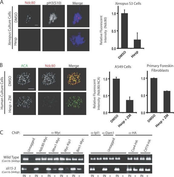
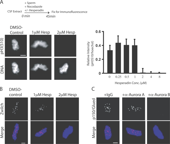
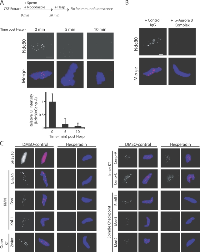
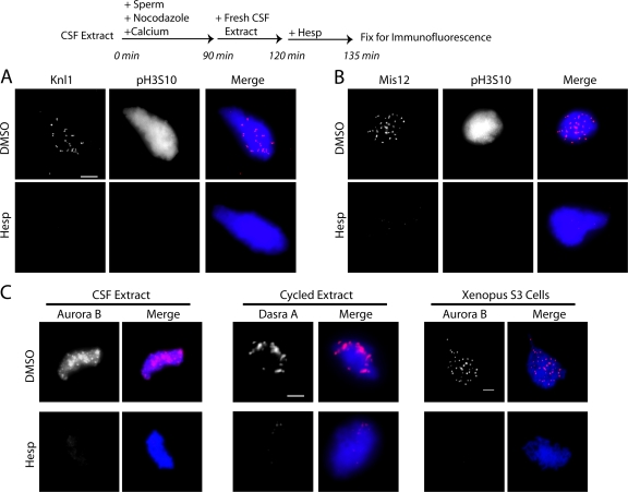
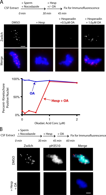
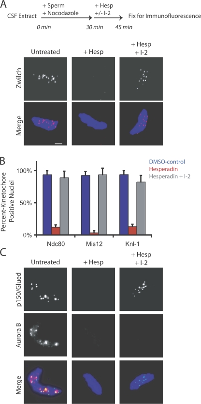
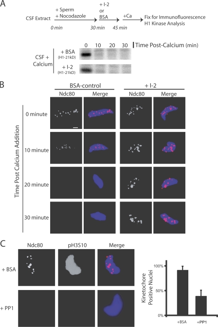
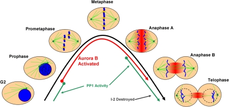
The outer kinetochore binds microtubules to control chromosome movement. Outer kinetochore assembly is restricted to mitosis, whereas the inner kinetochore remains tethered to centromeres throughout the cell cycle. The cues that regulate this transient assembly are unknown. We find that inhibition of Aurora B kinase significantly reduces outer kinetochore assembly in Xenopus laevis and human tissue culture cells, frog egg extracts, and budding yeast. In X. leavis M phase extracts, preassembled kinetochores disassemble after inhibiting Aurora B activity with either drugs or antibodies. Kinetochore disassembly, induced by Aurora B inhibition, is rescued by restraining protein phosphatase 1 (PP1) activity. PP1 is necessary for kinetochores to disassemble at the exit from M phase, and purified enzyme is sufficient to cause disassembly on isolated mitotic nuclei. These data demonstrate that Aurora B activity is required for kinetochore maintenance and that PP1 is necessary and sufficient to disassemble kinetochores. We suggest that Aurora B and PP1 coordinate cell cycle-dependent changes in kinetochore assembly though phosphorylation of kinetochore substrates.


Figures

Figure 1.
Figure 1.
Aurora B inhibition compromises kinetochore assembly in X. laevis and human tissue culture cells and in budding yeast. (A) Asynchronous X. laevis S3 cells were treated with hesperadin and fixed for immunofluorescence. Cells were immunostained with antibodies to Ndc80 and phosphorylated serine 10 on histone H3 (pH3S10). The graph shows levels of Ndc80 staining at kinetochores in control and hesperadin-treated cells (n = 60 kinetochores in three different cells per condition). (B) A549 human lung epithelial cells and primary human foreskin fibroblasts were treated with either DMSO or hesperadin and ZM447439 (Hesp + ZM) together. Cells were fixed and processed for immunofluorescence with ACA and Ndc80 antibodies. The amount of Ndc80 staining at kinetochores relative to ACA staining on the same kinetochore was determined and is graphed (n = 60 kinetochores in three cells per condition). Bars, = 5 μm. (C) Wild type or sli15-3 yeast cells were grown at a restrictive temperature of 37° for 3 h and kinetochore assembly was assessed by ChIP for Ndc80, Mtw1, Kip1, Bim1, Ipl1, Dam1, Cse4, and Ctf19. The outer kinetochore proteins are displaced from kinetochores in sli15-3 cells. IN and + indicate PCR performed on either total input or immunoprecipitated DNA, respectively. Centromeres on chromosome 16 (Cen16) were amplified. Graphs show mean plus standard deviation.
Figure 2.
Figure 2.
Aurora B activity is required for kinetochore assembly in X. laevis CSF extracts. (A and B) Hesperadin was titrated into X. laevis CSF egg extracts. Extracts were supplemented with nocodazole and sperm nuclei and, after 45 min, fixed and processed for immunofluorescence (A, top). (A) Nuclei stained with antibodies to pH3S10 and the nuclear stain Hoechst33342. The intensity of pH3S10 staining relative to the nuclear stain is graphed (right). Graphs show mean plus standard deviation. (B) Nuclei immunostained for the kinetochore protein Zwilch. Kinetochore assembly is inhibited at 2 μM hesperadin. (C) Kinetochores were assembled in CSF extracts preincubated with either anti–Aurora A or B antibodies. Nuclei were stained with antibodies to dynactin subunit p150Glued. Bars, 5 μm.
Figure 3.
Figure 3.
Aurora B activity is required for kinetochore maintenance in CSF extracts. (A) Kinetochore maintenance was assessed after hesperadin treatment in extracts containing nuclei with preassembled kinetochores. The relative intensity of Ndc80 to Cenp-A immunostaining on the same kinetochore was determined and graphed (bottom; n = 60 individual kinetochores on four nuclei per condition). Graphs show mean plus standard deviation. (B) The addition of anti–Aurora B antibodies caused preassembled kinetochores to disassemble in an identical manner. (C) A panel of antibodies was used to assess the sensitivity of outer kinetochore proteins to hesperadin treatment in extracts with preassembled kinetochores. The outer kinetochore proteins Dsn1, Knl1, Ndc80, and Zwint and the checkpoint proteins BubR1, Mad1, and Mad2 are displaced after hesperadin treatment. The inner kinetochore proteins Cenp-A and Cenp-C are unaffected. Bars, 5 μm.
Figure 4.
Figure 4.
Aurora B activity is required for kinetochore maintenance in cycled extracts. (A and B) Cycled X. laevis extracts containing replicated chromatids and assembled kinetochores were tested for their sensitivity to hesperadin (or DMSO). After 15 min in hesperadin, Knl1 (A) and Mis12 (B) were displaced from kinetochores. (C) Maintenance reactions were repeated in CSF and cycled egg extracts. Aurora B complex proteins were displaced from chromatin by the addition of hesperadin after assembly. X. laevis S3 cells also lost Aurora B staining on chromatin after treatment with hesperadin. Bars, 5 μm.
Figure 5.
Figure 5.
Phosphatases oppose Aurora B in maintaining an assembled kinetochore. (A) Hesperadin and okadaic acid were added alone or together and Zwilch maintenance at kinetochores was examined after 15 min. The addition of 1 μM okadaic acid rescued the effect of hesperadin on kinetochore maintenance. The percentage of nuclei staining positive for the kinetochore marker Zwilch is graphed (n > 100 nuclei per condition). (B) Kinetochores were assembled on sperm nuclei for 30 min in extracts. Hesperadin was added to turn off Aurora B and, after 15 min, 1 μM okadaic acid was also added to restrain phosphatase activity. Neither Zwilch nor pH3S10 staining returned to chromatin after 15 min in okadaic acid. Bars, 5 μm.
Figure 6.
Figure 6.
PP1 opposes Aurora B in the assembly pathway of the kinetochore. Hesperadin and the PP1-specific inhibitor I-2 were added alone or in combination in a kinetochore maintenance assay. (A) Zwilch is retained at kinetochores after coaddition of hesperadin and I-2. (B) The percentage of nuclei staining positive for the kinetochore proteins Ndc80, Mis12, and Knl1 after treatment with DMSO, hesperadin, or hesperadin and I-2 together is graphed (n > 100 nuclei per condition). Graphs show mean plus standard deviation. (C) Nuclei from the same experiment were stained with antibodies to p150Glued and Aurora B. Aurora B is not retained at kinetochores after treatment with hesperadin and I-2 but kinetochores are still intact, as assessed by p150 Glued staining. Bar, 5 μm.
Figure 7.
Figure 7.
PP1 is both necessary and sufficient to disassemble kinetochores on nuclei in extracts. (A and B) Kinetochores were assembled for 30 min in CSF extracts. I-2 or BSA was added to extracts and, 15 min later, the extract was driven out of M phase. (A) Both BSA- and I-2–treated extracts exited mitosis biochemically after the addition of calcium, as judged by histone H1 kinase activity. (B) In I-2–treated extracts, Ndc80 was retained at the kinetochore for 30 min after M phase exit. In control BSA-treated extracts, Ndc80 staining was reduced by 10 min and was undetectable by 20 min. (C) Nuclei with assembled kinetochores were isolated, washed, and mixed with purified PP1 enzyme. The addition of PP1 caused the dephosphorylation of H3S10 and the loss of kinetochore staining on the nuclei compared with BSA-treated nuclei. In control BSA-treated samples, 92% of nuclei stained positive for Ndc80. In PP1-treated samples, 39% of nuclei stained positive for Ndc80 (n > 100 nuclei per condition). Graphs show mean plus standard deviation. Bar, 5 μm.
Figure 8.
Figure 8.
Model of Aurora B and PP1 activity relative to kinetochore assembly in the mitosis. G2 and the mitotic phases (prophase, prometaphase, metaphase, anaphases A and B, and telophase) are depicted. Microtubules are displayed in green, chromosomes in blue, kinetochores in yellow, and active Aurora B kinase in red. The times at which Aurora B (red lines) and PP1 (green lines) are activated are shown relative to the morphological changes of mitosis. In prophase, Aurora B is activated and begins to move from chromosome arms to centromeres. By prometaphase, activated Aurora B is fully localized to the inner centromere and outer kinetochore proteins are assembled. Complete chromosome alignment occurs in metaphase, at which point the spindle checkpoint signal is extinguished and sister chromatids separate and begin poleward anaphase movements. Aurora B dissociates from the inner centromere in anaphase and relocalizes to overlapping spindle midzone microtubules, where it remains active in a gradient of kinase activity that spreads from out from the spindle midzone (fading red). Kinetochores disassemble as they move outside of the zone of Aurora B activity in anaphase B. PP1 is initially activated in anaphase, and its small protein inhibitor I-2 is further degraded in anaphase B and telophase. Active PP1, in the absence of Aurora B, leads to outer kinetochores disassembly in telophase before the ensuing G1.

Similar articles

Cited by

References

    1. Adams, R.R., H. Maiato, W.C. Earnshaw, and M. Carmena. 2001. Essential roles of Drosophila inner centromere protein (INCENP) and aurora B in histone H3 phosphorylation, metaphase chromosome alignment, kinetochore disjunction, and chromosome segregation. J. Cell Biol. 153:865–880. - PMC - PubMed
    1. Biggins, S., and A.W. Murray. 2001. The budding yeast protein kinase Ipl1/Aurora allows the absence of tension to activate the spindle checkpoint. Genes Dev. 15:3118–3129. - PMC - PubMed
    1. Brautigan, D.L., C.L. Shriner, and P.A. Gruppuso. 1985. Phosphorylase phosphatase catalytic subunit. Evidence that the Mr = 33,000 enzyme fragment is derived from a native protein of Mr = 70,000. J. Biol. Chem. 260:4295–4302. - PubMed
    1. Brautigan, D.L., J. Sunwoo, J.C. Labbe, A. Fernandez, and N.J. Lamb. 1990. Cell cycle oscillation of phosphatase inhibitor-2 in rat fibroblasts coincident with p34cdc2 restriction. Nature. 344:74–78. - PubMed
    1. Cheeseman, I.M., S. Anderson, M. Jwa, E.M. Green, J. Kang, J.R. Yates III, C.S. Chan, D.G. Drubin, and G. Barnes. 2002. Phospho-regulation of kinetochore-microtubule attachments by the Aurora kinase Ipl1p. Cell. 111:163–172. - PubMed

Publication types

MeSH terms