. 2008 Nov;1779(11):712-9.
doi: 10.1016/j.bbagrm.2008.03.007. Epub 2008 Apr 1.

TNF-alpha stimulation inhibits siRNA-mediated RNA interference through a mechanism involving poly-(A) tail stabilization

Affiliations

TNF-alpha stimulation inhibits siRNA-mediated RNA interference through a mechanism involving poly-(A) tail stabilization

Johann Mols et al. Biochim Biophys Acta. 2008 Nov.

Abstract

The control of mRNA stability is a complex biological process that involves numerous factors, including microRNA (miRNA) and short interfering RNA (siRNA). Here, we show that short interfering RNA (siRNA) and microRNA share some similarities in their response to cellular stress. miR16 expedites the degradation of mRNAs containing AU-rich elements (ARE) in their 3' untranslated region (UTR). si20 is an siRNA designed to target a non-ARE sequence in the TNF 3'UTR. We found that both si20 and miR16/ARE-mediated degradation of mRNAs can be inhibited by stimulating cells with different stresses. By analyzing TNF-alpha stimulation-mediated stabilization of si20- and miR16-targeted mRNA, we show that this stabilization is not caused by modifying si20 and miR16 loading into Ago2 complexes, or mRNA targeting to Ago2, but by inhibiting mRNA deadenylation. This is the first report showing that a specific siRNA-mediated mRNA degradation can be regulated by inflammatory stimuli, and that deadenylation is involved in this siRNA-mediated mRNA decay.


Figures

Figure 1
Figure 1. ARE and ΔARE reporter constructs and their expression in mammalian cells
A)Schematic representation of the ARE and ΔARE constructs. The full length mouse TNF-α 3’UTR was added downstream of the rabbit β-globin gene (ARE reporter). The AU-rich elements in the TNF-α 3’UTR were deleted to generate the ΔARE reporter. Both the ARE and ΔARE reporters were constructed under the control of the cytomegalovirus (CMV) or Tet promoters. The quantitative RT-PCR detection system spans intron 2. si20-targeting site and AU-rich elements (ARE) are localized in the 3’UTR of the constructs. Bovine growth hormone poly (A) signal (BGH poly (A)) was used. B) HEK-293T cells were transfected with CMV–ΔARE (ΔARE) or CMV-ARE (ARE) plasmids and cultured for 40 hours. Total RNA was extracted with Trizol® reagent. Quantitative one-step RT-PCR assays were then run to measure ARE and ΔARE mRNA. ARE and ΔARE mRNA levels were normalized against GAPDH mRNA levels. Results are expressed as the mean of 4 independent experiments, the error bar represents the standard deviation. C) HEK-293T cells were cotransfected with CMV-ΔARE and pSuper-si20 (ΔARE si20), or with CMV-ΔARE and pSuper-si ctrl (ΔARE si ctrl). Samples were analyzed as in B). D) HeLa Tet-off cells were transfected with pcDNA3 and Tet-ΔARE (ΔARE); pcDNA3; Tet-ARE (ARE); or pSuper si20 and Tet-ΔARE (ΔARE/si20) and cultured for 40 additional hours. Doxycycline was then added to block transcription, and total RNA was isolated with Trizol. Reporter mRNA degradation was analyzed by Northern-Blotting and 18S ribosomal RNA was utilized to control RNA loading. Half-lives were analyzed using the phosphoimager. E) HeLa Tet-off cells were transfected with a total of 5 µg of plasmid DNA. The mass ratio of pSuper-si20 and Tet-ΔARE plasmids were 0, 0.1, 0.2, 0.5, 1, and 2. pcDNA3 was utilized to normalize a total of 5 µg plasmid per transfection. Cells were then cultured for 40 additional hours before total RNA was isolated using Trizol. Reporter mRNA was analyzed by Northern-Blotting and the ratios of 5’frag/Full were analyzed using the phosphoimager.
Figure 2
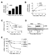
Figure 2. Inflammatory stimuli and other stresses interfere with ARE mRNA degradation
A) HEK-293T cells were transfected with CMV-ARE (ARE) and cultured for 40h. One hour before total RNA isolation, cells were treated with PBS, 50 ng/ml IFN-α, 50 ng/ml Il-1β, or with 50 ng/ml TNF-α. Quantitative one-step RT-PCR assays were used to measure ARE mRNA levels. ARE mRNA levels were normalized against GAPDH mRNA levels and increases were calculated using PBS-treated sample controls. Results are expressed as the mean of 3 independent experiments, with the error bar representing the standard deviation. B) HEK-293T cells were transfected with either CMV-ARE (ARE) or CMV-ΔARE (ΔARE) and cultured for 40 additional hours. Prior to RNA isolation, cells were treated with 50ng/ml TNF-α for 0.25, 0.5, 1, 2, and 4 hours. Quantitative one-step RT-PCR assays were performed and ARE and ΔARE mRNA increases were calculated using the time 0 point as 1. C) HeLa Tet-off cells were transfected with Tet-ARE and cultured for 40 additional hours. Then cells were treated with either PBS or 50 ng/ml TNF-α for 15 min. before doxycycline was added to block transcription. Samples were analyzed as in B. ARE mRNA levels were expressed as a percentage of the amount existing at time 0. D) As C, but the presence of ARE mRNA in the cells was analyzed by Northern-blotting. β-actin was used to ensure equal loading. E) As C, but HeLa Tet-off cells were pretreated with PBS (PBS), hydrogen peroxide (H2O2), MNNG, or anisomycin (Aniso) 30 min. before addition of doxycycline. Samples were analyzed as in B.
Figure 3
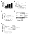
Figure 3. Inflammatory stimuli and other stresses interfere with si20-mediated mRNA degradation
A) HEK-293T cells were transfected with pSuper-si20 and CMV-ΔARE and cultured for 40 h. One hour before total RNA isolation, cells were treated with PBS, 50ng/ml IFN-α, 50ng/ml Il-1β or 50ng/ml TNF-α. Samples were analyzed as in 2A B) HEK-293T cells were transfected and cultured as in A. Prior to RNA isolation, cells were treated with 50ng/ml TNF-α for 0.25, 0.5, 1, 2, and 4 hours. Quantitative one-step RT-PCR assays were run to measure ΔARE mRNA levels in HEK-293T cells. ΔARE mRNA levels were normalized against GAPDH mRNA levels. ΔARE mRNA increases were calculated by using the time 0 point as 1. C) HeLa Tet-off cells were transfected with Tet-ΔARE and pSuper-si20 and cultured for 40h. Then cells were treated PBS or 50 ng/ml of TNF-α 15 min. prior to doxycycline treatment. Samples were analyzed as in B. ΔARE mRNA levels were expressed as a percentage of the amount existing at time 0. D) As C, but the presence of ΔARE mRNA in the cells was analyzed by Northern-blotting. Re-probing β-actin was used to ensure equal loading. E) As C, but HeLa Tet-off cells were pretreated with PBS (PBS), hydrogen peroxide (H2O2), MNNG, or anisomycin (Aniso) 30 min. before the addition of doxycycline. Samples were analyzed as in B. ΔARE mRNA levels were normalized against GAPDH mRNA levels. ΔARE mRNA levels were expressed as a percentage of the amount existing at time 0.
Figure 4
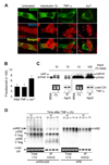
Figure 4. TNF-α stimulation does not affect Ago2’s localization to p-bodies and has no effect on siRNA/miRNA and mRNA loading into Ago2 complexes
A) HeLa cells transfected with Flag-Ago2 were treated with interleukin-1β, TNF-α or As3+ for 1 hour. Cells were fixed, permeabilized, and stained for Flag-Ago2 (green), TIA-1 (red), or Dcp1a (blue). Panels show merged pictures of TIA-1 and Dcp1a (2nd row) and merged pictures of Flag-Ago2, TIA-1, and Dcp1a (third row). B) Average p-body counts per cell (N = 60) from A). C) HEK-293T cells were cotransfected with Flag-Ago2 and pSupermiR16, or Flag-Ago2 and pSuper-si20. Cells were stimulated for 30 min. with or without TNF-α, lysed and subjected to anti-Flag immunoprecipitations. Total RNAs were extracted from the immunoprecipitates and subjected to primer extension to detect si20 and miR16 in Ago2 complexes, whereas aliquots were conserved for protein analysis by anti-Flag western-blots. Ribosomal RNAs were used as cell lysate loading controls, and were visualized on agarose gels by ethidium bromide staining. D) HEK-293T cells were cotransfected with Flag-Ago2 and CMV-ARE, or CMV-ΔARE and pSuper-si20. Cells were stimulated with TNF-α (for 0, 0.5, 1, 2, or 4 hours), lysed, and then subjected to anti-Flag immunoprecipitations. Total RNAs were extracted and subjected to Northern-blot analysis, and aliquots of the samples were conserved for protein analysis by anti-Flag western-blotting. Ribosomal RNAs were used as cell lysate loading control, and were visualized on agarose gels by ethidium bromide staining.
Figure 5
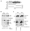
Figure 5. Capping and Poly(A) tail of si20 targeted ΔARE mRNAs after TNF-α stimulation
A)Schematic representation of the fragments generated by RNAse H during ΔARE mRNA poly(A) tail analysis. B) HEK-293T cells were transfected with CMV-31 ΔARE (ΔARE), or CMV-ΔARE plus pSuper-si20 (ΔARE/si20). Cells were treated with TNF-α for 0, 15, or 45 min. and total RNA was isolated. Cap-containing mRNAs were immunoprecipitated with K121 anti-2,2,7-trimethylguanosine agarose-conjugated mouse monoclonal antibodies. Northern-blotting was done with RNA extracted from cell lysates and after K121 immunoprecipitation. C) HeLa-Tet-off cells were transfected with Tet-ΔARE (ΔARE), or Tet-ΔARE plus pSuper-si20 (ΔARE/si20). Cells were treated for 15 min. with PBS or TNF-α prior to doxycycline addition for 0, 0.5, 1, and 2 hours. RNA was extracted and subjected to RNAse H treatment to visualize poly(A) tail length. As a control of deadenylation, oligo d(T)20 was added to remove the poly(A) tail. Northern-blots were used to analyze poly (A) tail distributions.

Similar articles

Cited by

References

    1. Khabar KS, Young HA. Post-transcriptional control of the interferon system. Biochimie. 2007;89:761–769. - PMC - PubMed
    1. Wang J, Fillebeen C, Chen G, Biederbick A, Lill R, Pantopoulos K. Iron-dependant degradation of apo-IRP1 by the ubiquitin-proteasome pathway. Mol. Cell. Biol. 2007;27:2423–2430. - PMC - PubMed
    1. Quattrone A, Pascale A, Nogues X, Zhao W, Gusev P, Pacini A, Alkon DL. Postranscriptional regulation of gene expression in learning by the neuronal ELAV-like mRNA-stabilizing proteins. Proc. Natl. Acad. Sci. USA. 2001;98:11668–116673. - PMC - PubMed
    1. Verrechia F, Mauviel A. Transforming growth factor-beta and fibrosis. World J. Gastroenterol. 2007;13:3056–3062. - PMC - PubMed
    1. Lubberts E, Koenders MI, Van den Berg WB. The role of T-cell interleukin-17 in conducting destructive arthritis: lessons from animal models. Arthritis Res. Ther. 2005;7:29–37. - PMC - PubMed

Publication types

MeSH terms

LinkOut - more resources