. 2008 Jun 13;283(24):16320-31.
doi: 10.1074/jbc.M801687200. Epub 2008 Apr 16.

Insulin-like growth factor-I stimulates Shc-dependent phosphatidylinositol 3-kinase activation via Grb2-associated p85 in vascular smooth muscle cells

Affiliations

Insulin-like growth factor-I stimulates Shc-dependent phosphatidylinositol 3-kinase activation via Grb2-associated p85 in vascular smooth muscle cells

Yashwanth Radhakrishnan et al. J Biol Chem. .

Abstract

Insulin-like growth factor-I (IGF-I) stimulates vascular smooth muscle cell proliferation and migration by activating both MAPK and phosphatidylinositol 3-kinase (PI3K). Vascular smooth muscle cells (VSMCs) maintained in 25 mm glucose sustain MAPK activation via increased Shc phosphorylation and Grb2 association resulting in an enhanced mitogenic response compared with cells grown in 5 mm glucose. PI3K plays a major role in IGF-I-stimulated VSMC migration, and hyperglycemia augments this response. In contrast to MAPK activation the role of Shc in modulating PI3K in response to IGF-I has not been determined. In this study we show that impaired Shc association with Grb2 results in decreased Grb2-p85 association, SHPS-1-p85 recruitment, and PI3K activation in response to IGF-I. Exposure of VSMCs to cell-permeable peptides, which contained polyproline sequences from p85 proposed to mediate Grb2 association, resulted in inhibition of Grb2-p85 binding and AKT phosphorylation. Transfected cells that expressed p85 mutant that had specific prolines mutated to alanines resulted in less Grb2-p85 association, and a Grb2 mutant (W36A/W193A) that attenuated p85 binding showed decreased association of p85 with SHPS-1, PI3K activation, AKT phosphorylation, cell proliferation, and migration in response to IGF-I. Cellular exposure to 25 mm glucose, which is required for Shc phosphorylation in response to IGF-I, resulted in enhanced Grb2 binding to p85, activation of PI3K activity, and increased AKT phosphorylation as compared with cells exposed to 5 mm glucose. We conclude that in VSMCs exposed to hyperglycemia, IGF-I stimulation of Shc facilitates the transfer of Grb2 to p85 resulting in enhanced PI3K activation and AKT phosphorylation leading to enhanced cell proliferation and migration.


Figures
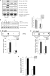
FIGURE 1.
FIGURE 1.
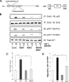
Shc mediates IGF-I-stimulated PI 3-kinase/AKT activation and cell migration in VSMCs in 25 mm glucose. A, confluent Shc-WT and Shc-3F cells were serum-starved for 14 h in DMEM-HG and then exposed to IGF-I (100 ng/ml) for 10 min. The extent of Shc phosphorylation was determined by immunoprecipitating p52Shc and then immunoblotting with an anti-phosphotyrosine antiserum. After stripping membranes were then reprobed with anti-Shc antibody to detect total Shc (upper panels). Similarly the lysates were immunoprecipitated with anti-Grb2 antibody and immunoblotted for p52Shc and p85. Membranes were then stripped and reprobed with anti-Grb2 antibody to detect total Grb2 (middle panels). Further lysates were immunoprecipitated with anti-SHPS-1 antibody and immunoblotted for p85. Subsequently the membranes were stripped and then reprobed with anti-SHPS-1 antibody to detect total SHPS-1 (lower panels). B, VSMCs expressing Shc-WT and Shc-3F were grown to confluency in DMEM-HG and then placed in serum-free medium for 14 h. Cultures were stimulated with or without IGF-I (100 ng/ml) for the time points indicated. Cell lysates were immunoblotted using the anti-phospho-AKT Ser-473 (top panel) and Thr-308 antibodies (middle panel). The blots were stripped and reprobed using anti-β-actin antibody. The bar graph shows the relative increase in AKT phosphorylation for at least three independent experiments and two independent transductions. Error bars represent mean ± S.E. **, p < 0.01; *, p < 0.05. C, confluent Shc-WT and Shc-3F cell cultures were grown to confluency in DMEM-HG, and maintained in this medium containing 0.2% serum for 14 h, and then treated with or without IGF-I (100 ng/ml) for 10 min. The anti-p85 immunoprecipitates were washed, and the pellets were incubated with PI3K and [32P]ATP as described under “Materials and Methods.” Radiolabeled PI3K was separated by thin layer chromatography. Representative autoradiograms are shown. The results are expressed as arbitrary scanning units, from three independent kinase assays and the error bars represent the mean ± S.E. D, confluent Shc-WT and Shc-3F cell cultures were serum-starved overnight and stimulated with IGF-I (50 ng/ml) for 10 min. The PI 3-kinase complex was immunoprecipitated using anti-Grb2 antibody, and the immunoprecipitates were analyzed for kinase activity as described under “Materials and Methods.” Radiolabeled PI3K is shown as PIP3. The bar graph shows the data expressed as arbitrary scan units (mean ± S.E.) (n = 3 independent experiments). ***, p < 0.001 when kinase activity in the presence of IGF-I is compared with that in nonstimulated Shc-WT cells; *, p < 0.05 when the kinase activity in the presence of IGF-I is compared between the Shc-WT and Shc-3F cells. E, migration of VSMCs expressing Shc-WT or the Shc-3F mutant. Shc-WT and Shc-3F cells were grown in 6-well dishes in DMEM-HG containing 10% FBS. After wounding they were allowed to migrate with or without IGF-I (100 ng/ml) in medium containing 0.2% FBS for 48 h. The total number of cells migrating past the wound line in the predetermined areas was counted, and the results shown are the mean ± S.E. of three independent experiments. *** indicates p < 0.001 and * indicates p < 0.05 when the number of cells migrating in response to IGF-I is compared with that in nonstimulated cells for the Shc-WT and Shc-3F cells, respectively. Cont, control; IB, immunoblot; IP, immunoprecipitation; 3F, Shc-3F; p-AKT, phospho-AKT.
FIGURE 2.
FIGURE 2.
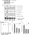
p85 polyproline region disruption attenuates Grb2-p85 association and AKT phosphorylation. A, schematic presentation of p85α subunit. The SH3 domain is located near the N terminus. The breakpoint cluster region is flanked by two proline-rich regions, P1 and P2, on either side. Following P2 is an SH2 domain, and another SH2 domain is located in the C-terminal region. Grb2 contains an SH2 domain and is flanked by two SH3 domains. The proline-rich region of p85 directly associates with the SH3 domain of the Grb2. Mutation of amino acid tryptophan at sites 36 and 193 to lysine in the SH3 domains of Grb2 impairs its association with p85 (modified from Kapeller et al. (19)). B, confluent VSMCs were serum-starved overnight in DMEM-HG and then incubated with or without cell-permeable Grb2-p85 peptides 264 and 265 (20 μg/ml) each for 2 h either individually or in combination. IGF-I (50 ng/ml) was added for 10 min. Cell lysates were immunoprecipitated with anti-Grb2 antibody and then immunoblotted with anti-p85 antibody (first panel). Blots were stripped and reblotted with anti-Grb2 antibody (second panel). Cell lysates were directly immunoblotted using anti-phospho-AKT Ser-473 (third panel) or Thr-308 (fourth panel) and for total AKT (bottom panel). C, VSMCs were plated at a density of 3 × 10⁴ cells/well in DMEM-HG + 2% FBS in 24-well plates and allowed to attach overnight. They were incubated with or without cell-permeable peptides 264 and 265 (20 μg/ml) together for 2 h before exposure to IGF-I (50 ng/ml) in DMEM + 0.2% platelet-poor plasma. Forty-eight hours after the addition of IGF-I, cell number was determined by trypan blue staining and counting. **, p < 0.01 when the cell number in response to IGF-I is compared with that in nonstimulated VSMCs and when the cell number in response to IGF-I is compared with that in cells treated with both peptides. Error bars represent the mean ± S.E. D, VSMCs were grown to confluency in DMEM-HG, wounded, and allowed to migrate with or without cell-permeable peptides 264 and 265 (20 μg/ml) added together for 2 h before stimulation with IGF-I (100 ng/ml) in medium containing 0.2% FBS for 48 h. The number of cells migrating in the predetermined areas was counted, and the results shown are the mean ± S.E. of three independent experiments. **, p < 0.01 when the number of migrating cells in response to IGF-I in cells is compared with that in nonstimulated VSMCs; *, p < 0.05 when the cell number in response to IGF-I is compared with that in cells treated with both peptides. IP, immunoprecipitation; IB, immunoblot; Cont, control; pAKT, phospho-AKT; Pep, peptide.
FIGURE 3.
FIGURE 3.
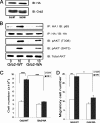
Disruption of p85 binding to Grb2 attenuates IGF-I-stimulated PI 3-kinase activation, AKT phosphorylation, and cellular responses. A, VSMCs expressing p85-WT, mutant p85-P1, and mutant p85-P2 were serum-starved overnight in DMEM-HG and analyzed for recombinant protein expression. Cell lysates were immunoblotted using an anti-HA antibody (top) and anti-p85 antibody (bottom). B, the p85-WT and p85-P1 and p85-P2 mutant cells were serum-starved overnight in DMEM-HG before stimulation with IGF-I (100 ng/ml) for 10 min. The lysates were immunoprecipitated with anti-Grb2 antibody and immunoblotted with anti-p85 antibody (first panel). The blots were stripped and reprobed with anti-Grb2 antibody (second panel). Similarly cell lysates were immunoblotted with anti-phospho-AKT Ser-473 (third panel) and Thr-308 (fourth panel) and for total AKT (fifth panel). Lysates were also immunoprecipitated with anti-SHPS-1 antibody and immunoblotted for p85 (sixth panel). Subsequently the membranes were stripped and then reprobed with anti-SHPS-1 antibody to detect total SHPS-1 (bottom panel). C, confluent p85-WT and p85-P1 cell cultures were serum-starved (in DMEM-HG, 0.2% FBS) overnight and stimulated with IGF-I (50 ng/ml) for 10 min. The PI 3-kinase complex was immunoprecipitated by using anti-p85 antibody, and the immunoprecipitates were analyzed for kinase activity. PI 3-kinase assay was performed three times, and a representative phosphorimage is shown. PIP3 indicates the radiolabeled phosphatidylinositol 3,4,5-trisphosphate. The origin denotes the location where the reaction products were spotted. D, p85-WT, p85-P1, and p85-P2 cells were plated at a density of 3 × 10⁴ cells/well in DMEM-HG, 2% FBS in 24-well plates for 16 h prior to exposure to IGF-I (50 ng/ml) in DMEM + 0.2% platelet-poor plasma. After 48 h, cell number was determined by trypan blue staining and counting. ***, p < 0.001 when the cell number after exposure to IGF-I in p85-WT cells is compared with that in nonstimulated p85-WT cells. * indicates p < 0.05 and ** indicates p < 0.01 when the cell number in response to IGF-I in p85-P1 and p85-P2 mutant cells is compared with that p85-WT cells. Error bars represent the mean ± S.E. E, cell migration in response to IGF-I in VSMCs expressing p85-WT and p85-P1 mutant cells. p85 wild type and mutant cells were grown to confluency in DMEM-HG, wounded, and allowed to migrate with or without IGF-I (100 ng/ml) in medium containing 0.2% FBS for 48 h. The number of cells migrating in the predetermined areas was counted, and the results shown are the mean ± S.E. of three independent experiments. ***, p < 0.001 when the number of p85WT cells migrating in response to IGF-I is compared with that in nonstimulated p85-WT cells. * indicates p < 0.05 and ** indicates p < 0.01 when the number of cells migrating in response to IGF-I is compared between p85-WT and p85-P1 mutant or p85-P2 mutant cells, respectively. pAKT, phospho-AKT; IB, immunoblot; IP, immunoprecipitation; Cont, control.
FIGURE 4.
FIGURE 4.
Grb2 mediates IGF-I-stimulated AKT activation, proliferation, and migration responses in VSMCs. A, VSMCs expressing Grb2-WT and VSMCs expressing Grb2-WA were serum-starved in DMEM-HG and analyzed for recombinant protein expression. Cell lysates were immunoblotted using an anti-HA antibody (top) and anti-Grb2 antibody (bottom). B, Grb2-WT- and the mutant (Grb2-WA)-expressing cells were serum-starved in DMEM-HG overnight, and then IGF-I (100 ng/ml) was added for 10 min. Cell lysates were immunoprecipitated with anti-HA antibody and immunoblotted with anti-p85 antibody (top panel). Blots were stripped and reblotted with anti-HA antibody (second panel). In addition, cell lysates were immunoblotted with anti-phospho-AKT antibody for Thr-308 (third panel) and Ser-473 (fourth panel) and for total AKT (bottom panel). Each experiment was performed at least three times. C, Grb2-WT and Grb2-WA mutant cells (3 × 10⁴) were plated in DMEM-HG + 2% FBS before exposure to IGF-I (50 ng/ml) in DMEM-HG + 0.2% platelet-poor plasma. Forty-eight hours after the addition of IGF-I, cell number was determined by trypan blue staining and counting. ***, p < 0.001 when the change in cell number in response to IGF-I in Grb2-WT cells is compared with that in the control or when the response to IGF-I in Grb2-WT cells is compared with that in Grb2-WA cells. Error bars represent mean ± S.E. D, Grb2 wild type and Grb2-WA mutant cells were grown to confluency in DMEM-HG, wounded, and allowed to migrate with or without IGF-I (100 ng/ml) in DMEM-HG containing 0.2% FBS for 48 h. The number of cells migrating in the predetermined areas was counted, and the results shown are the mean ± S.E. of three independent experiments. **, p < 0.01 when the number of Grb2-WT cells after IGF-I is compared with that in nonstimulated control; *, p < 0.05 when the number of migrating cells after IGF-I is compared between Grb2-WT and Grb2-WA cells. IP, immunoprecipitation; IB, immunoblot; pAKT, phospho-AKT; Cont, control.
FIGURE 5.
FIGURE 5.
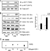
Hyperglycemia increases IGF-I-mediated AKT activation and Shc-Grb2-p85 association in porcine VSMCs. A, quiescent VSMC cultures were incubated in serum-free medium containing 5 (NG) or 25 (HG) mm glucose overnight. Cells were stimulated with IGF-I (100 ng/ml) for 10 min. After cell lysis aliquots containing equal amounts of protein were separated directly by SDS-PAGE and immunoblotted (IB) with anti-phospho-AKT-specific antibodies for phosphorylation of threonine 308 (upper panel) and serine 473 (middle panel) and also for total AKT (bottom panel). B, the extent of Shc association with Grb2 was determined by immunoprecipitating (IP) cell lysates with an anti-Grb2 antibody and then immunoblotting with an anti-Shc antibody. Similarly the extent of the p85 association with Grb2 was determined by immunoprecipitation using an anti-Grb2 antibody and then immunoblotting with an anti-p85 antibody. Membranes were then stripped and reprobed with anti-Grb2 antibody to detect total Grb2 in each sample. In addition, cell lysates were immunoblotted with anti-p85 (fourth panel) and anti-p52Shc antibodies (fifth panel). The blots were stripped and reprobed using anti-β-actin antibody to control for differences in loading (bottom panel). All experiments were repeated at least three times. Error bars represent mean ± S.E. **, p < 0.01. C, VSMCs cultured in normal glucose or high glucose were serum-starved (0.2% FBS) and stimulated with IGF-I (50 ng/ml) for 10 min. The PI 3-kinase complex was immunoprecipitated by using anti-Grb2 antibody, and the immunoprecipitates were analyzed for kinase activity. The PI 3-kinase assay was performed twice, and a representative phosphorimage is shown. PIP3 indicates the radiolabeled phosphatidylinositol 3,4,5-trisphosphate. p-AKT, phospho-AKT; Cont, control.
FIGURE 6.
FIGURE 6.
IGF-I-induced activation of PI 3-kinase/AKT and MAPK pathways in VSMCs. VSMCs maintained in HG conditions upon IGF-I stimulation show enhanced activation of the MAPK and PI3K/AKT pathways leading to increased proliferation and migration compared with cells grown in NG. IGF-I-stimulated Shc mediates activation of MAPK via Grb2-Sos binding. In HG conditions Shc-mediated, Grb2-associated p85 recruitment to SHPS-1 activates the PI3K/AKT pathway increasing VSMC proliferation and migration in response to IGF-I stimulation.

Similar articles

Cited by

References

    1. Clemmons, D. R. (2007) Nat. Rev. Drug Discov. 6 821–833 - PubMed
    1. Ling, Y., Maile, L. A., and Clemmons, D. R. (2003) Mol. Endocrinol. 17 1824–1833 - PubMed
    1. Ling, Y., Maile, L. A., Lieskovska, J., Badley-Clarke, J., and Clemmons, D. R. (2005) Mol. Biol. Cell 16 3353–3364 - PMC - PubMed
    1. Imai, Y., and Clemmons, D. R. (1999) Endocrinology 140 4228–4235 - PubMed
    1. Maile, L. A., Capps, B. E., Ling, Y., Xi, G., and Clemmons, D. R. (2007) Endocrinology 148 2435–2443 - PubMed

Publication types

MeSH terms

Substances