Review
. 2008 May;6(5):363-74.
doi: 10.1038/nrmicro1890.

Modification of intracellular membrane structures for virus replication

Affiliations
Review

Modification of intracellular membrane structures for virus replication

Sven Miller et al. Nat Rev Microbiol. 2008 May.

Abstract

Viruses are intracellular parasites that use the host cell they infect to produce new infectious progeny. Distinct steps of the virus life cycle occur in association with the cytoskeleton or cytoplasmic membranes, which are often modified during infection. Plus-stranded RNA viruses induce membrane proliferations that support the replication of their genomes. Similarly, cytoplasmic replication of some DNA viruses occurs in association with modified cellular membranes. We describe how viruses modify intracellular membranes, highlight similarities between the structures that are induced by viruses of different families and discuss how these structures could be formed.


Figures

Figure 1
Figure 1. Intracellular trafficking pathways and sites of membrane alterations that are induced by different viruses.
Schematic representation of a cell and different intracellular organelles. Proteins that are destined for secretion enter the secretory pathway by co-translational translocation into the endoplasmic reticulum (ER) (pink dots represent ribosomes). These proteins are then transported in a coatomer protein complex (COP) II-dependent way to the Golgi complex in a process that probably involves COPII-coated vesicles and membrane structures that are located in the intermediate compartment between the ER and the Golgi complex. Proteins can be recycled back to the ER using COPI-coated vesicles or can be transported through the Golgi complex. At the trans–Golgi network, they leave the Golgi and are transported to the plasma membrane. Endocytosis is initiated at the plasma membrane, and proteins are packed into clathrin-coated vesicles before being transported to early and late endosomes. From there, they are either recycled back to the plasma membrane or are degraded in lysosomes. The putative sites where different viruses modify intracellular membranes to assemble their replication complexes are indicated. EAV, equine arteritis virus; FHV, flock house virus; HCV, hepatitis C virus; KUNV, Kunjin virus; MHV, murine hepatitis virus; SARS-CoV, severe acute respiratory syndrome coronavirus; SFV, Semliki Forest virus; TMV, tobacco mosaic virus.
Figure 2
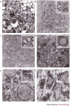
Figure 2. Electron microscopy (EM) images of the membrane alterations that are induced by different viruses.
a | Poliovirus-infected HeLa cells fixed 6–8 hours post infection. b | Hepatitis C virus-infected Huh7 cells. c | Dengue virus-infected Huh7 cells fixed 24 hours post infection. d | Severe acute respiratory syndrome coronavirus-infected Vero cells, showing a cluster of double-membrane vesicles (DMVs). The inset shows one DMV at a higher magnification. e | Vaccinia virus-infected cells, showing a replication site that is surrounded by the rough ER. The inset shows the ER membrane at a higher magnification, with the ribosomes on the outer membrane facing the cytoplasm. f | Semliki Forest virus-infected baby hamster kidney cells fixed 3 hours post infection, showing the typical cytopathic vacuoles that are induced upon infection. ER, endoplasmic reticulum; G, Golgi apparatus; M, mitochondrion; N, nucleus; RS, replication site. Part a is courtesy of K. Bienz and D. Egger, University of Basel, Switzerland; parts b and c are courtesy of R. Bartenschlager, University of Heidelberg, Germany; part d is courtesy of E. Snijder, M. Mommas and K. Knoops, Leiden University, The Netherlands; part e is reproduced, with permission, from Ref. © (2005) Blackwell Publishing; and part f is courtesy of T. Ahola and G. Balistrer, University of Helsinki, Finland.
Figure 3
Figure 3. Models for the formation of virus-induced double-membrane vesicles.
The protrusion and detachment model (a) proposes that part of the endoplasmic reticulum (ER) cisterna starts to bend, pinches off and then seals to form a double-membrane vesicle (DMV). Interactions between the lumenal domains of viral membrane proteins (coloured yellow in the ER membrane) could mediate the tight apposition of the two bilayers and induce curvature. In the double-budding model (b), a single-membrane vesicle buds into the lumen of the ER and then buds out again, and the membrane proteins could mediate inward as well as outward budding. Figure adapted, with permission, from Ref. © (1999) American Society for Microbiology.
Figure 4
Figure 4. Mechanisms of membrane-curvature induction.
Several mechanisms of membrane deformation by cellular proteins have been described. a | Integration of membrane proteins that have a conical shape induces curvature, as they act like a wedge that is inserted into the membrane. b | Amphipathic helices, stretches of alpha helices that have one polar and one hydrophobic side, are positioned flat on the membrane, with the hydrophobic side dipping into one of the two membrane layers: this causes destabilization of the membrane and membrane bending. c,d | Membrane bending can be induced by oligomerization of proteins that are integrated into, or associated with, cellular membranes. Proteins form a scaffold that makes the membrane bend. e | The lipid composition of a membrane can also induce membrane curvature. In this context, the head group, as well as the acyl chain of the membrane constituents, can have an effect on membrane curvature. Figure adapted, with permission, from Nature Ref. © (2005) Macmillan Publishers Ltd.
Figure 5
Figure 5. Hypothetical model for biogenesis and topology of the hepatitis C virus replication complex.
Upon release of viral genomic RNA into the cytoplasm of the infected cell (1), the viral genome is translated into a polyprotein that carries the structural (pink) and non-structural (green) proteins (2). The viral non-structural protein NS4B induces the formation of membrane alterations, which serve as a scaffold for the assembly of the viral replication complex (RC) (3). The RC consists of viral non-structural proteins, viral RNA and host cell factors. Within the induced vesicles, viral RNA is amplified via a negative-strand RNA intermediate. Figure adapted, with permission, from Ref. © (2005) American Society for Microbiology.

Similar articles

Cited by

References

    1. Klumperman J. Transport between ER and Golgi. Curr. Opin. Cell Biol. 2000;12:445–449. doi: 10.1016/S0955-0674(00)00115-0. - DOI - PubMed
    1. Kirchhausen T. Three ways to make a vesicle. Nature Rev. Mol. Cell Biol. 2000;1:187–198. doi: 10.1038/35043117. - DOI - PubMed
    1. Mackenzie JS, Gubler DJ, Petersen LR. Emerging flaviviruses: the spread and resurgence of Japanese encephalitis, West Nile and dengue viruses. Nature Med. 2004;10:S98–S109. doi: 10.1038/nm1144. - DOI - PubMed
    1. Moradpour D, Penin F, Rice CM. Replication of hepatitis C virus. Nature Rev. Microbiol. 2007;5:453–463. doi: 10.1038/nrmicro1645. - DOI - PubMed
    1. Moss B, et al. Fields Virology. 2001. pp. 2849–2883.