. 2008 Apr 21;181(2):213-26.
doi: 10.1083/jcb.200708210. Epub 2008 Apr 14.

Phosphorylation of SDT repeats in the MDC1 N terminus triggers retention of NBS1 at the DNA damage-modified chromatin

Affiliations

Phosphorylation of SDT repeats in the MDC1 N terminus triggers retention of NBS1 at the DNA damage-modified chromatin

Fredrik Melander et al. J Cell Biol. .

Abstract

DNA double-strand breaks (DSBs) trigger accumulation of the MRE11-RAD50-Nijmegen breakage syndrome 1 (NBS1 [MRN]) complex, whose retention on the DSB-flanking chromatin facilitates survival. Chromatin retention of MRN requires the MDC1 adaptor protein, but the mechanism behind the MRN-MDC1 interaction is unknown. We show that the NBS1 subunit of MRN interacts with the MDC1 N terminus enriched in Ser-Asp-Thr (SDT) repeats. This interaction was constitutive and mediated by binding between the phosphorylated SDT repeats of MDC1 and the phosphate-binding forkhead-associated domain of NBS1. Phosphorylation of the SDT repeats by casein kinase 2 (CK2) was sufficient to trigger MDC1-NBS1 interaction in vitro, and MDC1 associated with CK2 activity in cells. Inhibition of CK2 reduced SDT phosphorylation in vivo, and disruption of the SDT-associated phosphoacceptor sites prevented the retention of NBS1 at DSBs. Together, these data suggest that phosphorylation of the SDT repeats in the MDC1 N terminus functions to recruit NBS1 and, thereby, increases the local concentration of MRN at the sites of chromosomal breakage.


Figures
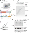
Figure 1.
Figure 1.
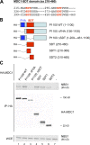
Interaction between NBS1 and MDC1 is mediated by the FHA domain of NBS1 and the first 500 amino acids of MDC1. (A) Endogenous MDC1 and NBS1 interact both before and after DNA damage. Lysates from U2OS cells either mock or γ irradiated (10 Gy for 1 h) were subjected to immunoprecipitation with an anti-MDC1 antibody and analyzed for the presence of NBS1 by immunoblotting. A nonimmune species-matched antibody was used as a control. Expression levels of NBS1 in whole cell extracts (bottom) indicate an equal input in each lane. (B) A schematic structure of MDC1 and NBS1. The red segment indicates the N-terminal region of MDC1 (amino acids 1–1,100) subjected to progressive deletion and interaction analysis with NBS1. (C) The N-terminal region of MDC1 spanning the first 500 amino acids efficiently interacts with cellular NBS1. The indicated HA-tagged fragments of the MDC1 N terminus were transfected into MDC1/shRNA cells (the numbers indicate the C-terminal amino acid of each fragment). After 24 h, lysates were prepared, subjected to immunoprecipitation with an anti-HA antibody, and analyzed by immunoblotting with antibodies to NBS1 (top) and HA (middle). The input into each reaction was controlled by NBS1 immunoblotting of the whole cell extracts as in A (bottom). (D) Efficient knockdown of endogenous MDC1. Lysates from U2OS cells and its derivative in which MDC1 is knocked down by stably integrated shRNA (MDC1/shRNA) were analyzed by immunoblotting with an MDC1-specific antibody (top). The NBS1 immunoblot serves as a loading control and indicates that the overall levels of NBS1 are not affected by the MDC1 knockdown (bottom). (E) The FHA domain of NBS1 interacts with the MDC1 N terminus. MDC1/shRNA cells were cotransfected with the HA-tagged MDC1 N terminus (p1100) and the following Myc-tagged variants of NBS1: WT (wild type), R28 (nonfunctional FHA domain), ΔC (C-terminal deletion without the MRE11 and ATM interaction domains), and 3A (alanine substitutions of serines 278, 343, and 397, the ATM phosphorylation sites). After 24 h, lysates were immunoprecipitated with an anti-HA antibody and analyzed by immunoblotting with anti-Myc (top) and anti-HA (middle) antibodies. The bottom panel is an immunoblot of whole cell extracts and shows the input of each of the Myc-tagged NBS1 proteins. WCE, whole cell extract; PST, Pro-Ser-Thr rich.
Figure 2.
Figure 2.
The SDT-rich region within the MDC1 N terminus promotes MDC1–NBS1 interaction. (A) The N-terminal part of MDC1 delineated by amino acids 218 and 455 contains six acidophilic Ser-Thr-Asp (SDT) repeats. (B) A schematic depiction of the MDC1 fragments used in C for the NBS1 binding assay. (C) The SDT region is necessary for and sufficient to promote interaction of the MDC1 N terminus with NBS1. MDC1/shRNA cells were transfected with the HA-tagged MDC1 fragments described in B. After 24 h, lysates were prepared, immunoprecipitated with an anti-HA antibody, and analyzed by immunoblotting with NBS1 (top) and HA (middle) antibodies. The bottom panel is an immunoblot of whole cell extracts and shows equal input of each NBS1 in each lane. WCE, whole cell extract.
Figure 3.
Figure 3.
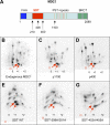
MDC1 N terminus is phosphorylated in vivo, and some of the prominent phosphorylation sites include the SDT clusters. (A) A schematic diagram of MDC1 and the position of the fragments used to identify phosphorylation sites in vivo. (B–G) Examples of SDT-associated Ser/Thr phosphorylations in vivo. U2OS cells either untransfected (B) or transfected with the indicated HA-tagged MDC1 constructs (C–G) were labeled with 32P for 4 h. Lysates from labeled cells were immunoprecipitated with anti-MDC1 (B) or anti-HA (C–G) antibodies, and the isolated proteins were subjected to tryptic digestion. The resulting peptides were separated by electrophoresis followed by thin-layer chromatography and analyzed by phosphorimager. The spots indicated by arrows mark prominent phosphopeptides derived from Ser299/Thr301 (thin arrows) and Ser453/Thr455 (bold arrows). Open circles mark the loading spots, and numbers indicate the charge of the phosphopeptides. Dotted arrows indicate the absence of a phosphopeptide spot.
Figure 4.
Figure 4.
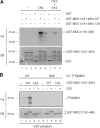
Phosphorylation of the SDT clusters by CK2 promotes MDC1–NBS1 interaction in vitro. (A) CK2 phosphorylates the SDT repeats of MDC1 in vitro. GST-MDC1 fragment spanning the entire SDT region (WT; amino acids 181–480) or its derivative in which all 12 potential CK2 sites were mutated to alanines (12A) was purified from E. coli and subjected to an in vitro kinase assay using a recombinant CK2 and 32P-labeled ATP. Where indicated, 10 μM of the DMAT CK2 inhibitor (CK2i) was added to the reaction. After separation by SDS-PAGE, the gel was stained with Coomassie blue to validate the equal input of the GST-MDC1 fragments (bottom) and was dried and analyzed by phosphorimager (top). (B) The purified SDT region of MDC1 interacts with NBS1 in a phosphorylation- and CK2-dependent manner. GST-MDC1 fragments (as in A) were phosphorylated or not phosphorylated with recombinant CK2 (using nonradioactive ATP), captured on glutathione–Sepharose beads, and incubated with in vitro–translated 35S-labeled NBS1 WT or its derivative with a point mutation within the FHA domain (R28) for 2 h. Bound complexes were resolved by SDS-PAGE. The gel was stained with Coomassie blue to validate equal input of the GST-MDC1 proteins (bottom), and the bound NBS1 was analyzed by phosphorimager (top). CB, Coomassie blue.
Figure 5.
Figure 5.
Inhibition of CK2 reduces phosphorylation of the SDT repeats in vivo. (A) Treatment of cells with a CK2 inhibitor reduces phosphate incorporation into the SDT-containing fragment of MDC1. U2OS cells were transfected with the HA-tagged fragment of MDC1 spanning the entire SDT region (amino acids 210–460) and labeled with 32P for 4 h. Where indicated, 10 μM of the DMAT CK2 inhibitor (CK2i) was added to the culture medium for the entire labeling period. Lysates from labeled cells were immunoprecipitated with an anti-HA antibody and, after resolving the proteins on SDS-PAGE, were analyzed by the phosphorimager for the extent of 32P incorporation (top). The equal input of proteins in each lane was subsequently verified by immunoblotting with the anti-HA antibody (bottom). (B) Inhibition of CK2 impairs phosphate incorporation into the dominant SDT repeats. The bands corresponding to the labeled SDT fragments from A were excised from the gel and subjected to tryptic digestion. The resulting phosphopeptides were separated by electrophoresis followed by thin-layer chromatography and analyzed by phosphorimager. Note that the 32P incorporation into two prominent spots representing Ser299/Thr301 (thin arrows) and Ser453/Thr455 (bold arrows) is reduced to ∼50% in cells treated with CK2 inhibitor (CK2i). Dotted circles indicate spots whose labeling did not appreciably change after CK2 inhibition; the intensity of the 32P signal associated with these spots was taken as a baseline to estimate the decrease in phosphate incorporation into the SDT repeats. Numbers indicate the charge of the phosphopeptides.
Figure 6.
Figure 6.
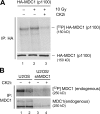
MDC1 associates with CK2 activity. (A) MDC1 N terminus copurifies with CK2 activity. U2OS cells were transfected with HA-tagged MDC1 N terminus (p1100). After 24 h, the cells were left untreated or were subjected to 10 Gy IR for 1 h. Cells were then lysed, and the MDC1 fragment was immunoprecipitated with an anti-HA antibody and assayed for the ability to undergo autophosphorylation in an in vitro kinase assay. Where indicated, the kinase reaction was supplemented with 10 μM of the DMAT CK2 inhibitor (CK2i). After SDS-PAGE and transfer to nitrocellulose, the extent of MDC1 phosphorylation was analyzed by phosphorimager (top), and the equal input of MDC1 in each reaction was validated by immunoblotting with the anti-HA antibody (bottom). (B) Endogenous MDC1 associates with CK2 activity. Lysates from asynchronously growing U2OS or U2OS/shMDC1 cells were subjected to immunoprecipitation with an MDC1-specific antibody and assayed for autophosphorylation of endogenous MDC1 (top). The equal input of MDC1 in each reaction was validated by immunoblotting with the anti-MDC1 antibody (bottom). Where indicated, 500 nM of the DMAT CK2 inhibitor (CK2i) was added to the kinase reaction.
Figure 7.
Figure 7.
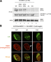
Disruption of the SDT phosphoacceptor sites impairs MDC1–NBS1 interaction and abrogates the retention of NBS1 at the DSB-modified chromatin. (A) Disruption of the SDT sites progressively inhibits MDC1–NBS1 interaction. U2OS cells were transfected with the HA-tagged MDC1 fragments spanning the intact N terminus (WT) and its derivatives where the indicated residues were substituted to alanines. The 4A fragment combines alanine substitutions of Ser299, Thr301, Ser453, and Thr455. In the 12A fragment, all of the 12 potential CK2 sites within the SDT region were mutated to alanines. After 24 h, lysates were prepared, immunoprecipitated with an anti-HA antibody, and analyzed by immunoblotting with the anti-NBS1 (top) and anti-HA (middle) antibodies. The immunoblotting of NBS1 from the whole cell extracts (bottom) validates an equal input of the lysate in each reaction. WCE, whole cell extract. (B) Disruption of the SDT sites impairs the sustained retention of NBS1 at the sites of DNA damage. MDC1/shRNA cells were transiently transfected with expression plasmids coding for the WT or the SDT-deficient 4A and 12A phosphorylation-deficient mutants of MDC1, respectively (the same mutations as in A but generated in the context of siRNA-insensitive full-length HA-MDC1). After 24 h, cells were subjected to laser microirradiation to generate local DSBs. After an additional 1 h, the cells were fixed and immunostained with antibodies to HA (to locate the DSB tracks) and to endogenous NBS1. The arrow indicates the residual amount of NBS1 retained in the DSB tracks in cells containing the MDC1-4A mutant. Bar, 10 μm.
Figure 8.
Figure 8.
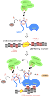
A hypothetical model of the MDC1–MRN interaction before and after DNA damage. In undamaged cells, CK2 targets the SDT-rich N terminus of MDC1. Phosphorylated SDTs are recognized by the FHA domain of NBS1 and allow dynamic interaction between MDC1 and the MRN complex (top). DSBs trigger the phosphorylation of H2AX (middle) followed by its recognition by the MDC1-associated BRCT domains (bottom). The resulting concentration of phosphorylated SDT repeated in the DSB-flanking chromatin allows immediate recruitment of the MRN complex to this compartment. CK2 (potentially together with another acidophilic kinase; indicated by an asterisk) may then stimulate MRN retention at the DSB sites by phosphorylating additional SDT repeats. Alternatively, the SDT-associated phosphates might be stabilized by inhibition of a DSB-associated protein phosphatase (PPase). Both mechanisms are compatible with a dynamic MRN exchange between distinct DSB-generated subcompartments but prevent its dilution in the undamaged nucleoplasm. See Discussion for an additional explanation.

Similar articles

Cited by

References

    1. Bartek, J., and J. Lukas. 2007. DNA damage checkpoints: from initiation to recovery or adaptation. Curr. Opin. Cell Biol. 19:238–245. - PubMed
    1. Beausoleil, S.A., M. Jedrychowski, D. Schwartz, J.E. Elias, J. Villen, J. Li, M.A. Cohn, L.C. Cantley, and S.P. Gygi. 2004. Large-scale characterization of HeLa cell nuclear phosphoproteins. Proc. Natl. Acad. Sci. USA. 101:12130–12135. - PMC - PubMed
    1. Bekker-Jensen, S., C. Lukas, F. Melander, J. Bartek, and J. Lukas. 2005. Dynamic assembly and sustained retention of 53BP1 at the sites of DNA damage are controlled by Mdc1/NFBD1. J. Cell Biol. 170:201–211. - PMC - PubMed
    1. Bekker-Jensen, S., C. Lukas, R. Kitagawa, F. Melander, M.B. Kastan, J. Bartek, and J. Lukas. 2006. Spatial organization of the mammalian genome surveillance machinery in response to DNA strand breaks. J. Cell Biol. 173:195–206. - PMC - PubMed
    1. Bekker-Jensen, S., K. Fugger, J.R. Danielsen, I. Gromova, M. Sehested, J. Celis, J. Bartek, J. Lukas, and N. Mailand. 2007. Human Xip1 (C2orf13) is a novel regulator of cellular responses to DNA strand breaks. J. Biol. Chem. 282:19638–19643. - PubMed

Publication types

MeSH terms