. 2008 Jul;7(7):1362-77.
doi: 10.1074/mcp.M800079-MCP200. Epub 2008 Apr 11.

Monitoring protein-protein interactions between the mammalian integral membrane transporters and PDZ-interacting partners using a modified split-ubiquitin membrane yeast two-hybrid system

Affiliations

Monitoring protein-protein interactions between the mammalian integral membrane transporters and PDZ-interacting partners using a modified split-ubiquitin membrane yeast two-hybrid system

Serge M Gisler et al. Mol Cell Proteomics. 2008 Jul.

Abstract

PDZ-binding motifs are found in the C-terminal tails of numerous integral membrane proteins where they mediate specific protein-protein interactions by binding to PDZ-containing proteins. Conventional yeast two-hybrid screens have been used to probe protein-protein interactions of these soluble C termini. However, to date no in vivo technology has been available to study interactions between the full-length integral membrane proteins and their cognate PDZ-interacting partners. We previously developed a split-ubiquitin membrane yeast two-hybrid (MYTH) system to test interactions between such integral membrane proteins by using a transcriptional output based on cleavage of a transcription factor from the C terminus of membrane-inserted baits. Here we modified MYTH to permit detection of C-terminal PDZ domain interactions by redirecting the transcription factor moiety from the C to the N terminus of a given integral membrane protein thus liberating their native C termini. We successfully applied this "MYTH 2.0" system to five different mammalian full-length renal transporters and identified novel PDZ domain-containing partners of the phosphate (NaPi-IIa) and sulfate (NaS1) transporters that would have otherwise not been detectable. Furthermore this assay was applied to locate the PDZ-binding domain on the NaS1 protein. We showed that the PDZ-binding domain for PDZK1 on NaS1 is upstream of its C terminus, whereas the two interacting proteins, NHERF-1 and NHERF-2, bind at a location closer to the N terminus of NaS1. Moreover NHERF-1 and NHERF-2 increased functional sulfate uptake in Xenopus oocytes when co-expressed with NaS1. Finally we used MYTH 2.0 to demonstrate that the NaPi-IIa transporter homodimerizes via protein-protein interactions within the lipid bilayer. In summary, our study establishes the MYTH 2.0 system as a novel tool for interactive proteomics studies of membrane protein complexes.


Figures
F<sc>ig</sc>. 1.
Fig. 1.
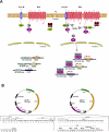
Principle of the MYTH 2.0 system. A, schema of the MYTH 2.0 basic principle to monitor protein-protein interactions. The MYTH 2.0 format differs from its previously described counterpart in that the reporters are placed N-terminal instead of C-terminal to the bait. The bait needs to be firmly attached to the membrane, whereas membrane anchorage of the prey is not mandatory. For a transmembrane prey protein, a default orientation of its termini is not pivotal provided that NubG resides within the cytosol. Upon association of TF-Cub-Bait with NubG-Prey, reconstitution of Ub from C- and N-terminal halves (i.e. Cub and NubG) leads to UBP-dependent proteolytic liberation (open scissors) of the transactivator TF fused to Cub and consequent activation of the reporter genes. TF is a chimera of the LexA DNA-binding domain with the VP16 activation domain. The TF triggers the induction of exogenous HIS3 and lacZ reporter genes that allow for the colorimetric detection of β-galactosidase (lacZ) and prototrophic growth without histidine (HIS3). C, C terminus; N, N terminus; PM, plasma membrane; Prey S, soluble prey; Prey M, membrane-associated prey. B, novel bait vectors (pCLB-1 and pTLB-1) for the MYTH 2.0 system. Both vectors are LEU2-based low copy number (CEN/ARS) vectors bearing either a weak yeast CYC1 (pCLB-1) or a strong TEF1 (pTLB-1) promoter. A TF (LexA-VP16) is followed by the Cub domain and the MCS. The foreign cDNA sequence encoding a transmembrane bait protein of interest is introduced into the MCS in-frame to the TF-Cub portion. Also shown in the MCS are unique PstI, Nco1, Stu1, SacII, Nco47III, AstII, and BglII restriction sites. *, stop codons.
F<sc>ig</sc>. 2.
Fig. 2.
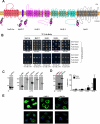
Characterization of various mammalian TF-Cub-Baits in yeast. A, membrane topology of TF-Cub-Baits (NaPi-IIa, MAP17, NHE-1, NHE-3, and NaS1) with N-terminally tagged TF-Cub fusion. B, proper insertion of TF-Cub-Baits into the membrane as exemplified by genetic NubG/NubI experiments. Yeast cells expressing TF-Cub-Baits of NaPi-IIa, MAP17, NHE-1, NHE-3 (pTLB-1 context), and NaS1 (pCLB-1 context) were transformed with control constructs of either yeast Ost1p (ER) or Fur4p (plasma membrane) fused to NubG and NubI. Yeast growth was challenged on minimal SD medium depleted of Trp and Leu (SD−WL) or Trp, Leu, Ade, and His (SD−WLAH) by spotting three independent transformants on the different media. A lack of growth in the presence of the two yeast integral membrane proteins fused to NubG confirms that TF-Cub-tagged mammalian renal baits are not self-activating. In contrast, growth on the same plates in the presence of either NubI construct indicates that the chimeric bait proteins are expressed, and the proteins are properly inserted into the membrane. C, targeting of TF-Cub-Baits to the membrane of yeast. Full-length proteins NaPi-IIa, NHE-1, NHE-3, and NaS1 were expressed in yeast as TF-Cub-Baits from pTLB-1. Lysates thereof were subjected to two consecutive ultracentrifugations to isolate the cytosolic fraction, C, from the detergent-soluble membrane fraction, M. An equal volume from each supernatant was used for the detection of VP16 from the TF-Cub-Bait proteins in immunoblots (IB). The localization of the endogenous ER membrane protein Alg5 to the detergent-soluble membrane fraction validates the centrifugation procedure (n = 2). D, functional phosphate transport activity of TF-Cub-NaPi-IIa in yeast. Yeast strain PM971 with a deficiency of two high affinity Pi transporters was transformed with TF-Cub-NaPi-IIa. In this strain, the bait was properly expressed as a post-translationally modified full-length and single form (left panel, arrowhead). Timed 32Pi uptake was measured at 30 °C on Pi-starved PM971 cells. Relative net Na+-dependent transport of Pi was calculated by subtracting the Na+-independent component (∼40%) from the total Pi uptake (right panel). Values are means of triplicate. The experiment was repeated twice with similar results (n = 3). E, localization of TF-Cub-Baits in yeast. Fixed and acetone-treated spheroplasts of yeast were processed for confocal microscopy using transporter-specific primary and FITC-conjugated secondary antibodies (green). Nonspecific immunofluorescence is shown in the lower panels from vector-transformed cells (n = 2). Confocal planes were based on the strongest 4`,6-diamidino-2-phenylindole stain (blue) of the nuclei, the centers of which are designated by arrows. Secondary antibodies alone yielded no signals (not shown). PM, plasma membrane.
F<sc>ig</sc>. 3.
Fig. 3.
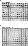
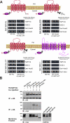
Proteomics analysis of NaPi-IIa C terminus binding to PDZ proteins. Arrays of 95 related class I PDZ domains from various proteins were spotted on nylon membranes and overlaid with the GST-fused C terminus of NaPi-IIa (A) or its truncation thereof for the very last amino acids, TRL (B). Interactions were visualized by a horseradish peroxidase-linked antibody directed against the GST moiety as described previously (41). The PDZ domains spotted in each bin are listed in Table I.
F<sc>ig</sc>. 4.
Fig. 4.
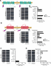
Applicaton of NHE-3, NaPi-IIa, NHE-1, and MAP17 to the MYTH 2.0 system. TF-Cub-Baits (pTLB-1) derived from full-length NHE-3 or its C-terminal truncation for THM (ΔTHM) (A), NaPi-IIa or its C-terminal truncation for TRL (ΔTRL) (B), NHE-1 (C), and MAP17 (D) were co-expressed with the following NubG-HA-Preys: PDZK1, NHERF-1, NHERF-2, CAL, NaPi-IIa, NHE-3, Ost1-NubG (negative control), and Ost1-NubI (positive control). Reconstitution of split Ub was determined by yeast growth on minimal SD−Trp/−Leu (SD−WL) medium. The interactions of baits with preys were analyzed by growth on selective SD−Trp/−Leu/−Ade/−His (SD−WLAH) medium supplemented with 3-AT (left and middle panels) as described in Fig. 2B. Quantification of MYTH reporter activity stemmed from ONPG liquid tests on permeabilized cells and was depicted against Miller units to normalize cell densities (right panels). The error bars show the standard deviations determined from triplicates. PM, plasma membrane.
F<sc>ig</sc>. 5.
Fig. 5.
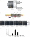
Characterization of NaS1 using the MYTH 2.0 assay. A, analysis of interactions between TF-Cub-NaS1 (in pCLB-1 backbone) and NubG-HA-Preys. Reporter gene activity of co-transformants was detected in triplicates by growth on minimal selective SD-Trp/-Leu/-Ade/-His (SD−WLAH) medium (left panel) and by ONPG liquid tests (right panel) as delineated in Fig. 4. B, mapping the PDZ-binding domain of NaS1. Five deletion constructs of TF-Cub-NaS1 were generated in pCLB-1 and subjected to yeast dot tests on selective SD-Trp/-Leu/-Ade/-His (SD−WLAH) medium after co-transformation with NubG-HA-Preys PDZK1, NHERF-1, NHERF-2, or CAL. Specificity was corroborated based on positive growth with Ost1-NubI and negative growth with Ost1-NubG. C, impact of NHERF isoforms on NaS1 transport in a Xenopus oocyte expression system. Oocytes were injected with either 50 nl of water (control), 5 ng of human NaS1 cRNA, 2.5 ng of either human NHERF-1/-2 cRNAs, or combinations as depicted. Transport measurements were determined by [35S]sulfate uptake and counted by liquid scintillation spectrometry. Data are shown as mean ± S.E. for 7–10 oocytes per condition and are representative of three similar experiments. S.E. bars not visible were smaller than the symbols. *, p < 0.05 when compared with NaS1-injected oocytes. PM, plasma membrane; SD−WL, SD−Trp/−Leu.
F<sc>ig</sc>. 6.
Fig. 6.
Homotropic dimerization of NaPi-IIa. A, MYTH 2.0 dot assays. TF-Cub-Baits in pTLB-1 harboring full-length NaPi-IIa, its C-terminal truncation for TRL (ΔTRL) or the entire tail (ΔCT), and full-length NHE-1 as a negative control were co-transformed in a yeast reporter strain with either NubG-HA-Preys of NaPi-IIa, NHE-3, Ost1-NubG, or Ost1-NubI. Prototrophic cells were rescued on minimal SD−Trp/−Leu/−Ade/−His (SD−WLAH) plates supplemented with 3-AT to suppress leakage of the respective baits. Rescued colonies were blue due to induction of lacZ as determined by β-galactosidase replica colony lift experiments (not shown). B, immunoprecipitation from total membranes of X. laevis oocytes. Various cRNAs (MYC-NaPi-IIa, its corresponding TRL ablation, or MYC-NaS1) were co-injected with cRNA of HA-NaPi-IIa in oocytes. Total solubilized membranes of oocytes were processed for immunoprecipitation by anti-HA. The immunocomplexes were resolved by SDS-PAGE for immunoblotting with the antibodies shown. Results from two other experiments were identical (n = 3). The detection of type IIa homotropic dimers was abrogated when solubilized membranes of oocytes after a single injection of HA-NaPi-IIa were mixed with those of MYC-NaPi-IIa-injected oocytes and used for immunoprecipitation (IP). PM, plasma membrane; SD−WL, SD−Trp/−Leu.

Similar articles

Cited by

References

    1. Hung A. Y., and Sheng, M. ( 2002) PDZ domains: structural modules for protein complex assembly. J. Biol. Chem. 277, 5699–5702 - PubMed
    1. Runyon, S. T., Zhang, Y., Appleton, B. A., Sazinsky, S. L., Wu, P., Pan, B., Wiesmann, C., Skelton, N. J., and Sidhu, S. S. ( 2007) Structural and functional analysis of the PDZ domains of human HtrA1 and HtrA3. Protein Sci. 16, 2454–2471 - PMC - PubMed
    1. Hillier, B. J., Christopherson, K. S., Prehoda, K. E., Bredt, D. S., and Lim, W. A. ( 1999) Unexpected modes of PDZ domain scaffolding revealed by structure of nNOS-syntrophin complex. Science 284, 812–815 - PubMed
    1. Bezprozvanny, I., and Maximov, A. ( 2001) Classification of PDZ domains. FEBS Lett. 509, 457–462 - PubMed
    1. Hernando, N., Wagner, C. A., Gisler, S. M., Biber, J., and Murer, H. ( 2004) PDZ proteins and proximal ion transport. Curr. Opin. Nephrol. Hypertens. 13, 569–574 - PubMed

Publication types

LinkOut - more resources