. 2008 Apr 17;3(4):253-62.
doi: 10.1016/j.chom.2008.03.002.

Human cytomegalovirus protein UL38 inhibits host cell stress responses by antagonizing the tuberous sclerosis protein complex

Affiliations

Human cytomegalovirus protein UL38 inhibits host cell stress responses by antagonizing the tuberous sclerosis protein complex

Nathaniel J Moorman et al. Cell Host Microbe. .

Abstract

Human cytomegalovirus proteins alter host cells to favor virus replication. These viral proteins include pUL38, which prevents apoptosis. To characterize the mode of action of pUL38, we modified the viral genome to encode an epitope-tagged pUL38 and used rapid immunoaffinity purification to isolate pUL38-interacting host proteins, which were then identified by mass spectrometry. One of the cellular proteins identified was TSC2, a constituent of the tuberous sclerosis tumor suppressor protein complex (TSC1/2). TSC1/2 integrates stress signals and regulates the mammalian target of rapamycin complex 1 (mTORC1), a protein complex that responds to stress by limiting protein synthesis and cell growth. We showed that pUL38 interacts with TSC1 and TSC2 in cells infected with wild-type cytomegalovirus. Furthermore, TSC1/2 failed to regulate mTORC1 in cells expressing pUL38, and these cells exhibited the enlarged size characteristic of cytomegalovirus infection. Thus, pUL38 supports virus replication at least in part by blocking cellular responses to stress.


Figures
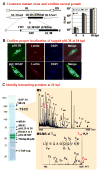
Figure 1
Figure 1
Process for identification of pUL38 binding partners during HCMV infection. (A) Construct mutant virus with tagged protein and confirm its normal growth. The top line of the diagram to the left locates the UL38 ORF within the unique long (UL) domain of the viral genome; the second line shows the location of the UL38 ORF between UL37 exons; the third line illustrates the fusion of a TAP tag to the C-terminus of pUL38 and the position of a FLP recombinase target (FRT) sequence introduced with the TAP sequence in BADinUL38TAP. Arrowheads mark the C-termini of ORFs. Yields (IU, infectious units determined by assay for HCMV IE1 protein fluorescence at 48 hpi) were determined for BADwt and BADinUL38TAP at 0 (immediately after virus adsorption), 5 and 10 days post infection (dpi) of fibroblasts (0.01 pfu/cell). (B) Confirm proper localization of the tagged protein at 24 h post infection (hpi). pUL38 and pUL38TAP localizations (green) were determined after infection with BADwt or BADinUL38TAP, respectively. For wild-type pUL38, anti-pUL38 antibody was used for immunofluorescence, while IgG was used to visualize pUL38TAP. Lectin HPA (red) stains the Golgi and DAPI (blue) stains nuclear DNA. Uninfected cells surround infected cells. (C) Identify interacting proteins at 24 hpi. pUL38TAP-interacting proteins were isolated by immunoaffinity purification, separated by gel electrophoresis and identified by sequential MS and MS/MS analysis. The positions of key captured proteins as well as pUL38TAP and free TAP tag (CTAP) are indicated. *, non-specific contaminants. ‡, because of the similarity of the sequences of RbAp48 and RBAp46, either or both may be present.
Figure 2
Figure 2
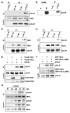
Confirmation of the interaction between pUL38 and the TSC1/2 protein complex. Fibroblasts were mock-infected (M), infected with BADwt (wt) (3 pfu/cell) or 293T cells were transfected with indicated expression vectors. Results are representative of two independent experiments. (A) pUL38 co-precipitates with TSC2. Cell lysates were prepared at 48 h post mock or BADwt infection, subjected to immunoprecipitation (IP) using rabbit polyclonal antibody to TSC2 (α-TSC2), control preimmune rabbit IgG (C Ab), or beads with no antibody (No Ab), and precipitated proteins were analyzed by Western blot (WB) using the indicated antibodies. As controls, the levels of TSC2 and pUL38 were monitored by Western blot assay. (B) Two pUL38 isoforms co-precipitate with TSC2. To improve the separation of pUL38 isoforms present at 48 h post infection, electrophoresis was performed using a larger format polyacrylamide gel. Left lanes, Western blots (WB) were preformed on mock- and infected lysates using antibody to pUL38; right lanes, Western blots were performed after immunoprecipitation (IP) from lysates using α-TSC2 or preimmune rabbit IgG (C Ab). (C) TSC1 co-precipitates with pUL38. Extracts were prepared at 48 h after mock or BADwt infection, and immunoprecipitations were performed using antibody to pUL38 (α-pUL38), a non-specific monoclonal antibody (C Ab) or beads with no antibody (No Ab). (D) pUL38 co-precipitates with TSC1. Extracts were prepared at 48 h after mock or BADwt infection, and immunoprecipitations were performed using antibody to TSC1 (α-TSC1), a non-specific monoclonal antibody (C Ab) or beads with no antibody (No Ab). (E) pUL38 co-precipitates with FLAG-TSC2 but not FLAG-TSC1. Cells were transfected with vectors expressing the indicated proteins, extracts were prepared 48 h later and immunoprecipitations were performed by using FLAG epitope-specific antibody. (F) pUL38 co-precipitates with EGFP-TSC2ΔHBD. Cells were transfected with vectors expressing the indicated proteins, extracts were prepared 48 h later and immunoprecipitations were performed by using GFP-specific antibody. (G) TSC1 and TSC2 co-immunoprecipitate with pUL38. Lysates were prepared at indicated times after mock or BADwt infection (h post infection, hpi), immunoprecipitated using antibody to TSC2, and assayed by Western blot using antibodies to the indicated proteins. Lysates were also assayed directly by western blot.
Figure 3
Figure 3
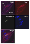
Co-localization of TSC1 and TSC2 in infected fibroblasts. Immunofluorescent analysis at 24 h post infection with BADinUL99GFP, a variant that contains a GFP tag at the C-terminus of the UL99 ORF. The field contains both an uninfected (Uninf) and infected cell (Inf) identified by GFP expression. Antibodies were employed to identify TSC2 (purple) and TSC1 (red), and DNA was stained with DAPI.
Figure 4
Figure 4
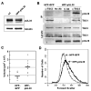
Fibroblasts expressing pUL38 are larger than normal fibroblasts. (A) Three isoforms of pUL38 are expressed in the absence of infection. Extracts of HFF-pUL38 cells and fibroblasts at 24 h post infection with BADwt were assayed by Western blot using antibody to pUL38. α-tubulin was monitored as a loading control. (B) pUL38 interacts with TSC2 in the absence of infection. Extracts were prepared from HFF-pUL38 or control HFF-GFP cells, immunoprecipitations (IP) were performed using rabbit antibody to TSC2 (α-TSC2) or control preimmune rabbit antibody (C Ab), and western blot (WB) assays utilized the indicated antibodies. The results are representative of two independent experiments. (C) Assay of cell volume by quantification of fluorescent intensity in HFF-GFP versus HFF-pUL38 cells. Cells were loaded with calcein green AM, complete z-stacks (0.3 μm slices) were collected for individual, fluorescent cells, and volumes were calculated. 10 well-isolated cells of each type were analyzed, and bars mark average cell volumes. (D) Assay of relative cell volume by forward scatter of HFF-GFP versus HFF-pUL38 cells. Forward scatter of ∼5×10⁵ cells per sample was measured by flow cytometry.
Figure 5
Figure 5
HCMV pUL38 is sufficient to prevent inhibition of the mTORC1 kinase by stress. pUL38 and tubulin were monitored as controls, and results are representative of 2 or 3 independent experiments. (A) Subconfluent HFF-pUL38 (+pUL38) or control HFF-GFP cells (-pUL38) were cultured with (+S) or without serum (-S) for 12 h. Some −S cultures were switched to PBS for 1 h or 2 h, after which cells were harvested, lysates prepared, and protein was analyzed by Western blot assay using antibodies specific for rpS6 phosphorylated at S235/236 and total rpS6. (B) Subconfluent +pUL38 and −pUL38 cells were maintained in serum-free medium for 12 h, and the medium was replaced with PBS for 2 h in the presence or absence of Rapamycin (Rapa, 20 nM). Proteins were assayed by Western blot using antibodies specific for indicated phosphorylated proteins or that recognize proteins irrespective of phosphorylation state. (C) Subconfluent +pUL38 and −pUL38 cells were maintained in serum-free medium (-S) for 12 h, then AICAR (AMPK activator, 5mM) was added to a portion of the cultures and cells were harvested 1, 3 or 6 h later. Assays were as described in panel A. (D) Subconfluent +pUL38 and −pUL38 cells were maintained in serum-free medium for 12 h, then AICAR with or without rapamycin was added to a portion of the cultures and cells were harvested 6 h later. Assays were as described in panel A. (E) Subconfluent +pUL38 and −pUL38 cells were maintained in serum-free medium for 12 h, then AICAR with or without rapamycin was added to a portion of the cultures and cells were harvested 6 h later. Proteins were assayed by Western blot using antibodies specific for acetyl CoA carboxylase (ACC) phosphorylated at S79 and total ACC.
Figure 6
Figure 6
Phosphorylation of rpS6 in response to stress is decreased following infection with a pUL38-deficient virus. As controls, pUL38 and tubulin were monitored. The results are representative of two independent experiments. (A) Fibroblasts were maintained for 12 h in serum-free medium, infected with BADwt (1 pfu/cell) or with BADdlUL38 at an equivalent number of genomes per cell, and re-fed with serum-free medium. At 48 h post infection, cells were harvested, lysates prepared and analyzed by Western blot using antibodies specific for indicated phosphorylated proteins or that recognize proteins irrespective of phosphorylation state. (B) Fibroblasts were treated as in panel (A), and, at the indicated h post infection (hpi), cells were harvested and analyzed by Western blot using antibodies specific for total rpS6 and rpS6 phosphorylated at ser235/236.
Figure 7
Figure 7
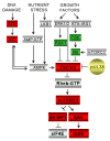
HCMV influences multiple cellular pathways that communicate with mTORC1 through TSC1/2. Activities that are known to be enhanced or inhibited by HCMV infection are indicated by green or red boxes, respectively. Gray boxes mark activities not known to be modified by HCMV. The “core” TSC1/2-mTOR pathway is rendered in larger, bold print: TSC1/2, tuberous sclerosis complex; Rheb-GTP, ras homologue enriched in brain protein; mTORC1, mammalian target of rapamycin complex 1; S6K, p70 S6 kinase; 4E-BP1, eukaryotic initiation factor 4E binding protein 1; rpS6, ribosomal protein S6. Some additional activities that impact the core pathway: ATM, ataxia-telangiectasia mutated protein; p53, p53 tumor suppressor protein; LKB1, Peutz-Jeghers syndrome protein; ERK1/2, extracellular signal-regulated kinase 1 and 2; RSK1, p90 ribosomal S6 kinase 1; IRS1, insulin receptor substrate protein; PI3K, phosphoinositide 3-kinase; Akt, protein kinase B; AMPK, AMP kinase.

Similar articles

Cited by

References

    1. Bowen NJ, Fujita N, Kajita M, Wade PA. Mi-2/NuRD: multiple complexes for many purposes. Biochimica et biophysica acta. 2004;1677:52–57. - PubMed
    1. Brunak S, Engelbrecht J, Knudsen S. Prediction of human mRNA donor and acceptor sites from the DNA sequence. J Mol Biol. 1991;220:49–65. - PubMed
    1. Casavant NC, Luo MH, Rosenke K, Winegardner T, Zurawska A, Fortunato EA. Potential role for p53 in the permissive life cycle of human cytomegalovirus. J Virol. 2006;80:8390–8401. - PMC - PubMed
    1. Castillo JP, Yurochko AD, Kowalik TF. Role of human cytomegalovirus immediate-early proteins in cell growth control. J Virol. 2000;74:8028–8037. - PMC - PubMed
    1. Coats S, Flanagan WM, Nourse J, Roberts JM. Requirement of p27Kip1 for restriction point control of the fibroblast cell cycle. Science. 1996;272:877–880. - PubMed

Publication types

MeSH terms