. 2008 May 1;121(Pt 9):1455-65.
doi: 10.1242/jcs.020362. Epub 2008 Apr 8.

Inhibition of beta-catenin signaling causes defects in postnatal cartilage development

Affiliations

Inhibition of beta-catenin signaling causes defects in postnatal cartilage development

Mo Chen et al. J Cell Sci. .

Abstract

The Wnt/beta-catenin signaling pathway is essential for normal skeletal development because conditional gain or loss of function of beta-catenin in cartilage results in embryonic or early postnatal death. To address the role of beta-catenin in postnatal skeletal growth and development, Col2a1-ICAT transgenic mice were generated. Mice were viable and had normal size at birth, but became progressively runted. Transgene expression was limited to the chondrocytes in the growth plate and articular cartilages and was associated with decreased beta-catenin signaling. Col2a1-ICAT transgenic mice showed reduced chondrocyte proliferation and differentiation, and an increase in chondrocyte apoptosis, leading to decreased widths of the proliferating and hypertrophic zones, delayed formation of the secondary ossification center, and reduced skeletal growth. Isolated primary Col2a1-ICAT transgenic chondrocytes showed reduced expression of chondrocyte genes associated with maturation, and demonstrated that VEGF gene expression requires cooperative interactions between BMP2 and beta-catenin signaling. Altogether the findings confirm a crucial role for Wnt/beta-catenin in postnatal growth.


Figures

Fig. 1
Fig. 1
X-Gal staining in E14.5 and E16.5 TOP-gal transgenic embryos. (A) X-Gal staining of E14.5 whole embryos shows that cartilage, skull, vertebral column, ribs and long bones are stained positive for X-Gal. (B,C) Chondrocytes in the ribs and forelimb of E14.5 transgenic embryos show strong X-Gal-positive staining. (D-F) In E16.5 embryos, (E) proliferating (red arrowhead) and (F) hypertrophic chondrocytes (black arrow) and osteoblasts (red arrows) in the metaphysis show positive X-Gal staining. The results indicate that β-catenin signaling is active in proliferating and hypertrophic chondrocytes during cartilage development.
Fig. 2
Fig. 2
Skeletal growth is reduced in Col2a1-ICAT transgenic mice. (A) Structure of the transgene construct and the priming sites for genotyping the Col2a1-ICAT transgenic mice. (B) PCR results; genotyping of Col2a1-ICAT transgenic mice. Mice 1, 2, 4 and 5 (lanes 1, 2, 4 and 5) are positive for Flag-ICAT transgene expression (P, positive control; N, negative control). (C) Body weight of 2-week-old and 4-week-old transgenic mice and WT littermates. A significant reduction in body weight was observed in Col2a1-ICAT transgenic mice. (D) Radiographic analysis showing the delay in the longitudinal growth of 1-week-old, 2-week-old and 4-week-old Col2a1-ICAT transgenic mice compared with their WT littermates. (E,F) Alizarin Red and Alcian Blue staining showed that skeletal development was relatively normal in 1-day-old new born Col2a1-ICAT transgenic mice. By contrast, a significant reduction in postnatal skeletal growth was observed in 2-week-old Col2a1-ICAT transgenic mice.
Fig. 3
Fig. 3
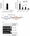
Expression of the Flag-ICAT transgene in Col2a1-ICAT transgenic mice. (A,B) Primary sternal chondrocytes were isolated from Col2a1-ICAT transgenic mice and their WT littermates. Expression of Flag-ICAT protein was detected in western blot analysis and immunostaining using the anti-Flag M2 antibody in Col2a1-ICAT transgenic chondrocytes. (C) Expression of the Flag-ICAT transgene in growth plate chondrocytes was examined by immunostaining using tissue sections from knee joints of 2-week-old WT and Col2a1-ICAT transgenic mice. The ICAT transgene was specifically expressed in proliferating chondrocytes in the growth plate of Col2a1-ICAT transgenic mice and in articular chondrocytes lining the joint surface. (D) ICAT inhibits canonical Wnt signaling in chondrocytes of Col2a1-ICAT transgenic mice. Primary chondrocytes isolated from 3-day-old WT and Col2a1-ICAT transgenic mice were transfected with TOP-flash reporter and treated with or without Wnt3a (100 ng/ml) for 24 hours. Wnt3a stimulated the reporter activity in WT but not in Col2a1-ICAT transgenic chondrocytes. (E) To determine the specificity of the Flag-ICAT transgene expression, total RNA was extracted from multiple tissues and the expression of Flag-ICAT was examined by PCR. The expression of Flag-ICAT was detected in ribs (strong expression) and brain (weak expression) but not in other tissues. (F) ICAT does not alter cell adhesion. Primary chondrocytes isolated from Col2a1-ICAT transgenic mice and WT littermates were placed in 24-well plates and grown to confluence. Cells were then trypsinized with 0.1% trypsin containing either 1 mM CaCl2 (TC) or 1 mM EDTA (TE) and incubated at 37°C for 30 minutes. Cells were pipetted gently five times with 10 ml of PBS and the cell clusters were counted. The degree of adhesion was expressed by determining the ratio of cell clusters in TC and TE containing solutions (TC:TE) for each cell type. A similar experiment was also performed using chondrocytes isolated from b-cateninfx/fx mice and infected with adenovirus expressing Cre recombinase (Ad-Cre) or GFP (Ad-GFP). Results showed that cell adhesion was not changed in Col2a1-ICAT transgenic mice.
Fig. 4
Fig. 4
Col2a1-ICAT transgenic mice have delayed appearance of the secondary ossification center (SOC) and altered growth plate morphology. (A) Development of the growth plate in postnatal mice was analyzed by histology staining with Alcian Blue/Hemotoxylin and Orange G. In WT mice, the formation of epiphysal SOC was initiated at 1 week of age and was well developed at age 2 weeks. Col2a1-ICAT transgenic mice display an obvious delay in the formation of SOC at 1 and 2 weeks of age. (B) Tissue sections stained with Safranin O and Fast Green show that the length of both the proliferating and hypertrophic zones was reduced in 2-week-old Col2a1-ICAT transgenic mice. The hypertrophic chondrocyte columns were disorganized in Col2a1-ICAT transgenic mice. (C-F) Histomorphometric measurements show that the (C) distance from articular surface to hypertrophic zone, (D) growth plate length, and the (E,F) lengths of the proliferating (E) and hypertrophic (F) zones are significantly decreased in 2-week-old Col2a1-ICAT transgenic mice compared with their WT littermates. *P<0.05, unpaired t-test, n=6.
Fig. 5
Fig. 5
Chondrocyte proliferation and differentiation are decreased and apoptosis is increased in Col2a1-ICAT transgenic mice. (A,B) Ki-67 and DAPI double staining was performed using anti-Ki-67 antibody in tissue sections collected from 2-week-old Col2a1-ICAT transgenic mice and their WT littermates. The proliferation rate was determined by counting the numbers of Ki-67-positive cells at the proliferating zone divided by the DAPI-positive cell number. The proliferation of growth plate chondrocytes is decreased by 24% in 2-week-old Col2a1-ICAT transgenic mice. *P<0.05, unpaired t-test, n=3. (C) The expression of cyclin D1, cyclin D2, cyclin A, PCNA, total and phosphorylated β-catenin and phosphorylated Rb was examined by western blotting. The expression of cyclin D1, cyclin D2 and PCNA was significantly decreased in primary chondrocytes derived from Col2a1-ICAT transgenic mice compared with those derived from WT mice. By contrast, the protein levels of cyclin A, total and phosphorylated β-catenin and phosphorylated Rb was not significantly changed. (D) Total RNA was extracted from primary sternal chondrocytes isolated from Col2a1-ICAT transgenic mice and WT littermates. The expression of chondrocyte differentiation marker genes, such as collagen type X (colX), Vegf, Mmp13 and Alp was determined by real-time reverse transcriptase (RT)-PCR and was normalized to β-actin levels. The expression of chondrocyte marker genes was significantly reduced in Col2a1-ICAT transgenic mice. *P<0.05, unpaired t-test, n=3. (E) The apoptosis of growth-plate chondrocytes was determined by TUNEL staining using 2-week-old WT and Col2a1-ICAT transgenic mice. Increased cell apoptosis was observed in the hypertrophic region of the growth plate in Col2a1-ICAT transgenic mice.
Fig. 6
Fig. 6
Wnt3a and β-catenin activate BMP signaling. (A) Primary chondrocytes were treated with or without Wnt3a (100 ng/ml) for 24 hours and the expression of Bmp2 and Bmp4 was examined by real-time RT-PCR. Wnt 3a upregulates mRNA expression of Bmp2 and Bmp4 in chondrocytes. (B) RCJ3.1C5.18 chondrocytes were transfected with the BMP signaling reporter (12×SBE-Luc) and control vector, and treated with BMP2 (100 ng/ml), BIO (1 μg/ml) or Wnt3a (100 ng/ml). To determine the effect of β-catenin on BMP signaling, RCJ3.1C5.18 cells were also co-transfected with BMP signaling reporter and constitutively active β-catenin (S33Y). Luciferase activity was measured using cell lysates 48 hours after transfection. β-catenin, BIO and Wnt3a stimulated BMP-reporter activity in chondrocytes. BMP2 was used as a positive control. (C) BMP signaling is required for Wnt3a-induced colX expression. Primary chondrocytes isolated from WT mice were treated with Wnt3a (100 ng/ml) for 4 and 6 days with or without noggin (300 ng/ml). Type X collagen (colX) mRNA levels were measured by real-time RT-PCR and were normalized to β-actin levels. The expression of colX was completely inhibited by the BMP antagonist noggin. (D) Expression of Bmp2 and Bmp4 mRNA was examined by real-time RT-PCR using primary chondrocytes isolated from Col2a1-ICAT transgenic mice and WT littermates after cells were cultured for 2 days. Expression of Bmp2 and Bmp4 was significantly decreased in Col2a1-ICAT transgenic mice. *P<0.05, unpaired t-test, n=4 (A-D).
Fig. 7
Fig. 7
β-catenin signaling is required for BMP2 to activate Vegf and Mmp13 expression. (A-C) Primary chondrocytes isolated from Col2a1-ICAT transgenic mice and WT littermates were treated with BMP2 (100 ng/ml) for 48 hours. The expression of colX, Vegf and Mmp13 was determined by real-time RT-PCR and was normalized to β-actin levels. ALP activity was measured using cell lysates from the same cells. BMP2 stimulated the expression of all marker genes in WT chondrocytes and rescued (A) the colX expression and (B) ALP activity in chondrocyte isolated from Col2a1-ICAT transgenic mice. However, BMP failed to induce the expression of (C) VEGF and (D)Mmp13 in Col2a1-ICAT transgenic chondrocytes. *P<0.05, unpaired t-test, n=3. (E) RCJ3.1C5.18 chondrogenic cells were treated with BMP2 (100 ng/ml) for 2 hours with or without the BMP antagonist noggin (300 ng/ml) or Wnt signaling inhibitor Dkk1 (1 μg/ml). Total RNA was extracted and VEGF expression determined by real-time RT-PCR normalized to β-actin levels. The induction of VEGF expression by BMP2 was completely inhibited by noggin and Dkk1. (F) RCJ3.1C5.18 cells were treated with BIO (1 μg/ml) for 2 hours with or without noggin or Dkk1. The expression of VEGF mRNA levels were measured and normalized to β-actin levels. BIO induced VEGF expression within 2 hours. Noggin partially inhibited BIO-induced VEGF expression. *P<0.05, unpaired t-test, compared to the untreated group, n=3. **P<0.05, unpaired t-test, compared with BMP2 or BIO treatment groups, n=3. (G,H) To further determine changes in VEGF and MMP13 expression in vivo in Col2a1-ICAT transgenic mice, we performed immunostaining using the anti-VEGF and anti-MMP13 antibodies. The results showed that the area and intensity of the expression of VEGF and MMP13 proteins were reduced in Col2a1-ICAT transgenic mice.
Fig. 8
Fig. 8
β-catenin activates the Vegfa promoter. 2.0-kb Vegfa promoter fragment was cloned into pGL4 vector and deletion mutants were generated. (A) RCJ3.1C5.18 chondrogenic cells were co-transfected with constitutively active β-catenin expression plasmid and the Vegfa promoter. Cell lysates were extracted 48 hours after transfection and luciferase was activity measured. β-catenin significantly stimulated VEGF-a promoter activity. (B) RCJ3.1C5.18 chondrocytes were transfected with the deletion constructs of the Vegfa promoter and the β-catenin expression plasmid. Luciferase activity was measured 48 hours after transfection. β-catenin activates Vegfa promoter activity in 2.0-kb, 1.34-kb and 0.94-kb fragments but not in the 0.14-kb fragment. The results suggest that the β-catenin-responsive region is located in the −940 to −140 region of the Vegfa promoter. *P<0.05, unpaired t-test, n=3 (A,B). (C) The TCF/LEF sites (TREs) in the Vegfa promoter are shown. (D) Chromatin IP assay was performed using RCJ3.1C5.18 cells treated with or without BIO (1 μg/ml) for 2 hours. IP was performed using anti-β-catenin antibody, and anti-Flag antibody was used as a negative control. The DNA-protein complex was crosslinked and used as PCR template. The ChIP assay showed that β-catenin binds to the Vegfa promoter and that addition of BIO to the cultures enhanced β-catenin binding.
Fig. 9
Fig. 9
Col2a1-ICAT transgenic mice have reduced angiogenesis. (A) PECAM-1 was detected by immunostaining using an anti-PECAM-1 antibody in tissue sections from 2-week-old mice. PECAM-1 expression was significantly decreased in Col2a1-ICAT transgenic mice at the secondary ossification center area of the femoral epiphysis compared with WT mice. (B,C) Microfil perfusion was performed using 2-week-old Col2a1-ICAT transgenic mice and their WT littermates. Femurs (B) and liver (C) were imaged using microCT after decalcification of femurs. The images of vessel structure showed a reduced vessel networks in the cartilage tissue (B) but not liver (C) in Col2a1-ICAT transgenic mice. The results indicate that inhibition of Wnt signaling by ICAT leads to reduced angiogenesis during endochondral bone formation.

Similar articles

Cited by

References

    1. Akiyama H, Lyons JP, Mori-Akiyama Y, Yang X, Zhang R, Zhang Z, Deng JM, Taketo MM, Nakamura T, Behringer RR, et al. Interactions between Sox9 and beta-catenin control chondrocyte differentiation. Genes Dev. 2004;18:1072–1087. - PMC - PubMed
    1. Brault V, Moore R, Kutsch S, Ishibashi M, Rowitch DH, McMahon AP, Sommer L, Boussadia O, Kemler R. Inactivation of the beta-catenin gene by Wnt1-Cre-mediated deletion results in dramatic brain malformation and failure of craniofacial development. Development. 2001;128:1253–1264. - PubMed
    1. Daniels DL, Weis WI. ICAT inhibits beta-catenin binding to Tcf/Lef-family transcription factors and the general coactivator p300 using independent structural modules. Mol. Cell. 2002;10:573–584. - PubMed
    1. DasGupta R, Fuchs E. Multiple roles for activated LEF/TCF transcription complexes during hair follicle development and differentiation. Development. 1999;126:4557–4568. - PubMed
    1. Day TF, Guo X, Garrett-Beal L, Yang Y. Wnt/beta-catenin signaling in mesenchymal progenitors controls osteoblast and chondrocyte differentiation during vertebrate skeletogenesis. Dev. Cell. 2005;8:739–750. - PubMed

Publication types

MeSH terms

LinkOut - more resources