

The design of electrospun PLLA nanofiber scaffolds compatible with serum-free growth of primary motor and sensory neurons
- PMID: 18396117
- PMCID: PMC2922842
- DOI: 10.1016/j.actbio.2008.02.020
The design of electrospun PLLA nanofiber scaffolds compatible with serum-free growth of primary motor and sensory neurons
Abstract
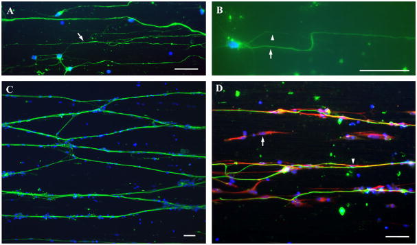
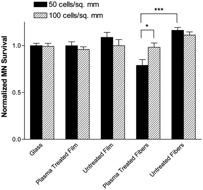
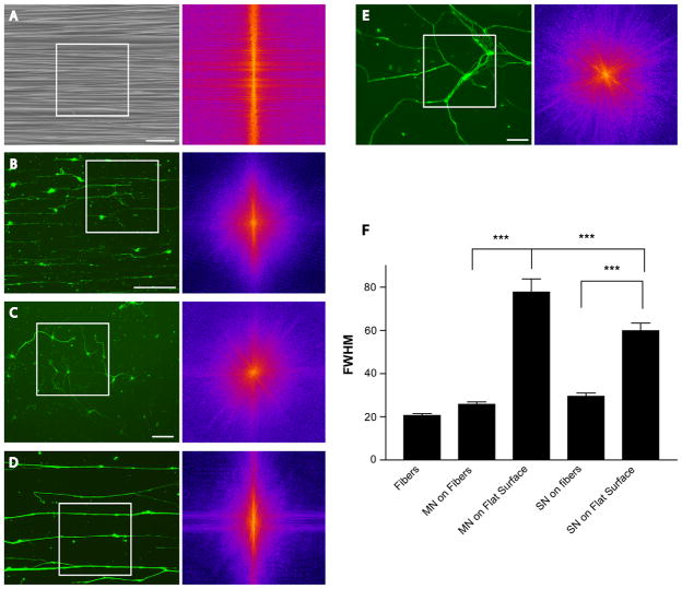
Aligned electrospun nanofibers direct neurite growth and may prove effective for repair throughout the nervous system. Applying nanofiber scaffolds to different nervous system regions will require prior in vitro testing of scaffold designs with specific neuronal and glial cell types. This would be best accomplished using primary neurons in serum-free media; however, such growth on nanofiber substrates has not yet been achieved. Here we report the development of poly(L-lactic acid) (PLLA) nanofiber substrates that support serum-free growth of primary motor and sensory neurons at low plating densities. In our study, we first compared materials used to anchor fibers to glass to keep cells submerged and maintain fiber alignment. We found that poly(lactic-co-glycolic acid) (PLGA) anchors fibers to glass and is less toxic to primary neurons than bandage and glue used in other studies. We then designed a substrate produced by electrospinning PLLA nanofibers directly on cover slips pre-coated with PLGA. This substrate retains fiber alignment even when the fiber bundle detaches from the cover slip and keeps cells in the same focal plane. To see if increasing wettability improves motor neuron survival, some fibers were plasma etched before cell plating. Survival on etched fibers was reduced at the lower plating density. Finally, the alignment of neurons grown on this substrate was equal to nanofiber alignment and surpassed the alignment of neurites from explants tested in a previous study. This substrate should facilitate investigating the behavior of many neuronal types on electrospun fibers in serum-free conditions.
Figures

Similar articles
-
The culture of primary motor and sensory neurons in defined media on electrospun poly-L-lactide nanofiber scaffolds.J Vis Exp. 2011 Feb 15;(48):2389. doi: 10.3791/2389. J Vis Exp. 2011. PMID: 21372783 Free PMC article.
-
Aligned electrospun nanofibers specify the direction of dorsal root ganglia neurite growth.J Biomed Mater Res A. 2007 Dec 1;83(3):636-45. doi: 10.1002/jbm.a.31285. J Biomed Mater Res A. 2007. PMID: 17508416
-
Creation of highly aligned electrospun poly-L-lactic acid fibers for nerve regeneration applications.J Neural Eng. 2009 Feb;6(1):016001. doi: 10.1088/1741-2560/6/1/016001. Epub 2008 Dec 22. J Neural Eng. 2009. PMID: 19104139
-
Varying the diameter of aligned electrospun fibers alters neurite outgrowth and Schwann cell migration.Acta Biomater. 2010 Aug;6(8):2970-8. doi: 10.1016/j.actbio.2010.02.020. Epub 2010 Feb 16. Acta Biomater. 2010. PMID: 20167292
-
Nebulized solvent ablation of aligned PLLA fibers for the study of neurite response to anisotropic-to-isotropic fiber/film transition (AFFT) boundaries in astrocyte-neuron co-cultures.Biomaterials. 2015 Apr;46:82-94. doi: 10.1016/j.biomaterials.2014.12.046. Epub 2015 Jan 17. Biomaterials. 2015. PMID: 25678118 Free PMC article.
Cited by
-
Fabrication of nanofiber scaffolds with gradations in fiber organization and their potential applications.Macromol Biosci. 2012 Oct;12(10):1336-41. doi: 10.1002/mabi.201200115. Epub 2012 Jul 30. Macromol Biosci. 2012. PMID: 22847852 Free PMC article.
-
Electrospun Fibers for Drug Delivery after Spinal Cord Injury and the Effects of Drug Incorporation on Fiber Properties.Cells Tissues Organs. 2016;202(1-2):116-135. doi: 10.1159/000446621. Epub 2016 Oct 5. Cells Tissues Organs. 2016. PMID: 27701153 Free PMC article.
-
Influence of nanofibers on growth and gene expression of human tendon derived fibroblast.Biomed Eng Online. 2010 Feb 17;9:9. doi: 10.1186/1475-925X-9-9. Biomed Eng Online. 2010. PMID: 20163724 Free PMC article.
-
Polymer nanofibrous structures: Fabrication, biofunctionalization, and cell interactions.Prog Polym Sci. 2010 Jul 1;35(7):868-892. doi: 10.1016/j.progpolymsci.2010.03.003. Prog Polym Sci. 2010. PMID: 20582161 Free PMC article.
-
Next generation of electrosprayed fibers for tissue regeneration.Tissue Eng Part B Rev. 2011 Apr;17(2):125-42. doi: 10.1089/ten.TEB.2010.0552. Epub 2011 Feb 20. Tissue Eng Part B Rev. 2011. PMID: 21210761 Free PMC article. Review.
References
-
- Corey JM, Lin DY, Mycek KB, Chen Q, Samuel S, Feldman EL, et al. Aligned electrospun nanofibers specify the direction of dorsal root ganglia neurite growth. J Biomed Mater Res A. 2007;83A:636. - PubMed
-
- Yang F, Murugan R, Wang S, Ramakrishna S. Electrospinning of nano/micro scale poly(L-lactic acid) aligned fibers and their potential in neural tissue engineering. Biomaterials. 2005;26:2603. - PubMed
-
- Schnell E, Klinkhammer K, Balzer S, Brook G, Klee D, Dalton P, et al. Guidance of glial cell migration and axonal growth on electrospun nanofibers of poly-epsilon-caprolactone and a collagen/poly-epsilon-caprolactone blend. Biomaterials. 2007;28:3012. - PubMed
-
- Peters A. The fine structure of the nervous system. New York: Oxford University Press; 1991.
Publication types
MeSH terms
Substances
Grants and funding
LinkOut - more resources
Full Text Sources
Other Literature Sources
