. 2008 Jun;82(11):5234-44.
doi: 10.1128/JVI.02497-07. Epub 2008 Apr 2.

Identification of a highly conserved, functional nuclear localization signal within the N-terminal region of herpes simplex virus type 1 VP1-2 tegument protein

Affiliations

Identification of a highly conserved, functional nuclear localization signal within the N-terminal region of herpes simplex virus type 1 VP1-2 tegument protein

F Abaitua et al. J Virol. 2008 Jun.

Abstract

VP1-2 is a large structural protein assembled into the tegument compartment of the virion, conserved across the herpesviridae, and essential for virus replication. In herpes simplex virus (HSV) and pseudorabies virus, VP1-2 is tightly associated with the capsid. Studies of its assembly and function remain incomplete, although recent data indicate that in HSV, VP1-2 is recruited onto capsids in the nucleus, with this being required for subsequent recruitment of additional structural proteins. Here we have developed an antibody to characterize VP1-2 localization, observing the protein in both cytoplasmic and nuclear compartments, frequently in clusters in both locations. Within the nucleus, a subpopulation of VP1-2 colocalized with VP26 and VP5, though VP1-2-positive foci devoid of these components were observed. We note a highly conserved basic motif adjacent to the previously identified N-terminal ubiquitin hydrolase domain (DUB). The DUB domain in isolation exhibited no specific localization, but when extended to include the adjacent motif, it efficiently accumulated in the nucleus. Transfer of the isolated motif to a test protein, beta-galactosidase, conferred specific nuclear localization. Substitution of a single amino acid within the motif abolished the nuclear localization function. Deletion of the motif from intact VP1-2 abrogated its nuclear localization. Moreover, in a functional assay examining the ability of VP1-2 to complement growth of a VP1-2-ve mutant, deletion of the nuclear localization signal abolished complementation. The nuclear localization signal may be involved in transport of VP1-2 early in infection or to late assembly sites within the nucleus or, considering the potential existence of VP1-2 cleavage products, in selective localization of subdomains to different compartments.


Figures

FIG. 1.
FIG. 1.
VP1-2 NT1 fragment purification and antibody generation. (a) Total protein staining of the soluble fraction of bacterial extracts expressing the GST-NT1 fusion protein (lane 1), the bound, purified GST-NT1 fusion protein (lane 2), or the final NT1 protein after specific protease cleavage and elution (lane 3). This last sample was then loaded onto a Mono Q column and fractionated as described in Materials and Methods. The Mono Q input and sample fractions are shown in panel b. Peak fractions were pooled and used to immunize rabbits. (c) Vero cells were mock infected or infected with the HSV-1 17 strain. At different times postinfection, samples were analyzed by Western blotting with the antibody raised against NT1, αVP1-2NT1r. The long arrow indicates full-length VP1-2, with cleavage products indicated by arrowheads.
FIG. 2.
FIG. 2.
Subcellular localization and fractionation of VP1-2 during virus infection. (a) Monolayers of Vero cells grown on coverslips were mock infected (panels I and VI) or infected with the HSV-1 17 strain (panels II to V and VII to X). At different times postinfection, coverslips were washed, fixed, and probed for VP1-2 using αVP1-2NT1r. Lower panels show higher-magnification images of the top row. Features of VP1-2 localization as discussed in the text are illustrated with different arrows, as follows: cytoplasmic perinuclear signal, arrow, e.g., panels VII and VIII; diffuse intranuclear localization, filled arrowhead, panel IX; nuclear punctate, long arrowheads. Cells exhibiting a diffuse localization are indicated by open arrowheads in the lower-magnification images in panels IV and V. (b) Monolayers of Vero cells were infected with a recombinant virus HSV-1 strain 17 expressing VP26-GFP and cells examined (18 hpi) for VP26 localization by GFP fluorescence and VP1-2 by immunostaining with αVP1-2NT1r. (c) Monolayers of Vero cells were infected with the HSV-1 17 strain harvested at 18 hpi and fractionated as described in Materials and Methods. Total extract (T) and cytoplasmic (C) and nuclear (N) fractions were separated on a 3 to 8% SDS-PAGE gradient gel and subsequently blotted with the specific anti-VP1-2 antibody. Fractionation was assessed using anti-lamin A/C and anti-AP-1 antibodies (lower panel) as markers for nuclear and Golgi compartments, respectively.
FIG. 3.
FIG. 3.
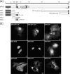
Cellular localization by immunofluorescence of transfected VP1-2 and VP1-2 fragments. (a) Schematic illustration of HSV-1 17 strain VP1-2 indicating positions of certain published features (including information on the PrV homologue), from the N-terminal end (NT) to the C-terminal end (CT): the deubiquitination enzymatic domain (DUB) (1 to 273), the putative NLS (425 to 444) region from the present work, the UL37 binding domain (UL37 BD) (579 to 609), putative leucine zipper sequences (L-zipper), a SalI fragment mapped as the tsB7 phenotype (770 to 1204), and the CT end involved in nuclear assembly (3104 to 3164). Subregions expressed from the various constructs are illustrated, including amino acid boundaries. (b) Monolayers of Vero cells were transfected with the plasmids showed in panel a. The cells were fixed with methanol 24 h after transfection and analyzed for localization using the monoclonal antibody against the SV5 N-terminal epitope tag. Representative images are shown for each fragment, including two examples of VP1-2 full length (VP1-2fl).
FIG. 4.
FIG. 4.
A functional NLS in VP1-2 transferable to a heterologous protein. (a) Schematic diagram illustrating the alignment of the N-terminal motif of VP1-2 from the three different herpesvirus families. Regions encompassing the highly conserved basic region of HSV VP1-2 were fused to the β-Gal gene, as illustrated in the bar diagrams below. Region 2 and region 4 were also analyzed, with a single substitution in a conserved lysine (K→S), represented as a solid line within the corresponding schematic and by a star on the sequence line. (b) Monolayers of Vero cells were transfected with the plasmids expressing the different regions of VP1-2 NT4 as β-Gal fusion proteins. Cells were fixed with methanol 24 h later and analyzed for β-Gal localization. Representative images are shown for each fusion region. Summary conclusions on the efficiency of the candidate regions are illustrated next to the schematic in a semiquantitative manner: +++, very efficient nuclear localization (at least as efficient as that of the SV40 NLS-β-gal vector); ++, mainly nuclear localization but also some weaker cytoplasmic distribution of the signal; -, no significant nuclear accumulation.
FIG. 5.
FIG. 5.
Effect of NLS deletion on localization of VP1-2. (a) COS cells were transfected with 1 μg of the pcDNA3-SV5-UL36fl (panels I and II) or pcDNA3-SV5-UL36ΔNLS (panels III and IV) expression vector. After 24 h, the coverslips were fixed and VP1-2 localization examined using the monoclonal antibody against the V5 tag. Panel b shows the distribution of the percentages of cells with localization patterns of VP1-2 or VP1-2ΔNLS, defined as follows: only cytoplasmic (C) or only nuclear (N); equal distribution in both compartments, C = N; cytoplasmic more abundant than nuclear, C>N; nuclear more abundant than cytoplasmic, C
FIG. 6.
FIG. 6.
The VP1-2 NLS is required for complementation of HSV-1ΔUL36 virus. (a) COS cells were transfected in triplicate with 1 μg of the pEGFP, pcDNA3-SV5-UL36fl, or pcDNA3-SV5-UL36ΔNLS vector. After 24 h posttransfection, the cells were infected with HSV-1 KΔUL36 at 5 PFU/cell and unabsorbed virus was inactivated by an acid wash (40 mM citric acid, 135 mM NaCl, 10 mM KCl, pH 3.0). At 16 h after infection, cells and media were harvested together, lysed by freeze/thawing three times, and the viral yield determined by plaque assay on HS30 cells. Results are plotted as virus yield in PFU/ml with the average and standard deviation for each series. (b) In parallel, cells were examined for expression levels by Western blotting of both proteins, VP1-2 and VP1-2ΔNLS, as described in Materials and Methods. Actin levels were determined as well as a loading control.

Similar articles

Cited by

References

    1. Batterson, W., and B. Roizman. 1983. Characterization of the herpes simplex virion-associated factor responsible for the induction of alpha genes. J. Virol. 46371-377. - PMC - PubMed
    1. Bucks, M. A., K. J. O'Regan, M. A. Murphy, J. W. Wills, and R. J. Courtney. 2007. Herpes simplex virus type 1 tegument proteins VP1/2 and UL37 are associated with intranuclear capsids. Virology 361316-324. - PMC - PubMed
    1. Coller, K. E., J. I. Lee, A. Ueda, and G. A. Smith. 2007. The capsid and tegument of the alphaherpesviruses are linked by an interaction between the UL25 and VP1-2 proteins. J. Virol. 8111790-11797. - PMC - PubMed
    1. Desai, P. J. 2000. A null mutation in the UL36 gene of herpes simplex virus type 1 results in accumulation of unenveloped DNA-filled capsids in the cytoplasm of infected cells. J. Virol. 7411608-11618. - PMC - PubMed
    1. Elliott, G., and P. O'Hare. 1999. Live-cell analysis of a green fluorescent protein-tagged herpes simplex virus infection. J. Virol. 734110-4119. - PMC - PubMed

Publication types

Substances

LinkOut - more resources