. 2008 Jun;28(11):3573-88.
doi: 10.1128/MCB.00087-08. Epub 2008 Mar 31.

Human U2 snRNA genes exhibit a persistently open transcriptional state and promoter disassembly at metaphase

Affiliations

Human U2 snRNA genes exhibit a persistently open transcriptional state and promoter disassembly at metaphase

Thomas Pavelitz et al. Mol Cell Biol. 2008 Jun.

Abstract

In mammals, small multigene families generate spliceosomal U snRNAs that are nearly as abundant as rRNA. Using the tandemly repeated human U2 genes as a model, we show by footprinting with DNase I and permanganate that nearly all sequences between the enhancer-like distal sequence element and the initiation site are protected during interphase whereas the upstream half of the U2 snRNA coding region is exposed. We also show by chromatin immunoprecipitation that the SNAPc complex, which binds the TATA-like proximal sequence element, is removed at metaphase but remains bound under conditions that induce locus-specific metaphase fragility of the U2 genes, such as loss of CSB, BRCA1, or BRCA2 function, treatment with actinomycin D, or overexpression of the tetrameric p53 C terminus. We propose that the U2 snRNA promoter establishes a persistently open state to facilitate rapid reinitiation and perhaps also to bypass TFIIH-dependent promoter melting; this open state would then be disassembled to allow metaphase chromatin condensation.


Figures
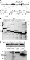
FIG. 1.
FIG. 1.
DNase I-hypersensitive sites in the U2 snRNA genes mapped by genomic blotting. (A) Upper, restriction map of the 6.1-kb U2 tandem repeat unit. The three DNase I-hypersensitive sites 1, 2, and 3 identified in panel B are shown. Restriction sites, from left to right, are HindIII, AseI, NdeI, HincII, and BstBI. Lower, enlarged view of the U2 snRNA coding region showing LM-PCR oligonucleotide sets. Key features are the DSE and PSE and the 3′-end formation signal (“3′” box). Restriction sites, from left to right, are StuI, HincII, BstBI, SfaNI, MseI, ApaLI, AflIII, and Bsu36I. Large arrows indicate LM-PCR oligonucleotide sets, each consisting of a primer extension, PCR, and labeling oligonucleotide. Smaller arrows indicate additional labeling primers; primer 2 was used with oligonucleotide set 1, primer 5b with set 5a. (B) Identification of DNase I-hypersensitive sites in the U2 tandem repeat unit by indirect end labeling. HT1080 cells were treated with DNase I in vivo. Genomic DNA was digested with AseI, redigested with the indicated restriction enzymes, and resolved by native agarose gel electrophoresis, and blots were probed with the AseI/NdeI fragment. The secondary restriction enzymes also generate unique, apparently single-copy bands which are unaffected by DNase I digestion; these may be orphan U2 repeat units or previously characterized junction fragments where the U2 tandem repeat meets flanking DNA (85). (C) Deletion of the DSE or PSE abolishes DNase I hypersensitivity of U2 genes. HT1080 cells and derivatives containing artificial tandem arrays of U2 minigenes (3) were treated with DNase I as in panel B. Genomic DNA was digested with AflIII and resolved by electrophoresis through 0.8% agarose (natural gene assay) or 1.5% agarose (minigene assay), and blots were probed with the StuI/HincII fragment. Site 2 resolves into two bands on the higher-percentage gel. The three rightmost lanes represent the 1, 0.25, and 0.125 standard sample loads (gray triangle). Cell line mU2 42 has 10-fold as many minigenes as natural U2 genes (3). The faint unmarked bands seen for mU2 25 and mU2 42 were disregarded since these do not increase significantly with the DNase I concentration.
FIG. 2.
FIG. 2.
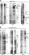
DNase I sensitivity of U2 snRNA genes in HT1080 cells assayed by LM-PCR. (A) Analysis of the U2 template strand. DNase I-hypersensitive site 2 (Fig. 1A) can be seen downstream from the DSE (label 2) and hypersensitive site 1 within the U2 coding region (label 3). The upstream boundary of the large PSE footprint (bracketed) is not visible here because primer set 3 lies just upstream from the PSE. (B) Analysis of the U2 nontemplate strand. The labeling primer used in the final two rounds of PCR is indicated below each panel; label 4 used a previously published primer set (19). DNA sequence features and positions are numbered relative to the U2 transcription start site at +1. Heavy and light vertical bars indicate the degree of DNase I sensitivity. V, genomic DNA treated in vivo with 50 to 100 U/ml DNase I, deproteinized, and digested with EcoRI, which does not cut within the U2 repeat unit (70, 100)]; N, naked genomic DNA treated in vitro with increasing DNase I; E, M, and A, untreated naked genomic DNA cut with EcoRI, MseI, or ApaLI. MseI and ApaI cut within the U2 coding region (Fig. 1A).
FIG. 3.
FIG. 3.
Summary of DNase I (Fig. 2) and permanganate sensitivity data (Fig. 4).
FIG. 4.
FIG. 4.
Permanganate sensitivity of U2 snRNA genes in HT1080 cells assayed by LM-PCR. In each panel, the three lanes from left to right are duplex genomic DNA treated with permanganate in vitro as a control for DNA breathing, single-stranded genomic DNA treated with permanganate in vitro as a control for context-dependent differences in reactivity (39) and to provide a T sequence ladder, and genomic DNA treated in vivo. An additional leftmost lane in two panels contains a 10-bp ladder. The labeling primer used in the final two rounds of PCR is indicated below each panel. Vertical bars indicate regions of DNA protection, sensitivity, or perturbation.
FIG. 5.
FIG. 5.
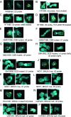
Metaphase fragility of the RNU1, RNU2, and RN5S loci in normal and mutant cells either treated with oxidizing agents or transduced with TAT-p53CTD. (A) WI38 lung fibroblasts immortalized with htert and probed for U2 genes. The RNU2 signals never split but are occasionally unequal in this and all other cell lines. (B) HT1080 fibrosarcoma line treated with MSB and probed for U2 genes. RNU2 often incompletely condensed and split. (C) HT1080 treated with H2O2 and probed for U2 genes. RNU2 often split. (D) HT1080 transduced with TAT-p53CTD and probed for U2 genes. RNU2 often incompletely condensed and/or split. (E) GM00739B primary CSB fibroblasts from a patient with severe CS, probed for U2 genes. RNU2 often incompletely condensed and split. (F) GM00739B probed for U1 genes. RNU1 often split. (G) GM01629 primary CSB fibroblasts from a patient with severe CS, probed for U2 genes. RNU2 often incompletely condensed and/or split. (H) GM10903, a primary fibroblast from a patient with a CSB mutation causing severe DeSanctis-Cacchione syndrome (DCS) probed for U1 genes (left) and U2 genes (right). RNU1 and RNU2 often incompletely condensed and/or split. (I) GM10903 probed for U1 genes. RNU1 often incompletely condensed and/or split. (J) MCF7 breast adenocarcinoma line expressing low levels of BRCA1, probed for U2 genes. RNU2 often incompletely condensed and/or split. (K) MCF7 probed for U1 genes. RNU1 often incompletely condensed and/or split. (L) MCF7 probed for 5S genes. RN5S often incompletely condensed, split, or separated from the telomere. (M) HCC1937, a breast carcinoma line homozygous for a C-terminal deletion that abolishes ubiquitylation of known BRCA1 targets (95), probed for U2 genes. RNU2 often incompletely condensed and/or split. (N) CAPAN1 pancreatic carcinoma line, a compound heterozygote for loss of BRCA2, probed for U2 genes. RNU2 often incompletely condensed and/or split. (O) CAPAN1 probed for U1 genes. RNU1 often incompletely condensed, split, or separated from the telomere. The CAPAN1 karyotype is complex, and RNU1 loci are found on different derivative chromosomes. (P) CAPAN1 probed for 5S genes. RNU5 often separated from the telomere.
FIG. 6.
FIG. 6.
Effect of U2 snRNA structure on fragility of artificial U2 tandem arrays. (A) Secondary structure of altered U2 snRNAs. The expanded Sm site AUUUUUUUUG between U2 stem-loops IIb and III is shown explicitly. Green, normal U2 sequence; red, artificial sequence, with stem length in base pairs, and tetraloops for stability. (B) Fragility of artificial U2 arrays. Note that the observed level of fragility varies from one independently derived line to another, presumably reflecting position effects, copy number, and/or the relative (and heritable) activity of resident and artificial arrays (3, 68). Tandem U2 gene copy numbers are in parentheses.
FIG. 7.
FIG. 7.
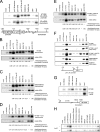
ChIP assays for factors bound to U2 genes at interphase and metaphase. (A) SNAPc and Pol II CTD phosphorylation on the U2 snRNA promoter (−264 to +15) and downstream flanking sequence (+791 to +1019) in HT1080 cells. The abundance of SNAPc on the promoter compared to the downstream fragment demonstrates that ChIP has sufficient resolution for these experiments, although chromatin shearing to the limit size of 500 bp leaves many fragments of >2 kb which reduce resolution on the small U snRNA genes. (B) SNAPc lost from the U2 promoter at metaphase in HT1080 cells. Quantification below ChIP panels is the ratio of mitotic to asynchronous signals when normalized to 50 ng input (upper row) or 1% input DNA (lower row). (C) Some SNAPc 43 but little SNAPc 190 remains on the U2 promoter in the CSB null cell line GM00739Bhtert, derived from patient CS1AN. Quantification is as in panel B. (D) Some SNAPc 43 but little SNAPc 190 remains on the U2 promoter at metaphase in HT1080 cells treated with a low concentration of actinomycin D to induce RNU2 fragility. Quantification is as in panel B. (E) Most SNAPc 43 but little SNAPc 190 remains on the U2 promoter at metaphase in HT1080 cells transduced with the tetrameric C-terminal domain of p53 (TAT-p53CTD) to induce RNU2 fragility. Quantification is as in panel B. (F) Neither SNAPc 43 nor SNAPc 190 is retained at metaphase on the promoter of the largest subunit of RNA polymerase II in untreated HT1080 cells, HT1080 treated with actinomycin D or transduced with TAT-p53CTD, or in mitotic GM00739Bhtert or WI38htert cells. (G) CSB can be detected on active rRNA transcription units (PCR primers amplify +3639 to +3798), but only the CSB N terminus is accessible to our antibody. (H) Neither CSB nor BRCA1 can be detected on the U2 snRNA promoter before or after treatment of asynchronous HT1080 cells with actinomycin D. Pisces is a polyclonal antibody directed against the C terminus of CSB, and Jasmine is a polyclonal antibody against the N terminus (80); “comm” is a commercial polyclonal antibody against CSB (Santa Cruz).

Similar articles

Cited by

References

    1. Akman, S. A., J. H. Doroshow, and M. Dizdaroglu. 1990. Base modifications in plasmid DNA caused by potassium permanganate. Arch. Biochem. Biophys. 282202-205. - PubMed
    1. Ares, M., Jr., J. S. Chung, L. Giglio, and A. M. Weiner. 1987. Distinct factors with Sp1 and NF-A specificities bind to adjacent functional elements of the human U2 snRNA gene enhancer. Genes Dev. 1808-817. - PubMed
    1. Bailey, A. D., Z. Li, T. Pavelitz, and A. M. Weiner. 1995. Adenovirus type 12-induced fragility of the human RNU2 locus requires U2 small nuclear RNA transcriptional regulatory elements. Mol. Cell. Biol. 156246-6255. - PMC - PubMed
    1. Bailey, A. D., T. Pavelitz, and A. M. Weiner. 1998. The microsatellite sequence (CT)n·(GA)n promotes stable chromosomal integration of large tandem arrays of functional human U2 small nuclear RNA genes. Mol. Cell. Biol. 182262-2271. - PMC - PubMed
    1. Baillat, D., M. A. Hakimi, A. M. Naar, A. Shilatifard, N. Cooch, and R. Shiekhattar. 2005. Integrator, a multiprotein mediator of small nuclear RNA processing, associates with the C-terminal repeat of RNA polymerase II. Cell 123265-276. - PubMed

Publication types

MeSH terms