. 2008 Mar 28:5:30.
doi: 10.1186/1742-4690-5-30.

Extracellular ATP reduces HIV-1 transfer from immature dendritic cells to CD4+ T lymphocytes

Affiliations

Extracellular ATP reduces HIV-1 transfer from immature dendritic cells to CD4+ T lymphocytes

Corinne Barat et al. Retrovirology. .

Abstract

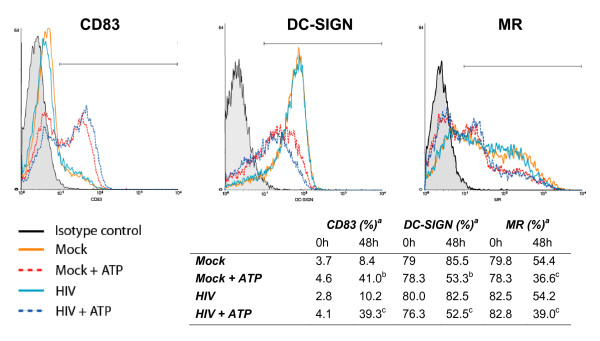
Background: Dendritic cells (DCs) are considered as key mediators of the early events in human immunodeficiency virus type 1 (HIV-1) infection at mucosal sites. Previous studies have shown that surface-bound virions and/or internalized viruses found in endocytic vacuoles of DCs are efficiently transferred to CD4+ T cells. Extracellular adenosine triphosphate (ATP) either secreted or released from necrotic cells induces a distorted maturation of DCs, transiently increases their endocytic capacity and affects their migratory capacity. Knowing that high extracellular ATP concentrations are present in situations of tissue injury and inflammation, we investigated the effect of ATP on HIV-1 transmission from DCs to CD4+ T lymphocytes.

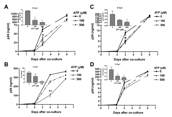
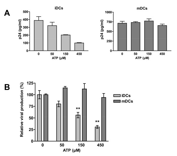
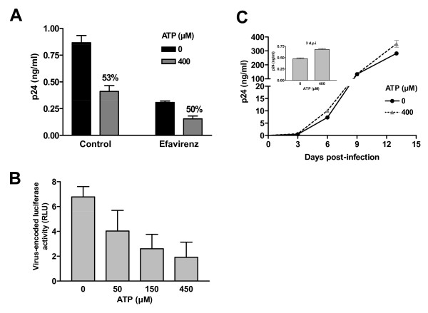
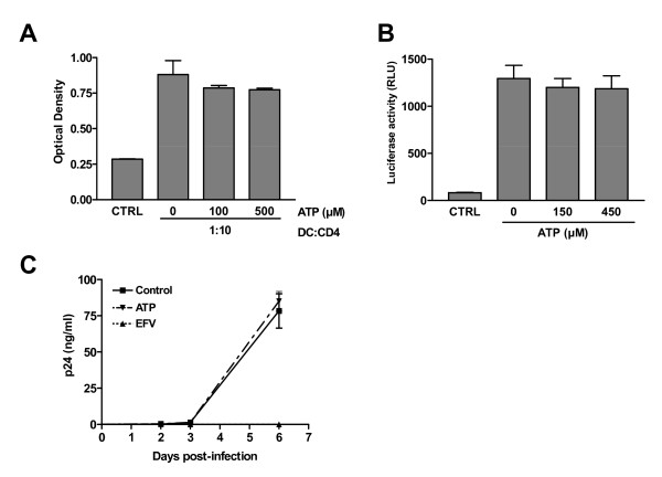
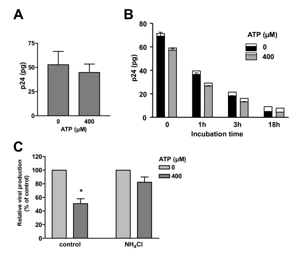
Results: In this study, we show that extracellular ATP reduces HIV-1 transfer from immature monocyte-derived DCs (iDCs) to autologous CD4+ T cells. This observed decrease in viral replication was related to a lower proportion of infected CD4+ T cells following transfer, and was seen with both X4- and R5-tropic isolates of HIV-1. Extracellular ATP had no effect on direct CD4+ T cell infection as well as on productive HIV-1 infection of iDCs. These observations indicate that extracellular ATP affects HIV-1 infection of CD4+ T cells in trans with no effect on de novo virus production by iDCs. Additional experiments suggest that extracellular ATP might modulate the trafficking pathway of internalized virions within iDCs leading to an increased lysosomal degradation, which could be partly responsible for the decreased HIV-1 transmission.

Conclusion: These results suggest that extracellular ATP can act as a factor controlling HIV-1 propagation.


Figures

Figure 1
Figure 1
Extracellular ATP reduces HIV-1 transfer from iDCs to autologous CD4+ T cells. iDCs were initially pulsed with R5-tropic NL4-3Balenv either produced upon transient transfection of 293T cells (A) or following acute infection of CD4+ T cells (B). In some experiments, iDCs were pulsed with X4-tropic viruses consisting of the laboratory variant NL4-3 produced upon transfection of 293T cells (C) or the clinical strain 92HT599 amplified in PBMCs (D). Pulsing was performed in absence or presence of the indicated concentrations of ATP for 60 min at 37°C. Next, iDCs were washed extensively to remove input virus and excess ATP and co-cultured with autologous CD4+ T cells at a 1:3 ratio. Virus production was assessed by measuring the cell-free p24 contents at the listed time points. Virus production at day 2 following initiation of the co-culture is depicted in the small inserts (upper left part of each panel). The data shown represent the means ± standard deviations of triplicate samples and are representative of four independent experiments for panels A, C and D, and three for panel B. Statistical analysis was performed on the results from all experiments. Asterisks denote statistically significant data (*, P < 0.05; **, P < 0.01).
Figure 2
Figure 2
ATP affects virus transfer from iDCs but not mDCs. iDCs and mDCs from the same donor were first pulsed for 60 min at 37°C with R5-tropic JR-CSF either in absence or presence of ATP at the listed concentrations. Next, cells were washed extensively to remove input virus and co-cultured with autologous CD4+ T lymphocytes at a 1:3 ratio. Viral production was assessed at day 2 following initiation of the co-culture. Results from one representative experiment with triplicate samples are presented in panel A. The data shown in panel B represent the means ± standard deviations calculated from two independent experiments and are expressed as relative viral production compared to untreated cells. Asterisks denote statistically significant data (**, P < 0.01).
Figure 3
Figure 3
HIV-1 is transferred to a lower number of CD4+ T cells in presence of ATP. iDCs were either left untreated (mock) or pulsed for 60 min at 37°C with NLHSA-IRES (20 ng of p24/105 cells) in absence or presence of ATP (400 μM). Next, cells were extensively washed to remove input virus and co-cultured for 3 days with autologous CD4+ T cells. Cells were then stained for CD3 (FITC labeling) and HSA (R-PE labeling) and analyzed by flow cytometry. The percentage of HSA-expressing cells in the CD3-positive population is shown for three independent experiments.
Figure 4
Figure 4
Extracellular ATP affects the early transfer phase. (A) iDCs were initially either left untreated or treated with 50 nM efavirenz. Next, cells were either left untreated or treated with ATP before addition of NL4-3Balenv. Finally, cells were washed and co-cultured with autologous CD4+ T cells for 2 days before assessing viral production. The percentage of inhibition is shown above the appropriate bars. (B) Transfer studies were carried out using recombinant luciferase-encoding viruses pseudotyped with the JR-FL envelope. Luciferase activity was assessed at 3 days following initiation of the co-culture. (C) iDCs were pulsed for 60 min with NL4-3Balenv in absence or presence of ATP (400 μM). Next, iDCs were extensively washed to remove input virus and cultured in complete culture RPMI medium supplemented with GM-CSF and IL-4. Cell-free supernatants were collected and assayed for the p24 content at the indicated days post-infection. Data shown represent the means ± standard deviations of triplicate samples and are representative of three (panels A and C) or two (panel B) independent experiments.
Figure 5
Figure 5
ATP is neither affecting the T-cell stimulatory capacity of iDCs nor virus production in CD4+ T cells. (A) iDCs were either left untreated or treated for 60 min at 37°C with the indicated concentrations of ATP. Next, cells were extensively washed and co-cultured for 3 days with heterologous CD4+ T cells at a 1:10 ratio. Cellular proliferation was finally assessed using a colorimetric MTS assay. (B) iDCs were either left untreated or treated for 60 min at 37°C with the indicated concentrations of ATP. Next, iDCs were extensively washed and co-cultured with autologous CD4+ T cells that were previously pulsed with VSV-G-pseudotyped luciferase reporter viruses. Luciferase activity was assessed at 48 h following initiation of the co-culture. (C) CD4+ T cells were infected with NL4-3Balenv either in absence or presence of ATP (400 μM). Cells treated with the antiviral compound efavirenz (EFV) were used as controls. Virus production was monitored by measuring the cell-free p24 contents over time. Data shown represent the means ± standard deviations of triplicate samples and are representative of three independent experiments.
Figure 6
Figure 6
ATP affects virus survival within iDC. iDCs were initially pulsed for 60 min at 37°C with NL4-3Balenv in absence or presence of ATP (400 μM). Next, cells were washed extensively, treated with trypsin to remove uninternalized viruses and then either lysed immediately (A) or incubated at 37°C for the indicated time periods (B). The viral contents found either intracellularly and in cell-free supernatant were assessed using a p24 test. The data shown represent the means ± standard deviations of triplicate samples and are representative of three independent experiments. In panel B, the upper part of each bar represents p24 recovered in the supernatant, whereas the intracellular p24 content is shown in the lower part of each bar. (C) iDCs were either left untreated or pretreated with NH4Cl (20 mM) for 30 min. Next, iDCs were pulsed for 60 min at 37°C with NL4-3Balenv in absence or presence of the indicated concentrations of ATP. Cells were washed extensively to remove input virus and excess ATP and co-cultured with autologous CD4+ T cells at a 1:3 ratio. Viral production was assessed two days following initiation of the co-culture. The data shown represent the means ± standard deviations calculated from four independent experiments with triplicate samples and are presented as relative viral production of ATP-treated cells compared to control cells for each condition. The asterisk denotes statistically significant data (*, P < 0.05).
Figure 7
Figure 7
Surface expression of CD83, DC-SIGN and MR is affected by ATP. iDCs (5 × 10⁵) were either left untreated or treated for 60 min at 37°C with ATP (400 μM), NL4-3Balenv or ATP and NL4-3Balenv. Next, cells were extensively washed and either stained immediately or cultured for 48 h in complete medium before staining. Cells were stained with antibodies specific for CD83, DC-SIGN, or MR and analyzed by flow cytometry. Results from one representative experiment and flow data acquired after 48 h are presented. a %, Percentage of cells expressing the studied cell surface marker as defined by flow cytometry. Statistical analyses were performed on the results of three independent experiments (b, P < 0.01 ; c, P < 0.05).

Similar articles

Cited by

References

    1. Smed-Sorensen A, Lore K, Vasudevan J, Louder MK, Andersson J, Mascola JR, Spetz AL, Koup RA. Differential susceptibility to human immunodeficiency virus type 1 infection of myeloid and plasmacytoid dendritic cells. J Virol. 2005;79:8861–8869. doi: 10.1128/JVI.79.14.8861-8869.2005. - DOI - PMC - PubMed
    1. Turville SG, Cameron PU, Handley A, Lin G, Pohlmann S, Doms RW, Cunningham AL. Diversity of receptors binding HIV on dendritic cell subsets. Nat Immunol. 2002;3:975–983. doi: 10.1038/ni841. - DOI - PubMed
    1. Geijtenbeek TB, Kwon DS, Torensma R, van Vliet SJ, van Duijnhoven GC, Middel J, Cornelissen IL, Nottet HS, KewalRamani VN, Littman DR, Figdor CG, van Kooyk Y. DC-SIGN, a dendritic cell-specific HIV-1-binding protein that enhances trans-infection of T cells. Cell. 2000;100:587–597. doi: 10.1016/S0092-8674(00)80694-7. - DOI - PubMed
    1. Geijtenbeek TB, Torensma R, van Vliet SJ, van Duijnhoven GC, Adema GJ, van Kooyk Y, Figdor CG. Identification of DC-SIGN, a novel dendritic cell-specific ICAM-3 receptor that supports primary immune responses. Cell. 2000;100:575–585. doi: 10.1016/S0092-8674(00)80693-5. - DOI - PubMed
    1. Magerus-Chatinet A, Yu H, Garcia S, Ducloux E, Terris B, Bomsel M. Galactosyl ceramide expressed on dendritic cells can mediate HIV-1 transfer from monocyte derived dendritic cells to autologous T cells. Virology. 2007;362:67–74. doi: 10.1016/j.virol.2006.11.035. - DOI - PubMed

Publication types

Substances