

TGF-beta-induced Foxp3 inhibits T(H)17 cell differentiation by antagonizing RORgammat function
- PMID: 18368049
- PMCID: PMC2597437
- DOI: 10.1038/nature06878
TGF-beta-induced Foxp3 inhibits T(H)17 cell differentiation by antagonizing RORgammat function
Abstract
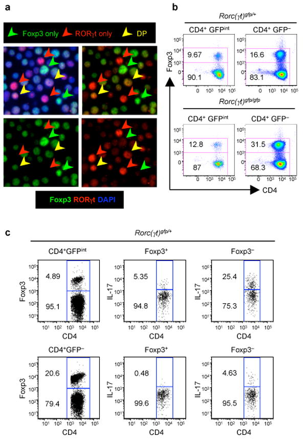
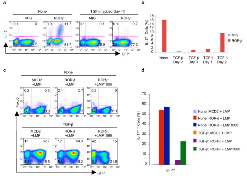
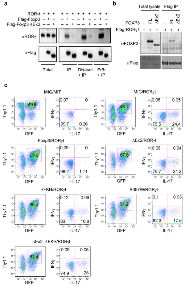
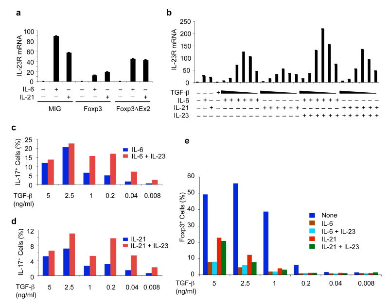
T helper cells that produce IL-17 (T(H)17 cells) promote autoimmunity in mice and have been implicated in the pathogenesis of human inflammatory diseases. At mucosal surfaces, T(H)17 cells are thought to protect the host from infection, whereas regulatory T (T(reg)) cells control immune responses and inflammation triggered by the resident microflora. Differentiation of both cell types requires transforming growth factor-beta (TGF-beta), but depends on distinct transcription factors: RORgammat (encoded by Rorc(gammat)) for T(H)17 cells and Foxp3 for T(reg) cells. How TGF-beta regulates the differentiation of T cells with opposing activities has been perplexing. Here we demonstrate that, together with pro-inflammatory cytokines, TGF-beta orchestrates T(H)17 cell differentiation in a concentration-dependent manner. At low concentrations, TGF-beta synergizes with interleukin (IL)-6 and IL-21 (refs 9-11) to promote IL-23 receptor (Il23r) expression, favouring T(H)17 cell differentiation. High concentrations of TGF-beta repress IL23r expression and favour Foxp3+ T(reg) cells. RORgammat and Foxp3 are co-expressed in naive CD4+ T cells exposed to TGF-beta and in a subset of T cells in the small intestinal lamina propria of the mouse. In vitro, TGF-beta-induced Foxp3 inhibits RORgammat function, at least in part through their interaction. Accordingly, lamina propria T cells that co-express both transcription factors produce less IL-17 (also known as IL-17a) than those that express RORgammat alone. IL-6, IL-21 and IL-23 relieve Foxp3-mediated inhibition of RORgammat, thereby promoting T(H)17 cell differentiation. Therefore, the decision of antigen-stimulated cells to differentiate into either T(H)17 or T(reg) cells depends on the cytokine-regulated balance of RORgammat and Foxp3.
Figures
Similar articles
-
The orphan nuclear receptor RORgammat directs the differentiation program of proinflammatory IL-17+ T helper cells.Cell. 2006 Sep 22;126(6):1121-33. doi: 10.1016/j.cell.2006.07.035. Cell. 2006. PMID: 16990136
-
Foxp3 inhibits RORgammat-mediated IL-17A mRNA transcription through direct interaction with RORgammat.J Biol Chem. 2008 Jun 20;283(25):17003-8. doi: 10.1074/jbc.M801286200. Epub 2008 Apr 23. J Biol Chem. 2008. PMID: 18434325
-
IL-6-dependent and -independent pathways in the development of interleukin 17-producing T helper cells.Proc Natl Acad Sci U S A. 2007 Jul 17;104(29):12099-104. doi: 10.1073/pnas.0705268104. Epub 2007 Jul 10. Proc Natl Acad Sci U S A. 2007. PMID: 17623780 Free PMC article.
-
Transcriptional regulation of Th17 cell differentiation.Semin Immunol. 2007 Dec;19(6):409-17. doi: 10.1016/j.smim.2007.10.011. Epub 2007 Nov 28. Semin Immunol. 2007. PMID: 18053739 Free PMC article. Review.
-
FOXP3 and the regulation of Treg/Th17 differentiation.Microbes Infect. 2009 Apr;11(5):594-8. doi: 10.1016/j.micinf.2009.04.002. Epub 2009 Apr 14. Microbes Infect. 2009. PMID: 19371792 Free PMC article. Review.
Cited by
-
Context and location dependence of adaptive Foxp3(+) regulatory T cell formation during immunopathological conditions.Cell Immunol. 2012 Sep;279(1):60-5. doi: 10.1016/j.cellimm.2012.09.009. Epub 2012 Oct 1. Cell Immunol. 2012. PMID: 23089195 Free PMC article. Review.
-
Jagged-1 signaling suppresses the IL-6 and TGF-β treatment-induced Th17 cell differentiation via the reduction of RORγt/IL-17A/IL-17F/IL-23a/IL-12rb1.Sci Rep. 2015 Feb 4;5:8234. doi: 10.1038/srep08234. Sci Rep. 2015. PMID: 25648768 Free PMC article.
-
IL-17A as a Potential Therapeutic Target for Patients on Peritoneal Dialysis.Biomolecules. 2020 Sep 24;10(10):1361. doi: 10.3390/biom10101361. Biomolecules. 2020. PMID: 32987705 Free PMC article. Review.
-
Regulatory T cells that co-express RORγt and FOXP3 are pro-inflammatory and immunosuppressive and expand in human pancreatic cancer.Oncoimmunology. 2015 Oct 29;5(4):e1102828. doi: 10.1080/2162402X.2015.1102828. eCollection 2016 Apr. Oncoimmunology. 2015. PMID: 27141387 Free PMC article.
-
Cutting Edge: a novel, human-specific interacting protein couples FOXP3 to a chromatin-remodeling complex that contains KAP1/TRIM28.J Immunol. 2013 May 1;190(9):4470-3. doi: 10.4049/jimmunol.1203561. Epub 2013 Mar 29. J Immunol. 2013. PMID: 23543754 Free PMC article.
References
-
- Weaver CT, Harrington LE, Mangan PR, Gavrieli M, Murphy KM. Th17: an effector CD4 T cell lineage with regulatory T cell ties. Immunity. 2006;24:677–688. - PubMed
-
- Weaver CT, Hatton RD, Mangan PR, Harrington LE. IL-17 family cytokines and the expanding diversity of effector T cell lineages. Annu Rev Immunol. 2007;25:821–852. - PubMed
-
- McKenzie BS, Kastelein RA, Cua DJ. Understanding the IL-23-IL-17 immune pathway. Trends Immunol. 2006;27:17–23. - PubMed
Publication types
MeSH terms
Substances
Grants and funding
LinkOut - more resources
Full Text Sources
Other Literature Sources
Molecular Biology Databases
Research Materials
