. 2008 Jun;19(6):2534-43.
doi: 10.1091/mbc.e07-12-1259. Epub 2008 Mar 26.

Spliceosomal small nuclear ribonucleoprotein particles repeatedly cycle through Cajal bodies

Affiliations

Spliceosomal small nuclear ribonucleoprotein particles repeatedly cycle through Cajal bodies

David Stanek et al. Mol Biol Cell. 2008 Jun.

Abstract

The Cajal body (CB) is a nuclear structure closely associated with import and biogenesis of small nuclear ribonucleoprotein particles (snRNPs). Here, we tested whether CBs also contain mature snRNPs and whether CB integrity depends on the ongoing snRNP splicing cycle. Sm proteins tagged with photoactivatable and color-maturing variants of fluorescent proteins were used to monitor snRNP behavior in living cells over time; mature snRNPs accumulated in CBs, traveled from one CB to another, and they were not preferentially replaced by newly imported snRNPs. To test whether CB integrity depends on the snRNP splicing cycle, two human orthologues of yeast proteins involved in distinct steps in spliceosome disassembly after splicing, hPrp22 and hNtr1, were depleted by small interfering RNA treatment. Surprisingly, depletion of either protein led to the accumulation of U4/U6 snRNPs in CBs, suggesting that reassembly of the U4/U6.U5 tri-snRNP was delayed. Accordingly, a relative decrease in U5 snRNPs compared with U4/U6 snRNPs was observed in CBs, as well as in nuclear extracts of treated cells. Together, the data show that particular phases of the spliceosome cycle are compartmentalized in living cells, with reassembly of the tri-snRNP occurring in CBs.


Figures

Figure 1.
Figure 1.
Mature snRNPs accumulate in Cajal bodies. To determine the age of snRNPs in CBs, SmB and SmD1 proteins that are stable components of snRNPs were tagged with E5-RFP. During maturation, E5-RFP changes its fluorescence from green to red in a time course of a couple hours. (A) HeLa cells were transfected with SmB-E5-RFP or SmD1-E5-RFP and fixed at different times after transfection. Ratio of red to green was measured in CBs (bars are SD) and in the nucleoplasm (lines). Red fluorescence in CBs increased ∼3 times over 22 h, indicating that mature snRNPs accumulate in CBs. (B) Cells were transiently transfected with SmB-E5-RFP, and 2 h before fixation they were treated with leptomycin B (LMB) that inhibits biogenesis of new snRNPs by blocking export of new snRNA from the nucleus. A 45% increase of red-to-green ratio after 38 h indicates import inhibition of the green (new) variant of SmB-E5-RFP. Bars are SE.
Figure 2.
Figure 2.
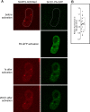
snRNPs newly imported from the cytoplasm represent only a minor fraction in CBs. (A) The SmD1 protein was tagged with PA-GFP and coexpressed in HeLa cells with SART3-HcDiRed as a marker of CBs. To distinguish between nuclear and cytoplasmic pool of snRNPs, nuclear snRNPs were specifically activated by short pulse of 405-nm laser line. Images were taken every 2 min for total 20 min. (B) Quantification of CB fluorescence. To avoid photobleaching effects, the ratio of fluorescent signals CB:nucleoplasm were determined immediately after activation and 20 min later. Values for individual CBs are plotted. Mean value is indicated by a solid line SD by a box. A small decrease in CB fluorescence indicates that within 20 min, snRNPs imported from the cytoplasm represent only small fraction of snRNPs in CBs.
Figure 3.
Figure 3.
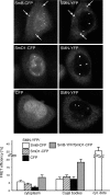
The SMN protein interacts with Sm proteins in the cytoplasm. To compare SMN–snRNP complexes in CBs and the cytoplasm, SMN-YFP was coexpressed with SmD1-CFP, SmB-CFP, or CFP alone as a negative control, and FRET was measured in the cytoplasm and in CBs by acceptor photobleaching method. The SMN-Sm FRET signal was two- to threefold higher over negative control in the cytoplasm but not in CBs. The SmB–SmD1 pair used as a positive control exhibited high FRET signal both in the cytoplasm and in CBs. In some cells, we observed cytoplasmic accumulation of SmB and SMN proteins (arrows) and high FRET signal was measured in these cytoplasmic inclusions. Ten measurements for each pair are averaged and are shown in the graph with SE bars.
Figure 4.
Figure 4.
snRNPs cycle between CBs. To observe movement of snRNPs between CBs and the nucleoplasm, SmB-PA-GFP was coexpressed with SART3-CFP and SmD1-PA-GFP with coilin-CFP. Sm-PA-GFPs were specifically activated in one CB (circle) by short pulse of 405-nm laser, and movement of activated molecules was observed for 5 min (also see Supplemental Videos). Activated molecules moved throughout the whole nucleoplasm and accumulated in other CBs in the same nucleus (arrows). The detection system was adjusted to detect very low signals of PA-GFP, but by using this setup we also detected cell autofluorescence in the cytoplasm (stars).
Figure 5.
Figure 5.
siRNA targeted depletion of hPrp22 and hNtr1. (A) Five different siRNAs against hPrp22 and three against hNtr1 protein were used. Cells were treated with siRNAs for 48 h, and mRNA levels of hPrp22 and hNtr1 were determined by RT-PCR. RT-PCR of 18S rRNA served as a loading control. (B) Extract from cells treated for 48 h with siRNAs was loaded on gel, and hNtr1 and hPrp22 protein levels were determined. Anti-tubulin antibody used as a loading control.
Figure 6.
Figure 6.
U4/U6-specific markers accumulate in CBs after hPrp22 and hNtr1 knockdown. (A) Cells treated with siRNA against hPrp22 (22–3) and hNtr1 (Ntr-27) for 48 h were fixed, and localization of snRNP-specific proteins was determined by antibody staining. To avoid distortion of CB morphology due to changes in intensity, the intensities of the images shown were adjusted to an equal maximum. This results in apparent fluorescent reduction in the nucleoplasm after siRNA treatments, which was not observed at raw images. NC, negative control siRNA. hSnu114, green; coilin, red. (B) Quantification of fluorescence is shown in the graph. Fluorescence ratio CB:nucleoplasm was calculated for each CB, and average with SE bars is shown (number of measured CBs indicated inside bars). *p < 0.0001 as determined by Student's t test with respect to cells treated with control siRNA.
Figure 7.
Figure 7.
Assembled U4/U6 snRNP accumulates in Cajal bodies after hPrp22 and hNtr1 knockdown. (A) Cells treated with siRNA against hPrp22 (22-3) were transfected with SART3-YFP and hPrp31-CFP, and FRET was measured by acceptor photobleaching in the nucleoplasm and CBs. (B) Quantification of SART3-YFP/hPrp31-CFP FRET measurements in cells treated with control (NC), hPrp22-3, or hNtr1-27 siRNAs. Average values of 10 measurements with SE bars are shown.
Figure 8.
Figure 8.
Mono-U5 snRNP is reduced after hPrp22 and hNtr1 knockdowns. HeLa cells were treated with 22-3 or Ntr-27 siRNA for 48 h, and nuclear extracts were centrifuged on 10–30% glycerol gradients. Parallel RNA gels were used for determination of snRNP complexes position in gradients. Proteins from individual fractions were isolated, and hSnu114 (marker of the U5 snRNP) and hPrp4 (marker of the U4/U6 snRNP) were detected. In siRNA-treated cells, the level of the free U5 snRNP decreased (fractions 8–10).

Comment in

  • Mol Biol Cell. 19:2349.

Similar articles

Cited by

References

    1. Achsel T., Brahms H., Kastner B., Bachi A., Wilm M., Luhrmann R. A doughnut-shaped heteromer of human Sm-like proteins binds to the 3′-end of U6 snRNA, thereby facilitating U4/U6 duplex formation in vitro. EMBO J. 1999;18:5789–5802. - PMC - PubMed
    1. Almeida F., Saffrich R., Ansorge W., Carmo-Fonseca M. Microinjection of anti-coilin antibodies affects the structure of coiled bodies. J. Cell Biol. 1998;142:899–912. - PMC - PubMed
    1. Arenas J. E., Abelson J. N. Prp 43: an RNA helicase-like factor involved in spliceosome disassembly. Proc. Natl. Acad. Sci. USA. 1997;94:11798–11802. - PMC - PubMed
    1. Bell M., Schreiner S., Damianov A., Reddy R., Bindereif A. p110, a novel human U6 snRNP protein and U4/U6 snRNP recycling factor. EMBO J. 2002;21:2724–2735. - PMC - PubMed
    1. Black D. L., Pinto A. L. U5 small nuclear ribonucleoprotein: RNA structure analysis and ATP-dependent interaction with U4/U6. Mol. Cell. Biol. 1989;9:3350–3359. - PMC - PubMed

Publication types

MeSH terms

Substances

LinkOut - more resources