. 2008 Mar 24;180(6):1205-18.
doi: 10.1083/jcb.200708115.

An endosomally localized isoform of Eps15 interacts with Hrs to mediate degradation of epidermal growth factor receptor

Affiliations

An endosomally localized isoform of Eps15 interacts with Hrs to mediate degradation of epidermal growth factor receptor

Ingrid Roxrud et al. J Cell Biol. .

Abstract

Down-regulation of activated and ubiquitinated growth factor (GF) receptors by endocytosis and subsequent lysosomal degradation ensures attenuation of GF signaling. The ubiquitin-binding adaptor protein Eps15 (epidermal growth factor receptor [EGFR] pathway substrate 15) functions in endocytosis of such receptors. Here, we identify an Eps15 isoform, Eps15b, and demonstrate its expression in human cells and conservation across vertebrate species. Although both Eps15 and Eps15b interact with the endosomal sorting protein Hrs (hepatocyte growth factor-regulated tyrosine kinase substrate) in vitro, we find that Hrs specifically binds Eps15b in vivo (whereas adaptor protein 2 preferentially interacts with Eps15). Although Eps15 mainly localizes to clathrin-coated pits at the plasma membrane, Eps15b localizes to Hrs-positive microdomains on endosomes. Eps15b overexpression, similarly to Hrs overexpression, inhibits ligand-mediated degradation of EGFR, whereas Eps15 is without effect. Similarly, depletion of Eps15b but not Eps15 delays degradation and promotes recycling of EGFR. These results indicate that Eps15b is an endosomally localized isoform of Eps15 that is present in the Hrs complex via direct Hrs interaction and important for the sorting function of this complex.


Figures

Figure 1.
Figure 1.
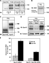
An Eps15-related protein is present in bilayered coats on endosomes. (A) HEp-2 cells incubated with EGF on ice and chased for 15 min at 37°C were prepared for immunoelectron microscopy and labeled using an antibody recognizing the C terminus of Eps15. (B) HeLa cells incubated with EGF on ice and chased for 15 min at 37°C were prepared for immunoelectron microscopy and labeled for Eps15 (15 nm gold) followed by labeling for Hrs (10 nm gold, arrows). The labeling for Eps15 localized to bilayered coats on endosome-like compartments as well as to the rim of coated pits at the plasma membrane (inset in A). The two micrographs shown in A are from the same cell. Coats are indicated by arrowheads. Bars, 100 nm.
Figure 2.
Figure 2.
Eps15b is an isoform of Eps15. (A) The EPS15 gene contains 25 annotated exons. Eps15 is produced from exons 1–25, whereas the Eps15b isoform originates from a transcriptional initiation in intron 12 continuing through exons 13–25. (B) The Eps15 protein contains several functional protein domains: three EH domains, a coiled-coil region, a DPF repeat domain, and two consecutive UIMs. The Eps15b isoform lacks the three EH domains and contains a short unique N-terminal region. The remaining part of the protein is identical to Eps15 and contains major parts of the coiled coil region in addition to the DPF repeat domain and the UIM motives. (C) Multiple sequence alignment of the predicted protein sequences for the putative Eps15b homologues in Homo sapiens (BX647676), Macaca fascicularis (AB172746), Mus musculus (BAC38796), Gallus gallus (CR407454), and Bos taurus (DV911715) compared with Eps15 from H. sapiens. The sequences in blue and red represent the N-terminal 32 Eps15b-specific sequences (conserved residues are shown in blue and unconserved residues in red), whereas the sequence in black indicates where Eps15b aligns with the Eps15 sequence. The asterisks indicate fully conserved amino acid residues in the Eps15b-specific region.
Figure 3.
Figure 3.
Eps15b migrates at 120 kD and is expressed in HeLa and HEp-2 cells. (A) In vitro translated Eps15b was compared with endogenous anti-Eps15 immunoreactive protein species from a HeLa cell lysate by SDS-PAGE followed by immunoblotting using anti-Eps15 from Santa Cruz Biotechnology. The total cell lysate pattern was reconfirmed with two additional antibodies generated against the C terminus of Eps15, anti-Eps15 No. 2 from Covenance, and anti-Eps15 No. 3 from Abcam. (B) HeLa cells were lysed and analyzed by SDS-PAGE and immunoblotting with the 6G4 antibody raised against the N-terminal region of Eps15. (C) Eps15 and Eps15R immunoreactive proteins were compared by SDS-PAGE and immunoblotting using anti-Eps15R followed by stripping of the membrane and reprobing with anti-Eps15. (D) HeLa cells were transfected with siRNA oligos targeting Eps15, Eps15, and Eps15b, or a nontargeting control RNA duplex as described in Materials and methods. The level of knockdown was assessed by Western analysis using anti-Eps15 and anti-tubulin. Numbers to the right of gel blots indicate molecular mass in kD. (E) The level of knockdown was additionally investigated by real-time PCR using Eps15- and Eps15b-specific primers.
Figure 4.
Figure 4.
Eps15b forms a complex with Hrs in vivo. (A) HEp-2 cells were lysed and immunoprecipitated with anti-Hrs as described in Materials and methods. The immunoprecipitate was analyzed by SDS-PAGE and immunoblotting with anti-Eps15 and anti-Hrs. (B) An immunoprecipitation experiment was performed with anti-Hrs as in A and analyzed using anti-Eps15R and anti-Hrs. (C) HeLa cells were depleted of Hrs by siRNA-mediated knockdown and analyzed by SDS-PAGE and immunoblotting with anti-Eps15, anti-Hrs, and anti-tubulin as a loading control. (D) A431 cells were subjected to size exclusion chromatography separating protein complexes after their molecular weight, and the resulting fractions were immunoblotted with anti-Eps15 and anti-Hrs. Numbers to the right of gel blots indicate molecular mass in kD.
Figure 5.
Figure 5.
Both Eps15 and Eps15b interact with Hrs in vitro, whereas only Eps15 shows strong interaction with AP2 in vivo. (A) In vitro translated 35S-labeled Eps15 and Eps15b were pulled down with recombinant GST-Hrs(1–500) or GST immobilized on glutathione-Sepharose beads, and the beads were analyzed by SDS-PAGE and autoradiography. (B) Hep-2 cells were lysed and immunoprecipitated with anti-adaptin as described in Materials and methods. The immunoprecipitate was analyzed by SDS-PAGE and immunoblotting using anti-Eps15 and anti-adaptin.
Figure 6.
Figure 6.
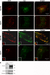
Eps15b is localized to Hrs-positive microdomains on early endosomes. HeLa cells grown on coverslips were transiently transfected with myc-Eps15b (A–C) or myc-Eps15 (D–F) for 20 h, permeabilized before fixation, and double labeled with anti-myc (green) and anti-EEA1 (red). Colocalization is indicated in yellow. HeLa cells grown on coverslips were transiently transfected with the constitutively active Rab5 mutant Rab5Q79L for 24 h, generating enlarged endosomes, followed by a transfection with myc-Eps15b (G–I) or myc-Eps15 (J–L) for an additional 24 hr. The cells were permeabilized before fixation and double labeled with anti-Hrs (red) and anti-myc (green). Colocalization is indicated in yellow. Insets show examples of endosomes that are sufficiently large to enable a distinction between different membrane domains. (M) HEp-2 cells were fractionated as described previously (Felberbaum-Corti et al., 2005) and the resulting early endosome–enriched fraction was analyzed by SDS-PAGE and immunoblotting with anti-Eps15, anti-EEA1, and anti–α-tubulin as loading controls. Bars, 10 μm.
Figure 7.
Figure 7.
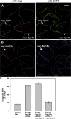
Overexpression of myc-Eps15b leads to an impairment of EGFR down-regulation independently of endocytosis. (A) HeLa cells grown on coverslips were transfected with myc-Eps15b or myc-Eps15 for 24 h and subjected to EGF stimulation for 15 or 120 min. The cells were permeabilized, fixed, and stained with anti-EGFR. EGFR degradation was analyzed by quantitating the amount of undegraded EGFR remaining in the cells at t = 120 min relative to t = 15 min by confocal immunofluorescence microscopy. Error bars represent standard errors (control cells, n = 18; Eps15-expressing cells, n = 10; and Eps15b-expressing cells, n = 11). (B) HeLa cells grown on coverslips were transfected with myc-Eps15b for 24 h and stimulated with EGF for 5 min. The EGFR remaining on the cell surface was removed by a 5-min incubation with stripping buffer followed by permeabilization, fixation, and staining with anti-EGFR. Internalized EGFR was quantitated by confocal immunofluorescence microscopy and transfected cells were detected with anti-myc. The figure shows the mean of three independent experiments and error bars represent standard deviations. (C–F) myc-Eps15b– or (G–J) myc-Eps15–expressing cells were subjected to the EGFR degradation assay as in A and triple-stained with anti-EEA1 (blue), anti-myc (red), and anti-EGFR (green). Triple colocalization is indicated in white. Arrows indicate transfected cells. Insets show magnified examples of colocalization.
Figure 8.
Figure 8.
Eps15b mediates down-regulation of the EGFR independently of endocytosis. (A) HeLa cells grown on coverslips were depleted of Eps15 and Eps15b by siRNA-mediated knockdown and stimulated with EGF for 5 min at 37°C to induce EGFR internalization. The EGFR remaining on the cell surface was removed by stripping buffer followed by permeabilization, fixation, and staining with anti-EGFR. Internalized EGFR was quantitated by confocal immunofluorescence microscopy. The figure shows the mean of three independent experiments and the error bars represent SEM. (B) Eps15/Eps15b-depleted HeLa cells grown on coverslips were subjected to EGF stimulation for 15, 60, 120, or 180 min. The cells were stained with anti-EGFR. EGFR degradation was analyzed by quantitating the amount of undegraded EGFR remaining in the Eps15/Eps15b knockdown and in cells treated with a control RNA duplex at each time point by confocal immunofluorescence microscopy. Error bars represent SEM (control: 15 min, n = 22; 60 min, n = 22; 120 min, n = 22; 180 min, n = 27; and Eps15+Eps15b siRNA: 15 min, n = 23; 60 min, n = 24; 120 min, n = 23; and 180 min, n = 21). (C) Internalization of [125I]EGF in HeLa cells depleted of Eps15/Eps15b. Eps15/Eps15b-depleted cells were seeded in 24-well plates and an internalization assay of [125I]EGF (1 ng/ml) was preformed as described in Materials and methods. Internalized [125I]EGF was calculated as counts per minute of internalized [125I]EGF divided by counts per minute of surface-localized [125I]EGF. The data represent six independent experiments with four parallels. The difference between control and both Eps15- and Eps15/Eps15b-depleted cells was statistically significant at time points 5, 7.5, and 10 min (P < 0.05). Error bars represent SEM. (D and E) Degradation (D) and recycling (E) of [125I]EGF in Eps15/Eps15b-depleted cells. Cells were incubated with [125I]EGF (1 ng/ml) on ice for 15 min then washed with PBS before preheated medium was added to the cells and chased for the indicated time period at 37°C. Upon incubation, the medium was divided into acid-soluble and -precipitable fractions representing degraded and recycled [125I]EGF, respectively. The cell-associated radioactivity represented internalized and cell-associated [125I]EGF. The data are shown as a percentage of total initially bound [125I]EGF and represent six independent experiments with four parallels. There was no statistically significant difference between control and Eps15-depleted cells with regard to degradation and recycling, but for Eps15b depleted cells, the difference to control cells was statistically significant (P < 0.05) for all time points (60, 120, and 180 min). Error bars represent SEM.
Figure 9.
Figure 9.
Eps15b is essential for EGFR degradation. HeLa cells were treated with Eps15- and Eps15b-specific siRNA oligonucleotides and at the same time transiently transfected with siRNA-resistant myc-Eps15 (A) or myc-Eps15b (B). The cells were stimulated with 50 ng/ml EGF for 15 min and then washed and incubated for 2 h in the presence of cycloheximide to allow degradation of internalized EGFRs. The cells were then prepared for confocal immunofluorescence microscopy and labeled with anti-EGFR (green) and anti-myc (red) antibodies. (A) Eps15- and Eps15b-depleted cells transfected with myc-Eps15 (arrows) showed inhibited EGFR degradation similar to Eps15/Eps15b-depleted neighboring cells. (B) Eps15- and Eps15b-depleted cells transfected with myc-Eps15b (arrows) showed normal degradation of EGFR as compared with Eps15/Eps15b-depleted neighboring cells in which the degradation was inhibited. White lines indicate the cell borders. Bar, 10 μm. (C) The amount of EGFR remaining after 2 h of chase was quantified as described in Materials and methods and is represented as percentage of the total amount of internalized EGFR after 15 min. Control cells were treated with a nontargeting RNA duplex. In total, 100–300 cells were measured for each treatment. Error bars show ± SEM from four independent experiments.
Figure 10.
Figure 10.
Differential functions of Eps15 and Eps15b in EGFR trafficking. Together with AP2, Epsin, and several other proteins, Eps15 mediates internalization of activated EGFRs. The endocytosed receptors and their ligands are then transported to early endosomes, where they are retained in the bilayered clathrin coat by Hrs, STAM, and Eps15b. They are further escorted into intraluminal vesicles of multivesicular endosomes by ESCRT I–III and finally degraded in lysosomes (not depicted).

Similar articles

Cited by

References

    1. Babst, M., D.J. Katzmann, E.J. Estepa-Sabal, T. Meerloo, and S.D. Emr. 2002. a. Escrt-III: An endosome-associated heterooligomeric protein complex required for mvb sorting. Dev. Cell. 3:271–282. - PubMed
    1. Babst, M., D.J. Katzmann, W.B. Snyder, B. Wendland, and S.D. Emr. 2002. b. Endosome-associated complex, ESCRT-II, recruits transport machinery for protein sorting at the multivesicular body. Dev. Cell. 3:283–289. - PubMed
    1. Bache, K.G., C. Raiborg, A. Mehlum, and H. Stenmark. 2003. STAM and Hrs are subunits of a multivalent ubiquitin-binding complex on early endosomes. J. Biol. Chem. 278:12513–12521. - PubMed
    1. Bache, K.G., S. Stuffers, L. Malerod, T. Slagsvold, C. Raiborg, D. Lechardeur, S. Walchli, G.L. Lukacs, A. Brech, and H. Stenmark. 2006. The ESCRT-III subunit hVps24 is required for degradation but not silencing of the epidermal growth factor receptor. Mol. Biol. Cell. 17:2513–2523. - PMC - PubMed
    1. Bean, A.J., S. Davanger, M.F. Chou, B. Gerhardt, S. Tsujimoto, and Y. Chang. 2000. Hrs-2 regulates receptor-mediated endocytosis via interactions with Eps15. J. Biol. Chem. 275:15271–15278. - PubMed

Publication types

MeSH terms

Substances