. 2008 Apr 1;180(7):4742-53.
doi: 10.4049/jimmunol.180.7.4742.

Homeostasis and effector function of lymphopenia-induced "memory-like" T cells in constitutively T cell-depleted mice

Affiliations

Homeostasis and effector function of lymphopenia-induced "memory-like" T cells in constitutively T cell-depleted mice

David Voehringer et al. J Immunol. .

Abstract

Naive T lymphocytes acquire a phenotype similar to Ag-experienced memory T cells as a result of proliferation under lymphopenic conditions. Such "memory-like" T (T(ML)) cells constitute a large fraction of the peripheral T cell pool in patients recovering from T cell ablative therapies, HIV patients under highly active antiretroviral therapy, and in the elderly population. To generate a model that allows characterization of T(ML) cells without adoptive transfer, irradiation, or thymectomy, we developed genetically modified mice that express diphtheria toxin A under control of a loxP-flanked stop cassette (R-DTA mice). Crossing these mice to CD4Cre mice resulted in efficient ablation of CD4 single-positive thymocytes, whereas double-positive and CD8 single-positive thymocytes were only partially affected. In the periphery the pool of naive (CD44(low)CD62L(high)) T cells was depleted. However, some T cells were resistant to Cre activity, escaped deletion in the thymus, and underwent lymphopenia-induced proliferation resulting in a pool of T(ML) cells that was similar in size and turnover to the pool of CD44(high)CD62L(low) "memory phenotype" T cells in control mice. CD4Cre/R-DTA mice remained lymphopenic despite the large available immunological "space" and normal Ag-induced T cell proliferation. CD4Cre/R-DTA mice showed a biased TCR repertoire indicating oligoclonal T cell expansion. Infection with the helminth Nippostrongylus brasiliensis resulted in diminished effector cell recruitment and impaired worm expulsion, demonstrating that T(ML) cells are not sufficient to mediate an effective immune response.


Figures
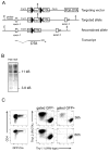
Figure 1
Figure 1. Generation of R-DTA mice and selective T cell ablation in vitro
A) Schematic representation of the targeting construct used to generate R-DTA mice which express the diphtheria toxin A (DTA) subunit under control of a loxP-flanked stop cassette in the ubiquitously expressed ROSA26 locus. Cre activity removes the stop cassette and allows DTA expression. B) Southern blot analysis of tail DNA from heterozygous (Het) offspring and wild type (WT) mice. The wild-type allele generates a band at 11 kB and the targeted allele at 3.8 kB. C) Efficient Cre-mediated killing of T cells from R-DTA mice in vitro. A mixed T cell culture with CD4+ T cells from control mice (Thy1.1+) and R-DTA mice (Thy1.2+) was stimulated for 2 days with plate-bound anti-TCR antibody and then transduced with a retrovirus expressing a GFP-Cre fusion protein. 36 and 60 hours after transduction the ratio of Thy1.1/Thy1.2 cells was analyzed among the transduced cells (GFP+) and non-transduced cells (GFP) by flow cytometry.
Figure 2
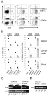
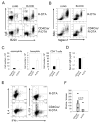
Figure 2. Analysis of thymocyte development in CD4Cre/R-DTA mice
A) Total numbers of double positive (DP), CD4 single positive (SP) and CD8 SP cells from pooled data of three independent experiments with 6 individual mice per group. B) Total thymocytes from CD4Cre or CD4Cre/R-DTA mice were stained with anti-CD4 and anti-CD8 antibodies and analyzed by flow cytometry. Contour-plots show expression levels of CD5 and CD24 on gated CD8 SP cells from CD4Cre mice (left) and CD4Cre/R-DTA mice (right).
Figure 3
Figure 3. Low numbers of peripheral T cells in CD4Cre/R-DTA mice
A) Mesenteric lymph node cells from CD4Cre/R-DTA and CD4Cre control mice were stained with anti-CD4, anti-CD8 and anti-CD62L antibodies and analyzed by flow cytometry. B) Blood, spleen and mesenteric lymph nodes of three individual mice were analyzed for the frequency of CD4+ and CD8+ T cells among total cells (left graphs) and the frequency of CD62L+ cells among CD4+ and CD8+ T cells (right graphs). Results are representative of several independent experiments. C) Semiquantitative RT-PCR analysis for Cre- and Hprt-expression of sorted CD4+ and CD8+ T cells from CD4Cre mice (left) and sorted CD4+ T cells from CD4Cre/R-DTA mice (right). D) Genomic PCR of sorted CD4+ and CD8+ T cells from CD4Cre and CD4Cre/R-DTA mice to determine the presence or absence of the loxP flanked neomycin resistance gene.
Figure 4
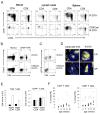
Figure 4. CD4Cre/R-DTA mice lack naïve T cells but show comparable numbers of memory phenotype T cells
A) Analysis of CD4+ and CD8+ T cell subsets in mesenteric lymph node, spleen and blood. Cells were stained with anti-CD4, anti-CD8, anti-CD62L and anti-CD44 and analyzed by four color flow cytometry. B) Analysis of Treg cells in thymus and mesenteric lymph nodes. The dot plots show the frequency of CD4+Foxp3+ Treg cells among total live cells. Results are representative of 3–4 independently analyzed mice. C) Frequency of CD4+ and CD4 NKT cells (αβTCR+ cells) in the liver. Dot plots are gated on NK1.1+ cells. D) Smaller T cell zones in lymph node and spleen of CD4Cre/R-DTA mice as compared to R-DTA control mice. Sections of spleen and mesenteric lymph nodes were stained with anti-Thy1.2 (yellow) and anti-B220 (blue) antibodies. Original magnification was 80x. E) Number of total (left) and CD44hi ‘memory phenotype’ (right) T cells in inguinal lymph nodes of six week old CD4Cre/R-DTA mice (filled bars) or R-DTA mice (open bars). Graphs show combined results from two independent experiments with four mice total per group. Error bars indicate standard deviation. F) The frequency of T cells in the peripheral blood of individual CD4Cre/R-DTA mice (filled symbols) and R-DTA mice (open symbols) was monitored over 32 weeks by flow cytometry.
Figure 5
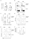
Figure 5. Remaining T cells in CD4Cre/R-DTA mice are functional and show normal homeostatic proliferation
A) Turnover of ‘memory phenotype’ T cells was analyzed by BrdU labeling in the drinking water for 7 days and staining for CD4, CD8, CD44 and BrdU. The graph shows the percentage of BrdU+ cells among CD44hi gated CD4+ or CD8+ T cells from three R-DTA mice (open bars) and four CD4Cre/R-DTA mice (filled bars). B) KLRG1 expression was analyzed on splenic CD4+ and CD8+ T cells of CD4Cre/R-DTA mice (upper panel) or R-DTA mice (lower panel) by staining for CD62L, KLRG1 and CD4 or CD8. Percentages indicate the frequency of KLRG1+ cells among CD62L cells. C) Expansion of T cells from CD4Cre/R-DTA or control mice in vitro. Single cell suspensions of spleen and mesenteric lymph nodes were cultured in vitro for 5 days in the presence of 20 ng/ml IL-2. The total number of CD4+ and CD8+ T cells was determined on indicated days after setup of the culture. The results show the mean from two individual mice per group. D) TCR-mediated in vitro proliferation. Single cell suspensions of spleen and mesenteric lymph nodes were labeled with CFSE, incubated with 0.2 μg/ml anti-TCR (H57) and 0.2 μg/ml anti-CD28 antibody in the presence of 20 ng/ml IL-2 and analyzed three days later. The experiment has been repeated with comparable results. E) Spontaneous in vivo proliferation. Single cell suspension from spleen and mesenteric lymph nodes of CD4Cre/R-DTA or control mice were labeled with CFSE and transferred into 600 rad irradiated recipient mice. 7 days later mice were analyzed for spontaneous proliferation of transferred T cells. F) Quantitative RT-PCR to determine the expression of IL-4 and IFN-γ in polarized T cell cultures from CD4Cre/R-DTA (filled bars) and control mice (open bars). Error bars show s.d. of triplicate samples.
Figure 6
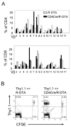
Figure 6. Biased Vβ repertoire indicates TCR repertoire incompleteness
A) The peripheral blood of two CD4Cre/R-DTA mice (filled bars) and two age- and sex-matched control R-DTA mice (open bars) was analyzed by staining for CD4, CD8 and individual TCR-Vβ domains. The relative increase/decrease of the frequency of certain Vβ domains in CD4Cre/R-DTA mice indicates a biased TCR repertoire. The experiment has been repeated with similar results. B) Sorted naïve CD4+ T cells from wild-type Thy1.1 mice were labeled with CFSE and transferred into non-irradiated R-DTA or CD4Cre/R-DTA mice. 7 days later mice were analyzed for proliferation of transferred CD4+ T cells which only occurs in hosts with an incomplete TCR repertoire.
Figure 7
Figure 7. Naïve CD4+ T cells from CD4Cre/R-DTA mice can be detected shortly after CD4-depletion or when the peripheral T cell pool has been replenished by wild-type T cells
A) The frequency of CD4+ T cells in the peripheral blood of CD4Cre/R-DTA mice was analyzed in two individual mice at different days after intraperitoneal administration of 200 μg anti-CD4 monoclonal antibody (GK1.5). The experiment was repeated once with similar results. B) Analysis of the frequency of naïve (CD62L+CD44lo) CD4+ T cells at 0, 7, 12 and 18 days after anti-CD4 depletion. C) Percentage of wild-type (Thy1.1+) CD4+ T cells among total donor-derived (Ly5.2+) CD4+ T cells in the peripheral blood at 4, 6 and 9 weeks after bone marrow reconstitution of lethally irradiated Ly5.1+ recipient mice with 106 total bone marrow cells from Thy1.2+Ly5.2+ CD4Cre/R-DTA mice and 105 total bone marrow cells from Thy1.1+Ly5.2+ wild-type mice. D) Left: frequency of total thymocytes derived from wild-type bone marrow (Thy1.1+) or CD4Cre/R-DTA bone marrow (Thy1.1). Middle: frequency of thymocyte subsets derived from wild-type precursors. Right: frequency of thymocyte subsets derived from CD4Cre/R-DTA precursors. Lower panel: Frequency of naïve (CD44lo) and activated/memory phenotype (CD44hi) CD4+ T cells from wild-type (Thy1.1+) and CD4Cre/R-DTA (Thy1.1) donors in spleen and lymph node. Data are representative of 4 individual mice. E) Frequency of naïve (CD44lo) CD4+ T cells derived from wild-type bone marrow or CD4Cre/R-DTA bone marrow in mixed bone marrow chimeras (MC) in comparison to the frequency of naïve CD4+ T cells in wild-type and CD4Cre/R-DTA mice.
Figure 8
Figure 8. Infection of CD4Cre/R-DTA mice with N. brasiliensis reveals the functionally impaired immune response
A) and B) Frequency of basophils (IgE+B220) and eosinophils (Sigelc-F+SSChi) in lung and peripheral blood on day 9 after N. brasiliensis infection. Dot plots are gated on CD4autofluorescence cells and are representative of 4 independent experiments. C) Total numbers of eosinophils, basophils and CD4 T cells in the lung of R-DTA (n=5) and CD4Cre/R-DTA (n=6) mice on day 9 after N. brasiliensis infection. The graph shows pooled results from 3 independent experiments. Error bars indicate standard deviation. Differences between groups were statistically highly significant (p<0.01, Student’s T-test). D) Serum IgE levels of N. brasiliensis-infected R-DTA (n=7) and CD4Cre/R-DTA (n=8) mice from 4 independent experiments. The difference is not significant (p=0.13, Student’s T-test). E) Intracellular cytokine staining of CD4+ T cells isolated from mesenteric lymph nodes of indicated mice on day 9 after N. brasiliensis infection. The experiment was repeated with comparable results. F) The number of adult worms in the small intestine was determined on day 9 after infection of R-DTA (n=6) or CD4Cre/R-DTA (n=8) mice. The graph shows pooled results from 3 independent experiments.

Similar articles

Cited by

References

    1. Fadel S, Sarzotti M. Cellular immune responses in neonates. Int Rev Immunol. 2000;19:173–193. - PubMed
    1. Modigliani Y, Coutinho G, Burlen-Defranoux O, Coutinho A, Bandeira A. Differential contribution of thymic outputs and peripheral expansion in the development of peripheral T cell pools. Eur J Immunol. 1994;24:1223–1227. - PubMed
    1. Schonland SO, Zimmer JK, Lopez-Benitez CM, Widmann T, Ramin KD, Goronzy JJ, Weyand CM. Homeostatic control of T-cell generation in neonates. Blood. 2003;102:1428–1434. - PubMed
    1. Steinmann GG, Klaus B, Muller-Hermelink HK. The involution of the ageing human thymic epithelium is independent of puberty. A morphometric study. Scand J Immunol. 1985;22:563–575. - PubMed
    1. Goronzy JJ, Weyand CM. T cell development and receptor diversity during aging. Curr Opin Immunol. 2005;17:468–475. - PubMed

Publication types

Substances