. 2008 Mar 10;180(5):989-1003.
doi: 10.1083/jcb.200708043.

Inhibition of "self" engulfment through deactivation of myosin-II at the phagocytic synapse between human cells

Affiliations

Inhibition of "self" engulfment through deactivation of myosin-II at the phagocytic synapse between human cells

Richard K Tsai et al. J Cell Biol. .

Abstract

Phagocytosis of foreign cells or particles by macrophages is a rapid process that is inefficient when faced with "self" cells that display CD47-although signaling mechanisms in self-recognition have remained largely unknown. With human macrophages, we show the phagocytic synapse at cell contacts involves a basal level of actin-driven phagocytosis that, in the absence of species-specific CD47 signaling, is made more efficient by phospho-activated myosin. We use "foreign" sheep red blood cells (RBCs) together with CD47-blocked, antibody-opsonized human RBCs in order to visualize synaptic accumulation of phosphotyrosine, paxillin, F-actin, and the major motor isoform, nonmuscle myosin-IIA. When CD47 is functional, the macrophage counter-receptor and phosphatase-activator SIRPalpha localizes to the synapse, suppressing accumulation of phosphotyrosine and myosin without affecting F-actin. On both RBCs and microbeads, human CD47 potently inhibits phagocytosis as does direct inhibition of myosin. CD47-SIRPalpha interaction initiates a dephosphorylation cascade directed in part at phosphotyrosine in myosin. A point mutation turns off this motor's contribution to phagocytosis, suggesting that self-recognition inhibits contractile engulfment.


Figures
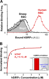
Figure 1.
Figure 1.
Species-specific binding of soluble human SIRPα to RBCs and CD47-coated beads. (A) Fresh human and sheep RBCs binding to soluble hSIRPα (4 μM of GST conjugate), as detected by FITC-anti-GST. “Bkgd” is obtained with RBCs plus antibody. (B) Affinity of hCD47-coated beads binding to soluble hSIRPα based on flow cytometry (see Fig. S2, available at http://www.jcb.org/cgi/content/full/jcb.200708043/DC1). Saturation binding fit gave the indicated dissociation constant, K d. Because this is a 3D binding constant relevant to binding in a narrow membrane gap between two cells, it is equivalent to K d ≈ 1 molecule/[10 nm x (10 μm)2], which is the concentration of free SIRPα that would half-saturate CD47 on a surface. The inset shows inhibition of soluble hSIRPα binding to hCD47 beads by using anti-CD47 F(ab′)2 generated from B6H12 antibody; similar inhibition is obtained with human RBCs.
Figure 2.
Figure 2.
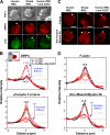
Signaling and cytoskeletal proteins at the phagocytic synapse depend on CD47. Human-derived THP-1 macrophages () were incubated for 10 min at 37°C with IgG-opsonized human RBCs or sheep RBCs (Target: HuRBC or ShRBC) for 10 min. Blocking is done with anti-CD47 F(ab′)2. After fixation, cells were stained for SIRPα and pTyr (A and B) or F-actin and nonmuscle myosin IIA (C and D). Phagocytic synapses are indicated with black arrowheads in DIC images, and in fluorescence images with either white or gray arrowheads, depending on enrichment. Bars, 10 μm. Protein localization was quantified for phagocytic synapses randomly selected in DIC images (n = 5, ± SD).
Figure 3.
Figure 3.
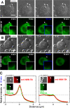
Time-lapse image of phagocytic synapse formation. Human-derived THP-1 macrophages stably transfected with GFP-NMM IIA were incubated at 37°C with IgG-opsonized sheep RBCs (A) or human-RBCs (B) for 10 min after an initial 4°C incubation. Time-lapse images in DIC and fluorescence microscopy were taken upon identification of a target cell adhered to the macrophage. Arrows indicate the site of target cell contact with magnified images of the phagocytic synapse. Bars, 10 μm. (C) Phagocytic synapses fixed after 10 min. Cells were immunostained for total NMM IIA and protein localization to the phagocytic synapse was quantified for randomly chosen GFP+ cells by normalization to cytoplasmic intensity of 1.0 (n = 5, ± SD).
Figure 4.
Figure 4.
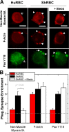
CD47 inhibits myosin localization similar to blebbistatin. Phagocytic synapses of THP-1 macrophages with IgG-opsonized sheep or human RBCs (37°C, 10 min) after preincubation with blebbistatin (50 μM, 10 min). DMSO control showed no effect. (A) Cells fixed and immunostained for nonmuscle myosin IIA, F-actin, paxillin-Y118. Bars, 10 μM. (B) Quantitation of protein localization to the phagocytic synapse relative to the cytoplasmic intensity set at 1.0 (n = 5 cells, ± SD).
Figure 5.
Figure 5.
Human macrophages and monocytes are inhibited by human CD47. (A) DIC images of THP-1 phagocytes plus CD47-blocked, IgG-opsonized human RBCs, using F(ab′)2 made from monoclonal B6H12. 45 min at 37°C is sufficient time for phagocytic internalization of RBCs, which are protected from hypotonic lysis. Bars, 10 mm. (B) Human peripheral blood monocyte plus CD47-blocked human RBCs after lysis, after 45 min of phagocytosis. Red: PKH26-labeled human RBCs. Green: lysed RBCs labeled with FITC-anti-Fc indicating the cell is not internalized. Human RBCs and sheep RBCs were phagocytosed by THP-1 cells (C) or human monocytes (D), showing phagocytosis increases with opsonization. Inset shows blebbistatin treatment of ShRBC showing decrease in phagocytosis. Phagocytosis is measured as the ratio of internalized RBCs per phagocyte, with 200 phagocytes counted in triplicate experiments (± SEM).
Figure 6.
Figure 6.
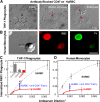
Human-CD47 is sufficient to inhibit phagocytosis. (A) Streptavidin beads coated with both anti-streptavidin IgG as the opsonin and biotinylated human CD47. Phagocytosis of beads (red) by THP-1 cells was assessed in DIC and fluorescence microscopy with non-ingested beads (green) visible with rabbit anti-streptavidin plus a second, goat anti–rabbit antibody. Bars, 10 μm. Beads ± hCD47 (at normal RBC density) were engulfed by THP-1 cells or human monocytes (B), demonstrating inhibition of phagocytosis by hCD47 unless the beads are blocked with anti-CD47. Inset bar graph compares to inhibited phagocytosis of uncoated beads after 10 min pretreatment of THP-1 cells with blebbistatin (50 μM). (C) Inhibition of phagocytosis depends on density of human CD47 on beads. All assays were conducted and analyzed as in Fig. 5. Phagocytosis inhibition occurs with an effective K i ≈ 20 molecules/μm2, which considerably exceeds the relevant dissociation constant for a 10-nm gap of K d,10 nm ≈ 1 molecule/(10 μm)2 (Fig. 1 B). This ratio of ∼1,000 as well as the known cell surface densities imply that almost all SIRPα and CD47 that diffuse into the gap will bind and thus enrich in the synapse (see Fig. 2, A and B). (D) Bar graph compares hCD47 inhibition of phagocytosis in peripheral blood monocytes.
Figure 7.
Figure 7.
Myosin increases the efficiency of phagocytosis. Phagocytosis of IgG-opsonized sheep RBCs was measured as a ratio of internalized RBCs per phagocyte, with 200 phagocytes counted in triplicate experiments (± SEM). The level of NMM IIA activity is normalized to wild-type levels in THP-1, with relative levels of knockdown or overexpression quantified by immunofluorescence and Western blot. Results for phagocytosis of the IgG-opsonized sheep RBCs were fit very well to a line; the intercept approximates the result for human RBCs. Inset bar graph shows blebbistatin inhibition of ShRBC phagocytosis by GFP-NMM IIA expressing wild-type THP-1. No compensating changes in NMM IIB were evident (see Fig. S4 B, available at http://www.jcb.org/cgi/content/full/jcb.200708043/DC1).
Figure 8.
Figure 8.
Species-specific signaling through SIRPα and ultimately to cytoskeletal proteins. (A) hCD47 was bound at varying densities to opsonized beads and phagocytosed by THP-1 macrophages. From macrophage lysates, SIRPα was immunoprecipitated and immunoblotted (inset) for quantitation of phosphotyrosine and total SIRPα for normalization. Fits of the data gave an effective signaling constant K s for each species that depends on the CD47 density; all densities are scaled by hCD47's inhibitory constant (K i) as determined in Fig. 6 at the same opsonization. (B) Phosphotyrosine decreases in Cbl, Syk, and FcγR within THP-1 during phagocytosis of IgG-opsonized human RBCs normalized to sheep RBCs. Whole-cell lysates were immunoblotted and densitometry was used to quantify suitable MW bands (also see Fig. S5, available at http://www.jcb.org/cgi/content/full/jcb.200708043/DC1). (C) Major phosphotyrosine differences in THP-1 during phagocytosis of IgG-opsonized human RBCs versus sheep RBCs. Whole-cell lysates were immunoblotted and densitometry was used to identify bands (a–c) that showed the largest relative differences in intensity for sheep or human RBC targets compared with THP-1 cells. For bands a–c, the plot shows the intensity obtained with phagocytosis of human RBCs relative to sheep RBCs. (triplicate experiments ± SEM). The list from MS analyses of bands a–c indicates top candidate phospho-proteins or else a directly detected sequence (for myosin). (D) Immunoprecipitation of NMM IIA from lysates followed by immunoblot for pTyr, confirming the major decrease in phosphotyrosine NMM IIA when THP-1 phagocytose human RBCs compared with sheep RBCs.
Figure 9.
Figure 9.
Phosphorylated myosin IIA important for efficient phagocytosis. THP-1 macrophages and NMM IIA knockdown THP-1 were used as is or else transfected with GFP-NMM IIA, GFP-ΔC170, GFP-Y277F, or GFP-Y1805F. (A) Phagocytosis of IgG-opsonized sheep RBCs at 37°C for 45 min, measured as the ratio of internalized RBCs per phagocyte with 200 phagocytes counted in triplicate experiments (± SEM). (B) Phagocytic synapses fixed after 10 min. Cells were stained for NMM IIA and protein localization to the phagocytic synapse was quantified for randomly chosen GFP + cells by normalization to cytoplasmic intensity of 1.0 (n = 5, ± SD).
Figure 10.
Figure 10.
Phagocytic synapse and CD47's signaling in cytoskeleton remodeling. IgG-opsonized target cell or particle lacking CD47 results binds FcγR which activates assembly of paxillin, F-actin, and nonmuscle myosin IIA at the synapse. In contrast, parallel interactions with CD47 signals through SIRPα to inhibit myosin assembly and contractile contributions to efficient phagocytosis.

Similar articles

Cited by

References

    1. Aderem, A., and D.M. Underhill. 1999. Mechanisms of phagocytosis in macrophages. Annu. Rev. Immunol. 17:593–623. - PubMed
    1. Allen, L.A., and A. Aderem. 1996. Molecular definition of distinct cytoskeletal structures involved in complement- and Fc receptor-mediated phagocytosis in macrophages. J. Exp. Med. 184:627–637. - PMC - PubMed
    1. Araki, N., M.T. Johnson, and J.A. Swanson. 1996. A role for phosphoinositide 3-kinase in the completion of macropinocytosis and phagocytosis by macrophages. J. Cell Biol. 135:1249–1260. - PMC - PubMed
    1. Arndt, P.A., and G. Garratty. 2004. Rh(null) red blood cells with reduced CD47 do not show increased interactions with peripheral blood monocytes. Br. J. Haematol. 125:412–414. - PubMed
    1. Baba, T., N. Fusaki, N. Shinya, A. Iwamatsu, and N. Hozumi. 2003. Myosin is an in vivo substrate of the protein tyrosine phosphatase (SHP-1) after mIgM cross-linking. Biochem. Biophys. Res. Commun. 304:67–72. - PubMed

Publication types

MeSH terms