. 2008 Jun 1;412(2):287-98.
doi: 10.1042/BJ20071512.

Negative-feedback regulation of FGF signalling by DUSP6/MKP-3 is driven by ERK1/2 and mediated by Ets factor binding to a conserved site within the DUSP6/MKP-3 gene promoter

Affiliations

Negative-feedback regulation of FGF signalling by DUSP6/MKP-3 is driven by ERK1/2 and mediated by Ets factor binding to a conserved site within the DUSP6/MKP-3 gene promoter

Maria Ekerot et al. Biochem J. .

Abstract

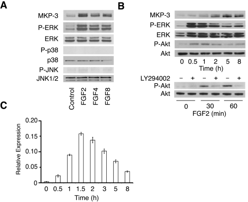
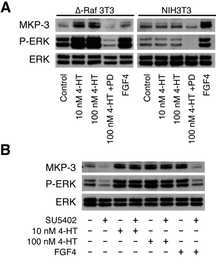
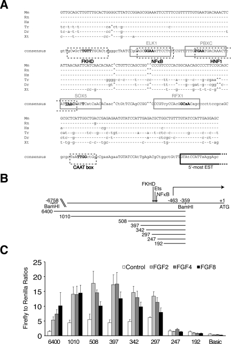
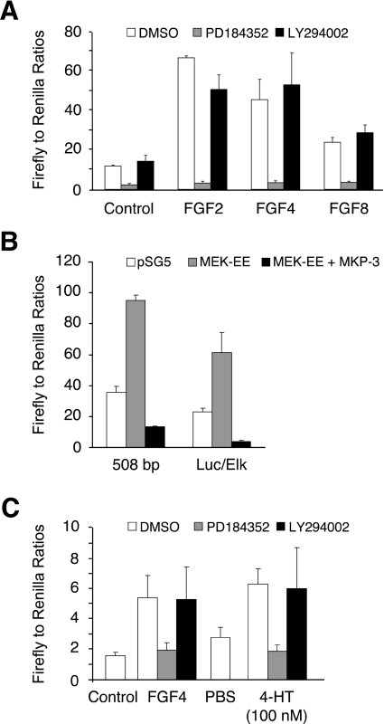
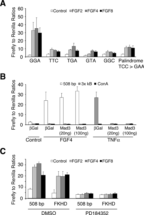
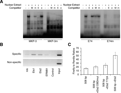
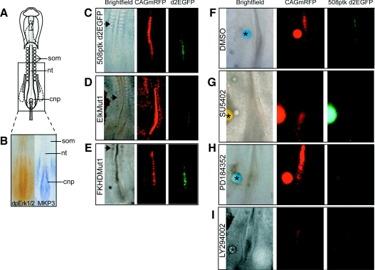
DUSP6 (dual-specificity phosphatase 6), also known as MKP-3 [MAPK (mitogen-activated protein kinase) phosphatase-3] specifically inactivates ERK1/2 (extracellular-signal-regulated kinase 1/2) in vitro and in vivo. DUSP6/MKP-3 is inducible by FGF (fibroblast growth factor) signalling and acts as a negative regulator of ERK activity in key and discrete signalling centres that direct outgrowth and patterning in early vertebrate embryos. However, the molecular mechanism by which FGFs induce DUSP6/MKP-3 expression and hence help to set ERK1/2 signalling levels is unknown. In the present study, we demonstrate, using pharmacological inhibitors and analysis of the murine DUSP6/MKP-3 gene promoter, that the ERK pathway is critical for FGF-induced DUSP6/MKP-3 transcription. Furthermore, we show that this response is mediated by a conserved binding site for the Ets (E twenty-six) family of transcriptional regulators and that the Ets2 protein, a known target of ERK signalling, binds to the endogenous DUSP6/MKP-3 promoter. Finally, the murine DUSP6/MKP-3 promoter coupled to EGFP (enhanced green fluorescent protein) recapitulates the specific pattern of endogenous DUSP6/MKP-3 mRNA expression in the chicken neural plate, where its activity depends on FGFR (FGF receptor) and MAPK signalling and an intact Ets-binding site. These findings identify a conserved Ets-factor-dependent mechanism by which ERK signalling activates DUSP6/MKP-3 transcription to deliver ERK1/2-specific negative-feedback control of FGF signalling.


Figures

Figure 1
Figure 1. The expression of DUSP6/MKP-3 mRNA and protein is inducible by FGF in NIH 3T3 cells
(A) NIH 3T3 cells were serum-starved overnight before exposure to FGF2, FGF4 or FGF8 (all at 30 ng/ml) for 5 h. Cells were then lysed, and proteins were analysed by SDS/PAGE and Western blotting using antisera against MKP-3, phospho-ERK (P-ERK), ERK, phospho-p38 (P-p38), p38, phospho-JNK (P-JNK) and JNK. (B) NIH 3T3 cells were serum-starved overnight and then exposed to FGF2 (30 ng/ml) for the indicated time before lysis and analysis of proteins by SDS/PAGE and Western blotting using antisera against MKP-3, phospho-ERK (P-ERK), ERK, phospho-Akt (P-Akt) or Akt (upper panels). NIH 3T3 cells were serum-starved overnight and then exposed to FGF2 (30 ng/ml) for the times indicated in either the absence or the presence of the PI3K inhibitor LY294002 (10 μM) before lysis and analysis of proteins by SDS/PAGE and Western blotting using antisera against phospho-Akt (P-Akt) or Akt (lower panel). (C) NIH 3T3 cells were serum-starved overnight before exposure to FGF2 (30 ng/ml) for the times indicated. Cells were then lysed, and cellular RNA was prepared. DUSP6/MKP-3 mRNA levels were then analysed using real-time PCR. Assays were performed in triplicate and relative DUSP6/MKP-3 mRNA levels are presented as means±S.E.M.
Figure 2
Figure 2. The induction of DUSP6/MKP-3 protein and mRNA by FGFs is blocked by chemical inhibitors of either the FGFR tyrosine kinase or MEK, but not by a specific inhibitor of PI3K activity in NIH 3T3 cells
NIH 3T3 cells were serum-starved overnight before exposure to FGF2, FGF4 or FGF8 (all at 30 ng/ml) for 5 h either in the absence (DMSO vehicle only) or presence of (A) the FGFR inhibitor SU5402 (50 μM), (B) the MEK inhibitor PD184352 (2 μM) or (C) the PI3K inhibitor LY294002 (10 μM). Cell lysates were analysed by SDS/PAGE and Western blotting using antisera against MKP-3, phospho-ERK (P-ERK), ERK, phospho-Akt (P-Akt) and Akt. (D) NIH 3T3 cells were serum-starved overnight before exposure to FGF2, FGF4 or FGF8 (all at 30 ng/ml) for 90 min in either the absence (DMSO vehicle only) or the presence of the indicated inhibitor. Cells were then lysed, and cellular RNA was prepared. DUSP6/MKP-3 mRNA levels were then analysed using real-time PCR. Assays were performed in triplicate and relative DUSP6/MKP-3 mRNA levels are presented as means±S.E.M.
Figure 3
Figure 3. ERK activation is both necessary and sufficient for the induction of DUSP6/MKP-3 protein in NIH 3T3 cells
(A) Either Δ-Raf-1:ER* NIH 3T3 cells (Δ-Raf 3T3) or NIH 3T3 cells were serum-starved overnight and then exposed to the indicated concentration of 4-HT in either the absence or the presence of PD184352 (PD; 2 μM). As a control, cells were exposed to FGF4 (30 ng/ml). After 5 h of incubation, cells were lysed, and proteins were analysed by SDS/PAGE and Western blotting using antisera against MKP-3, phospho-ERK (P-ERK) and ERK. (B) Δ-Raf-1:ER* NIH 3T3 cells were serum-starved overnight and then exposed to either the indicated concentration of 4-HT or FGF4 (30 ng/ml) in the absence or presence of the FGFR inhibitor SU5402 (50 μM). After 5 h of incubation, cells were lysed, and proteins were analysed by SDS/PAGE and Western blotting using antisera against MKP-3, phospho-ERK (P-ERK) and ERK.
Figure 4
Figure 4. Bioinformatic and functional analysis of the murine DUSP6/MKP-3 gene promoter
(A) DNA sequence alignment of a conserved region identified within the proximal promoter of the DUSP6/MKP-3 gene in vertebrates: Mm (Mus musculus), Rn (Rattus norvegicus), Hs (Homo sapiens), Tr (Takifugu rubripes), Dr (Danio rerio) and Xt (Xenopus tropicalis). Conserved transcription factor-binding sites are boxed, either grey-lined (forward strand) or black broken-lined (reverse strand) and identified as follows: FKHD (Forkhead family), ELK1 (Ets-binding site), NF-κB, PBXC (PBX1–Meis1 complexes), HNF1 (hepatic nuclear factor 1), SOX5 [Sox/SRY-sex/testis determining and related HMG (high-mobility group) box factors], RFX1 (regulatory factor X1), CAAT box (CAAT box: promoter element in some genes located approx. 75–80 bp upstream of the start site for transcription). Core bases are shown in bold, and, where these overlap, the nucleotides belonging to each site are identified to indicate orientation (either bold grey or hatched underlined). The putative transcriptional start site is also indicated (boxed in black). The latter is based on mapping the 5′-most extent of annotated ESTs (expressed sequence tags) for DUSP6/MKP-3. (B) Schematic diagram showing the 5′ boundaries of the DUSP6/MKP-3 promoter–reporter constructs employed in the deletion analysis. Nucleotides are numbered with the A of the ATG start codon designated as +1. The putative transcriptional start site (−463) is indicated as are the BamHI restriction enzyme sites used to subclone the longest (6400 bp) genomic fragment into the firefly luciferase reporter plasmid. The position of the conserved region containing the putative transcription factor-binding sites is also indicated. (C) The constructs indicated in (B) were co-transfected into NIH 3T3 cells along with pRL-TK Renilla to normalize for transfection efficiency. Cells were then starved overnight in 0.5% serum. The following day, cells were either left untreated or stimulated with the indicated FGF (all at 30 ng/ml) for an additional 24 h, before cells were lysed, and luciferase assays were performed. pGL3Basic acted as a negative (promoterless) control. Luciferase assays were performed in quadruplicate, and results are mean±S.E.M. firefly/Renilla ratios.
Figure 5
Figure 5. FGF-dependent DUSP6/MKP-3 promoter activity is mediated by activation of ERK and not by activation of the PI3K pathway
(A) The 508 bp DUSP6/MKP-3 reporter construct was co-transfected into NIH 3T3 cells along with pRL-TK Renilla to normalize for transfection efficiency. Cells were then starved overnight in 0.5% serum. The following day, cells were either left untreated or stimulated with the indicated FGF (all at 30 ng/ml) in either the absence or the presence of vehicle (DMSO), PD184352 (2 μM) or LY294002 (10 μM) for an additional 24 h. Cells were then lysed, and luciferase assays were performed in quadruplicate. (B) Either the 508 bp DUSP6/MKP-3 reporter construct or a plasmid encoding a GAL4–Elk1 fusion protein together with a GAL4-dependent luciferase reporter (Luc/Elk) was co-transfected into NIH 3T3 cells along with pRL-TK Renilla to normalize for transfection efficiency and either empty pSG5 expression vector or pSG5 encoding either a constitutively active mutant of MEK (MEK-EE) or DUSP6/MKP-3 itself. After 24 h, cells were lysed, and luciferase assays were performed in quadruplicate. (C) Δ-Raf-1:ER* NIH 3T3 cells were co-transfected with the 508 bp DUSP6/MKP-3 reporter construct and pRL-TK Renilla to normalize for transfection efficiency. Cells were then serum-starved overnight and either left untreated or treated with either FGF4 (30 ng/ml) or 4-HT (100 nM) in the presence or absence of vehicle (DMSO), PD184352 (2 μM) or LY294002 (10 μM). After 24 h, cells were lysed, and luciferase assays were performed in quadruplicate. Results are mean±S.E.M. firefly/Renilla ratios.
Figure 6
Figure 6. A conserved Ets-binding site is required for FGF-inducible DUSP6/MKP-3 promoter activity
(A) NIH 3T3 cells were co-transfected with either the wild-type 508 bp DUSP6/MKP-3 reporter construct or the indicated mutants together with pRL-TK Renilla to normalize for transfection efficiency. Cells were then starved overnight in 0.5% serum. The following day, cells were either left untreated or stimulated with the indicated FGF (all at 30 ng/ml). After 24 h, cells were lysed, and luciferase assays were performed in quadruplicate. (B) NIH 3T3 cells were co-transfected with the indicated reporter constructs along with either RSV-Mad3 (20 or 100 ng) or as a control RSV-βGal and pRL-TK Renilla to normalize for transfection efficiency. Cells were then serum-starved overnight (0.5% FBS) and either left untreated or exposed to either FGF4 (30 ng/ml) for 24 h or TNFα (10 ng/ml) for 6 h. Cells were then lysed, and luciferase assays were performed in quadruplicate. (C) NIH 3T3 cells were co-transfected with either the wild-type 508 bp DUSP6/MKP-3 reporter construct or a 508 bp reporter in which the Forkhead-binding site was mutated (FKHD) together with pRL-TK Renilla to normalize for transfection efficiency. Cells were then starved overnight in 0.5% FBS. The following day, cells were either left untreated or stimulated with the indicated FGF (all at 30 ng/ml) either in the absence (DMSO) or presence of PD184352 (2 μM). After 24 h, cells were lysed, and luciferase assays were performed in quadruplicate. Results are mean±S.E.M. firefly/Renilla ratios.
Figure 7
Figure 7. Ets-family transcription factors bind to the endogenous DUSP6/MKP-3 gene promoter
(A) EMSAs were performed using the following labelled oligonucleotide probes: wild-type MKP-3 or an Ets site mutant MKP-3 (MKP-3m), and wild-type canonical Ets-binding site (E74) or its corresponding Ets-binding site mutant (E74m). Labelled probes were then incubated with nuclear extract from NIH 3T3 cells in either the absence or the presence of the following unlabelled competitor DNAs: wild-type MKP-3 (M), mutant MKP-3 (m), wild-type E74 (E) or the corresponding Ets site mutant (e). Following incubation, mobility-shifts were visualized by electrophoresis and autoradiography. (B) ChIP assays were performed using an unrelated control antibody (HA) and antibodies specific for Ets1, Ets2 or ERM81. Specific PCR products corresponding to the region of the DUSP6/MKP-3 promoter containing the putative Ets-binding site and the non-specific control reactions are shown, as are the controls lacking template and the results of PCRs performed using the input chromatin as template. (C) NIH 3T3 cells were either transfected with the 508 bp DUSP6/MKP-3 reporter alone or together with either a deletion mutant of Ets2 lacking a functional DNA-binding domain (Ets2 DBDmut), a mutant Ets2 protein lacking a conserved MAPK phosphorylation site (Ets2 T72A) or wild-type Ets2. Following transfection, cells were cultured overnight in medium containing 10% FBS. Cells were then lysed, and luciferase assays were performed in triplicate. Results are mean±S.E.M. firefly/Renilla ratios.
Figure 8
Figure 8. The 508 bp DUSP6/MKP-3 promoter fragment directs transgene expression in an endogenous DUSP6/MKP-3 domain
(A) Schematic diagram of the HH10 chick embryo. The boxed area indicates approximate field in (BI). (B) Comparison of ERK1/2 phosphorylation and DUSP6/MKP-3 mRNA expression in the caudal region of the HH10 chick embryo. dpErk1/2, dual-phosphorylated ERK1/2. (C) HH10 chick embryo co-electroporated with a plasmid constitutively expressing mRFP (CAGmRFP) and the 508 bp fragment of the DUSP6/MKP-3 promoter driving expression of EGFP (508ptkd2EGFP) only within the endogenous DUSP6/MKP-3 domain. (D) Embryo co-electroporated with plasmid 508ptkd2EGFP in which the Ets-binding site is mutated (EtsMut1) and CAGmRFP shows reduced EGFP expression. (E) Co-electroporation of plasmid 508ptkd2EGFP in which the Forkhead-binding site is mutated (FKHDMut1) and CAGmRFP. (FH). Embryos were co-electroporated with the wild-type DUSP6/MKP-3 promoter fragment (508ptkd2EGFP) and CAGmRFP before beads were soaked in DMSO (F), 5 mM SU5402 (G), 20 mM PD184352 (H) or 20 mM LY294002 (I) were placed next to the electroporation site. Both SU5402 and PD184352 cause a local down-regulation of DUSP6/MKP-3 promoter activity. In contrast, neither LY294002 nor DMSO alone have any significant effect on DUSP6/MKP-3 promoter activity. Asterisks (*) indicates grafted bead, and the arrowhead indicates the position of the last formed somite. nt, neural tube; cnp, caudal neural plate; som, somite.

Similar articles

Cited by

References

    1. Camps M., Nichols A., Arkinstall S. Dual specificity phosphatases: a gene family for control of MAP kinase function. FASEB J. 2000;14:6–16. - PubMed
    1. Dickinson R. J., Keyse S. M. Diverse physiological functions for dual-specificity MAP kinase phosphatases. J. Cell Sci. 2006;119:4607–4615. - PubMed
    1. Groom L. A., Sneddon A. A., Alessi D. R., Dowd S., Keyse S. M. Differential regulation of the MAP, SAP and RK/p38 kinases by Pyst1, a novel cytosolic dual-specificity phosphatase. EMBO J. 1996;15:3621–3632. - PMC - PubMed
    1. Muda M., Theodosiou A., Rodrigues N., Boschert U., Camps M., Gillieron C., Davies K., Ashworth A., Arkinstall S. The dual specificity phosphatases M3/6 and MKP-3 are highly selective for inactivation of distinct mitogen-activated protein kinases. J. Biol. Chem. 1996;271:27205–27208. - PubMed
    1. Nichols A., Camps M., Gillieron C., Chabert C., Brunet A., Wilsbacher J., Cobb M., Pouyssegur J., Shaw J. P., Arkinstall S. Substrate recognition domains within extracellular signal-regulated kinase mediate binding and catalytic activation of mitogen-activated protein kinase phosphatase-3. J. Biol. Chem. 2000;275:24613–24621. - PubMed

Publication types

MeSH terms

Substances