. 2008 May;36(8):2530-46.
doi: 10.1093/nar/gkn096. Epub 2008 Mar 4.

Bioinformatic and functional analysis of RNA secondary structure elements among different genera of human and animal caliciviruses

Affiliations

Bioinformatic and functional analysis of RNA secondary structure elements among different genera of human and animal caliciviruses

Peter Simmonds et al. Nucleic Acids Res. 2008 May.

Abstract

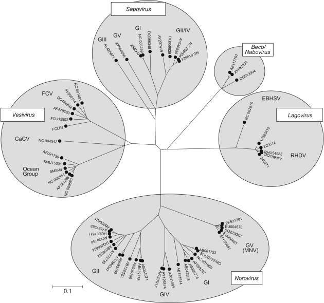
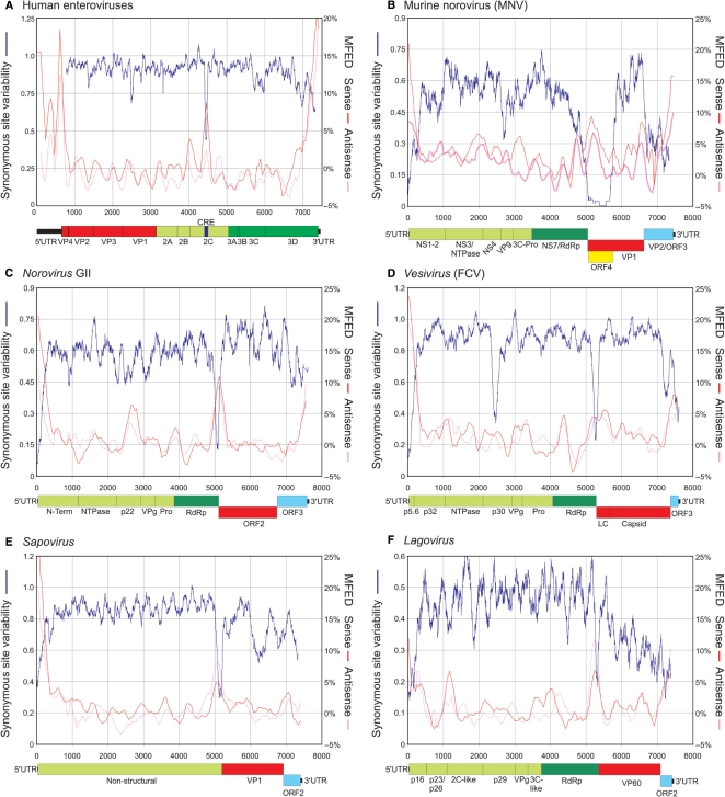
The mechanism and role of RNA structure elements in the replication and translation of Caliciviridae remains poorly understood. Several algorithmically independent methods were used to predict secondary structures within the Norovirus, Sapovirus, Vesivirus and Lagovirus genera. All showed profound suppression of synonymous site variability (SSSV) at genomic 5' ends and the start of the sub-genomic (sg) transcript, consistent with evolutionary constraints from underlying RNA structure. A newly developed thermodynamic scanning method predicted RNA folding mapping precisely to regions of SSSV and at the genomic 3' end. These regions contained several evolutionarily conserved RNA secondary structures, of variable size and positions. However, all caliciviruses contained 3' terminal hairpins, and stem-loops in the anti-genomic strand invariably six bases upstream of the sg transcript, indicating putative roles as sg promoters. Using the murine norovirus (MNV) reverse-genetics system, disruption of 5' end stem-loops produced approximately 15- to 20-fold infectivity reductions, while disruption of the RNA structure in the sg promoter region and at the 3' end entirely destroyed replication ability. Restoration of infectivity by repair mutations in the sg promoter region confirmed a functional role for the RNA secondary structure, not the sequence. This study provides comprehensive bioinformatic resources for future functional studies of MNV and other caliciviruses.


Figures

Figure 1.
Figure 1.
Phylogenetic analysis of the RNA-dependent RNA polymerase (RdRp)-encoding region of representative variants (up to five per genus or genogroup) of each genus of caliciviruses, shown as an unrooted tree. Sequences were translated and aligned using ClustalW with default settings, and a neighbour-joining phylogenetic tree constructed from pair wise translated amino acid distances. The scale bar indicates a distance of 0.1 (10% amino sequence divergence).
Figure 2.
Figure 2.
Scanning alignments of complete genome sequences of enteroviruses and caliciviruses for RNA secondary structure. Variability at synonymous sites (left y-axis) was computed at each codon position in alignments, plotted with a window size of 41 codons. MFED values (right y-axis) for sense and anti-sense RNA sequences were calculated for 200 base fragments, incrementing by 48 bases; values plotted represent mean values of five consecutive fragments. All nucleotide positions were calculated relative to reference sequences listed in methods; gene names, boundaries and other structural features followed the annotation provided for the reference sequences used for numbering. A higher resolution vector diagram of the figure is available from Supplementary Data (Figure S1).
Figure 3.
Figure 3.
RNA structure prediction for the first 400 bases of genome sequence alignments of (A) the first 800 bases of genome sequence alignments of human enteroviruses species A, and (B–F) the first 400 bases of genome sequence alignments of different calicivirus genera/groups. Pairing predicted by Alifold shown in lower right quadrant by PFOLD in the upper right. For both predictive algorithms, the size of the dots is proportional to the probability of pairing; predicted paired sites containing co-variant or semi-co-variant substitutions shown in green. Synonymous variability averaged over a window size of 21 codon shown as blue line (right-hand y-axis). A higher-resolution vector diagram of the figure is available from Supplementary Data (Figure S2).
Figure 4.
Figure 4.
Predicted RNA secondary structures using Alifold and PFOLD (see legend to Figure 2) in (A) 2C/CRE region human enterovirus species A and (B–F) regions of maximum SSSV in different calicivirus genera. For human enterovirus sequences, separate plots of synonymous variability were shown for species A–C. The positions of the CRE in 2C and the arrangement of NS (green) and S (red) gene reading frames in caliciviruses shown to scale on the x-axis, with the sub-genomic transcript shown in pink. A higher-resolution vector diagram of the figure is available from Supplementary Data (Figure S3).
Figure 5.
Figure 5.
(A–D) Predicted RNA secondary structures for 3′ ends of MNV, norovirus, vesivirus and lagovirus sequences, and (E, F) regions in the NS genes of MNV and Vesiviruses showing SSSV (Figure. 1B and D). A higher-resolution vector diagram of the figure is available from Supplementary Data (Figure S4).
Figure 6.
Figure 6.
Consensus RNA secondary structures upstream of the sub-genomic transcript (grey filled boxes; initiating anti-codons blocked) for the five calicivirus genera predicted by Alifold, shown in anti-sense orientation in a 3′ to 5′ left to right direction. Separate structure predictions were made for different human norovirus and sapovirus genogroups and for Newbury1- and NB-like beco/naboviruses because of their high sequence variability in this region (see text). The unpaired 6-base sequences found in all genera/groups between the predicted RNA structures and the start of the transcript are underlined.
Figure 7.
Figure 7.
Upper panels: Alifold predictions of RNA secondary structures in 5′, NS/S junction and 3′ terminal region of MNV for sequences in sense (lower right quadrant), and anti-sense (upper left quadrant). Lower figure: predicted RNA secondary structures for the prototype MNV-1 sequence (NC_008311), showing positions of introduced substitutions in mutants m50, m51, m53 and m54 to disrupt secondary structure (filled and unfilled circles) and compensatory changes in m53r to restore pairings (unfilled and filled squares; see Legend). SL8, SL29 and SL7330 were drawn in their sense orientation; SLa5045 in anti-sense orientation (see text). The position of naturally occurring insertions of pyrimidines in the 3′ terminal loop (SL7330) is indicated.
Figure 8.
Figure 8.
Replication ability of MNV with mutations that disrupted predicted RNA secondary structures in the 5′ end (m50, m51), NS/S junction (m53) and 3′ end (m54) of the genome (Figure 6) and MNV with compensatory mutations that restores base pairing in SLa5045. (A, C) Western blot analysis of NS7 levels during MNV recovery. Cells were infected with FPV-T7 and subsequently transfected with cDNA constructs containing WT or mutant MNV genomes under control of a T7 RNA polymerase promoter. Twenty-four hours post-transfection cells were harvested and the levels of NS7 determined by immunoblot using rabbit polyclonal anti-sera to MNV NS7. (B) Virus yield and plaque phenotype of viruses generated from either WT or mutant MNV cDNA constructs. Virus yields of mutants were expressed as log10 reductions in TCID50 per 35 mm dish from the WT control (mean value 50 100 ± 18 200). All virus recoveries were performed five times and mean reductions and standard deviations shown; the limit of detection was 60 TCID50/35 mm dish. (D) Virus yield and plaque phenotype of WT or mutant (m53 and m53r) cDNA constructs. Virus recoveries were performed five times with a mean recovery from the WT control of 14 160 ± 2500, and a limit of detection of 60 TCID50/35 mm dish.

Similar articles

Cited by

References

    1. Thiel HJ, Konig M. Caliciviruses: an overview. Vet. Microbiol. 1999;69:55–62. - PubMed
    1. Green KY, Ando T, Balayan MS, Berke T, Clarke IN, Estes MK, Matson DO, Nakata S, Neill JD, Studdert MJ, et al. Taxonomy of the caliciviruses. J. Infect. Dis. 2000;181(Suppl 2):S322–S330. - PubMed
    1. Oliver SL, Asobayire E, Dastjerdi AM, Bridger JC. Genomic characterization of the unclassified bovine enteric virus Newbury agent-1 (Newbury1) endorses a new genus in the family Caliciviridae. Virology. 2006;350:240–250. - PMC - PubMed
    1. Neill JD, Meyer RF, Seal BS. Genetic relatedness of the caliciviruses: San Miguel sea lion and vesicular exanthema of swine viruses constitute a single genotype within the Caliciviridae. J. Virol. 1995;69:4484–4488. - PMC - PubMed
    1. Smiley JR, Chang KO, Hayes J, Vinje J, Saif LJ. Characterization of an enteropathogenic bovine calicivirus representing a potentially new calicivirus genus. J. Virol. 2002;76:10089–10098. - PMC - PubMed

Publication types