. 2008 Mar 10;180(5):973-88.
doi: 10.1083/jcb.200707090. Epub 2008 Mar 3.

The Caenorhabditis elegans nephrocystins act as global modifiers of cilium structure

Affiliations

The Caenorhabditis elegans nephrocystins act as global modifiers of cilium structure

Andrew R Jauregui et al. J Cell Biol. .

Abstract

Nephronophthisis (NPHP) is the most common genetic cause of end-stage renal disease in children and young adults. In Chlamydomonas reinhardtii, Caenorhabditis elegans, and mammals, the NPHP1 and NPHP4 gene products nephrocystin-1 and nephrocystin-4 localize to basal bodies or ciliary transition zones (TZs), but their function in this location remains unknown. We show here that loss of C. elegans NPHP-1 and NPHP-4 from TZs is tolerated in developing cilia but causes changes in localization of specific ciliary components and a broad range of subtle axonemal ultrastructural defects. In amphid channel cilia, nphp-4 mutations cause B tubule defects that further disrupt intraflagellar transport (IFT). We propose that NPHP-1 and NPHP-4 act globally at the TZ to regulate ciliary access of the IFT machinery, axonemal structural components, and signaling molecules, and that perturbing this balance results in cell type-specific phenotypes.


Figures
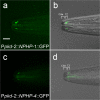
Figure 1.
Figure 1.
NPHP-1 and NPHP-4 localize to TZs of male-specific CEM cilia. 3D deconvolution images of NPHP-1∷GFP (a) and NPHP-4∷GFP (c) expressed only in male-specific CEM neurons and overlaid on Nomarski images (b and d). NPHP-1∷GFP and NPHP-4∷GFP localize to the CEM TZ but not the cilium proper (a–d). The CEM axoneme emanates from the TZ to the cuticle (territories of CEM cilium are indicated by brackets in b and d). Lateral view is shown. Bar, 10 μm.
Figure 2.
Figure 2.
PKD-2∷GFP localizes to abnormally formed CEM cilia in nphp-1 and nphp-4 mutants. 3D deconvolution images of a single CEM cilium in wild type (a–c), nphp-1 (d–f), and nphp-4 (g–i). See also Videos 1–3 (available at http://www.jcb.org/cgi/content/full/jcb.200707090/DC1). Cilium structure is labeled by β-tubulin (Ppkd-2∷tbb-4∷tdTomato; c, f, and i). PKD-2∷GFP localizes to the TZ and cilium proper in the wild type (a), to abnormally curled nphp-1 cilia (d), and to nphp-4 stunted cilia (g). Drawings show relationship between reporters in context of the male nose. Bar, 5 μm.
Figure 3.
Figure 3.
nphp-1 and nphp-4 mutants have misshaped, long, and stunted cilia. (A) CEM cilia are visualized using Ppkd-2∷tbb-4∷GFP. Dark gray indicates the frequency of misshaped curly CEM cilia; light gray indicates the frequency of stunted or mispositioned cilia. nphp-1 mutants have predominantly curly CEM cilia with some stunted cilia. nphp-4 and nphp-1;nphp-4 mutants have equal numbers of stunted and curly cilia. At least 50 animals are scored per genotype (∼200 individual CEM cilia are observed). Error bars indicate ±SEM. (B) CEM cilia were divided into wild-type, curly, and stunted categories. From z stack compressions of 3D wild-type and mutant CEM cilia; lengths were measured by tracing the cilium from the base of the TZ to the end of the axoneme. Curly cilia are significantly longer than the wild type. Stunted or misplaced cilia are slightly shorter than the wild type. The z distance of curls is not represented in this assay. p-values from a t test compare mutants to the wild type.
Figure 4.
Figure 4.
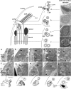
CEM, CEP and OLQ ciliary ultrastructure is abnormal in nphp-4 mutants. TEM of wild type (a–e) and nphp-4 (f–j) mutant animals in transverse thin sections. Black arrows, OLQ; white arrows, CEP; arrowheads, CEM. As depicted in the top drawings, the distal-most region of the CEM axoneme is narrow, curved outward, and exposed to the environment. Microtubule singlets are seen longitudinally near the opening (a). Traveling posteriorly, the distal segment aligns with the body axis and nears the edge of CEP socket cell (b). The CEM middle segment widens and is filled with microtubule outer singlets and dispersed inner singlets (c). The middle segment twists around the CEP away from the OLQ such that the CEM axoneme is furthest away from OLQ (d). CEM TZs are similar to CEP and OLQ TZs with 9 + 0 microtubule doublets (e). In nphp-4 mutants, OLQ distal segments are occasionally missing (f) but middle segments are present (g) with missing microtubules (g and h). CEM and OLQ TZs appear normal (i and j). CEP axonemes are irregular with large unidentified accumulations (g–i). Panel j is from a different section of the same worm illustrating well-formed OLQ and CEM TZs but apparently missing a CEP TZ, replaced by a large mass of TAM surrounded by microtubule singlets. CEM axonemes can lack distal segments (empty channel in f with no microtubules). Drawings at bottom illustrate nphp-4 defects. Bars: (top) 100 nm; (bottom) 0.5 μm.
Figure 5.
Figure 5.
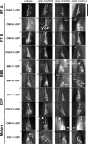
The localization of some IFT components is affected in nphp-4 mutants. Amphid channel cilia expressing various IFT reporters in wild-type and nephrocystin mutants. In nephrocystin mutants, CHE-11∷GFP (a1–a4), CHE-2∷GFP (not depicted), and OSM-5∷GFP (c1–c4) appear normal. In contrast, complex B polypeptide OSM-6∷GFP is dimmer in amphid cilia and fails to enter or is restricted from the distal segment of nphp-4 and nphp-1;nphp-4 (b1–b4). In b3, “Dim distal” segments are observed and in b4, distal segments are not visible, indicated by “No distal.” See Fig. S3 (available at http://www.jcb.org/cgi/content/full/jcb.200707090/DC1) for qualitative analysis of dim cilia. CHE-13∷GFP (d1-d4) is normal in nphp-1 mutants (d2) but is absent from the distal segment in 26% of nphp-4 animals (d3 and d4). d3 is an example of amphid channel cilia that appeared normal, and d4 depicts amphid channel cilia lacking distal expression of CHE-13∷GFP. BBS-7∷GFP is also restricted from nphp-4 and nphp-1;nphp-4 distal segments (e3 and e4) and slightly aggregates in middle segments (e3, arrow). In nphp-4, BBS-8∷GFP (f1–f4) was dim and barely detectable and “dim in cilia,” although clearly present in TZs (f3 and f4, arrows). Localization of both DYF-1∷GFP (g1–g3) and DYF-13∷GFP (g1–g4) appears normal. OSM-3∷GFP is considerably dimmer in nphp-4 and nphp-1;nphp-4 amphid cilia with bright signal in the distal dendrite (i3 and i4, arrowheads). KAP-1∷GFP localization also appears normal, with no distal localization (j1–j4). Similar results were observed in phasmid cilia (not depicted). Lateral view of the lips is shown, the anterior is to the top of each panel. Bar, 10 μm.
Figure 6.
Figure 6.
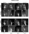
IFT-B polypeptide OSM-6∷GFP abnormally associates with kinesin-II in nphp-4 mutants. OSM-6∷GFP in amphid (a–d and i–l) and phasmid (e–h and m–p) cilia in various IFT and nphp-4 mutants. The IFT-B polypeptide OSM-6∷GFP enters ciliary distal segments in kap-1 and kap-1;bbs-7 single and double mutants (b and f and d and h). OSM-6∷GFP localizes to middle segments of osm-3 mutants (c and g) and is absent from missing distal segments. In kap-1;nphp-4 and kap-1;bbs-7;nphp-4 animals, OSM-6∷GFP severely aggregates in amphid cilia (j and l, arrowhead) and fails to enter distal segments (j and n and l and p). The arrow in panel n indicates dendritic accumulation of OSM-6∷GFP. No IFT is detected, though slight amounts of OSM-6∷GFP passively enter severely stunted cilia (n and p). In contrast, IFT is detected in nphp-4;osm-3 double mutants (k and o; Table I). Brackets indicate separation of middle (M), distal (D), and TZ regions of cilia. (a–d) Lateral view of the lips; the anterior is to the top. (e–h) Lateral view of the tail; posterior is to the top. Bar, 10 μm.
Figure 7.
Figure 7.
OSM-6∷GFP overexpression in nphp-4 mutants results in a severe SynDyf phenotype. Compression of a 3D deconvoluted z stack of amphid ciliated neurons filled with DiI. Wild type (a) and kap-1 mutants (b) fill with DiI. In contrast, osm-3 animals (c) lacking distal segments of cilia fail to fill with DiI. Some but not all amphid neurons fill with DiI in nphp-4 single (d) and kap-1;nphp-4 double mutants (e). Panel f is a 2.5× view of wild-type and nphp-4 neuron cell bodies that filled with dye. There is slight variability in the neurons that fail to fill between individual animals. The OSM-6∷GFP transgene in the nphp-4 mutant background causes a SynDyf defect (j–l). The mnIs17 OSM-6∷GFP transgene does not interfere with dye filling in the wild type (g–i) or kap-1 or nphp-1 mutants (not depicted), nor does OSM-6∷GFP interfere with ciliogenesis in any published reports (Collet et al., 1998; Snow et al., 2004; Ou et al., 2005). Bars: (a) 20 μm; (f) 5 μm.
Figure 8.
Figure 8.
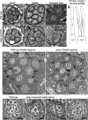
Amphid cilia are shortened and have abnormal microtubule structure in nphp-4 mutants. TEM of amphid channel cilia in the wild type (a–c, h, and j) and nphp-4 (d–f, i, and k–m). In nphp-4, some distal segments are shortened; panel d shows 6 out of the normal 10 distal segments that should be present. Middle segments (e, i, and m) are sometimes swollen or withered. Because of slight cilia bending, views of individual microtubules within a cilium are sometimes seen obliquely and seem distorted; when tilted in the microscope, those distortions go away. Occasionally the channel contains <10 cilia; in panel e, nine are present, suggesting that at least one cilium is completely absent past the TZ. Some middle segments have microtubule doublets abnormally converting prematurely to singlets lacking B tubules (e and i, arrowhead) or abnormal doublets with an inner seam break of the B tubule (i, arrows), whereas others appear normal. Such inner seam breaks are also found in wild-type cilia of ADL and ASI (h, arrows) but become much more common in other cilia in nphp-4 (i, k, and l). Schematic cartoon (g) displays the region of the middle segment where seam breaks are common in ADL (wild type and mutant) and in ASJ mutant cilia, seen from a lateral aspect. In wild-type and nphp-4 ADL cilia, all microtubules tend to have long continuous seam breaks. In ASJ, shorter, discontinuous seam breaks are scattered along the middle segment, but only in nphp-4 animals. Small spurs can also be seen on the outer surface of many A tubules within the middle segment, looking vaguely similar to Y-link features of the axoneme (compare c with k–m). Panel m shows a nphp-4 ADL cilium, just distal to the axoneme, where an increased number of microtubule doublets surround the outer edge (13 rather than 8–9), most showing B tubule seam breaks; some inner “singlets” even have partial B tubules. This represents a fused cilium (seen in 2/6 amphids in nphp-4); ADL normally forms two separate cilia (g). (i) An electron-dense mass that may represent jammed IFT particles. TZs are typically well-formed in nphp-4 mutants (f). Bars: (a) 0.5 μm; (h) 0.5 μm; (j) 100 nm.

Similar articles

Cited by

References

    1. Arts, H.H., D. Doherty, S.E. van Beersum, M.A. Parisi, S.J. Letteboer, N.T. Gorden, T.A. Peters, T. Marker, K. Voesenek, A. Kartono, et al. 2007. Mutations in the gene encoding the basal body protein RPGRIP1L, a nephrocystin-4 interactor, cause Joubert syndrome. Nat. Genet. 39:882–888. - PubMed
    1. Attanasio, M., N.H. Uhlenhaut, V.H. Sousa, J.F. O'Toole, E. Otto, K. Anlag, C. Klugmann, A.C. Treier, J. Helou, J.A. Sayer, et al. 2007. Loss of GLIS2 causes nephronophthisis in humans and mice by increased apoptosis and fibrosis. Nat. Genet. 39:1018–1024. - PubMed
    1. Badano, J.L., N. Mitsuma, P.L. Beales, and N. Katsanis. 2006. The ciliopathies: an emerging class of human genetic disorders. Annu. Rev. Genomics Hum. Genet. 7:125–148. - PubMed
    1. Bae, Y.K., H. Qin, K.M. Knobel, J. Hu, J.L. Rosenbaum, and M.M. Barr. 2006. General and cell-type specific mechanisms target TRPP2/PKD-2 to cilia. Development. 133:3859–3870. - PubMed
    1. Bargmann, C.I. 2006. Related comparative chemosensation from receptors to ecology. Nature. 444:295–301. - PubMed

Publication types

MeSH terms

Substances

LinkOut - more resources