. 2008 May;48(5):780-91.
doi: 10.1016/j.jhep.2007.12.020. Epub 2008 Feb 7.

Functional interaction between Wnt3 and Frizzled-7 leads to activation of the Wnt/beta-catenin signaling pathway in hepatocellular carcinoma cells

Affiliations

Functional interaction between Wnt3 and Frizzled-7 leads to activation of the Wnt/beta-catenin signaling pathway in hepatocellular carcinoma cells

Miran Kim et al. J Hepatol. 2008 May.

Abstract

Background/aims: The canonical Wnt signaling is frequently activated in human hepatocellular carcinoma (HCC). We previously demonstrated that upregulation of Frizzled-7 receptor (FZD7) in HCC was associated with nuclear accumulation of wild-type beta-catenin. Here, we investigated Wnt ligand(s) that may activate the Wnt/beta-catenin pathway through FZD7 in HCC cells.

Methods: To identify Wnt ligand expression, RT-PCR was performed in HCC cells. To evaluate the function of Wnt3 and FZD7 in HCC, we utilized Wnt3 overexpressing FOCUS HCC cells (FOCUS-Wnt3) and human tumors.

Results: In hepatitis B virus (HBV)-induced HCC, Wnt3 was upregulated in tumor and peritumoral tissues compared to normal liver and downstream beta-catenin target genes were also increased in these samples. Activation of the Wnt/beta-catenin pathway in FOCUS-Wnt3 cells was demonstrated by beta-catenin accumulation, enhanced TCF transcriptional activity and proliferation rate. The activation of Wnt/beta-catenin signaling in FOCUS-Wnt3 was abolished by a knockdown of FZD7 expression by siRNA. More important, a specific Wnt3-FZD7 interaction was observed by co-immunoprecipitation experiments, which suggest that the action of Wnt3 was mediated via FZD7.

Conclusions: These findings demonstrate a functional interaction between Wnt3 and FZD7 leading to activation of the Wnt/beta-catenin signaling pathway in HCC cells and may play a role during hepatocarcinogenesis.


Figures
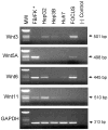
Fig. 1
Fig. 1
Expression of Wnt mRNAs in HCC cell lines. Wnt ligand mRNAs were detected in HCC cell lines using RT-PCR. Wnt3 and Wnt6 mRNAs were found in all HCC cell lines. Wnt5A was present only in FOCUS, whereas Wnt11 was detected in HepG2, Hep3B, and Huh7. The expression of glyceraldehydes-3-phosphate dehydrogenase gene (GAPDH) verified the quality of mRNA in each sample. * Wnt3, Wnt6, and Wnt11 from FB (fetal brain), whereas Wnt5A from FK (fetal kidney).
Fig. 2
Fig. 2
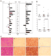
Expression of Wnt3, FZD7, and β-catenin target genes in human HBV-related HCC tissues. (A) The levels of Wnt3 and FZD7 mRNA measured by real-time RT-PCR. The black bars represent the mRNA levels in HCC tissues, and the white bars in corresponding peritumoral areas. Experiments were performed in duplicate, and data are expressed as a ratio to the mean value found in normal liver (N = 4). The Wnt3 mRNA (left panel) expression showed increased in HCCs (77%) and peritumoral tissues (59%). The FZD7 mRNA (right panel) expression was increased in both HCC and peritumoral tissues (59%) compared to normal liver. Within the paired samples, 11 of 17 (65%) showed increased expression of FZD7 mRNA in tumors compared with corresponding peritumoral tissues (P < 0.05). Tumors with a mutant β-catenin gene are marked with an asterisk. (B) Distribution of Wnt3 and FZD7 expression according to β-catenin status. T(mut); tumor with β-catenin mutation, T(WT); tumor with wild-type β-catenin, pT; peritumoral tissue. (C) Detection of Wnt3 and FZD7 proteins by immunohistochemistry. Representative example (case No. 6) of HCC (e–h) and peritumoral tissue (a–d) immunostained with either anti-Wnt3 Ab (b, f) or anti-FZD7 Ab (c, g). Panels (a, e) depict hematoxylin and eosin staining, and (d, h) represent negative controls (see Methods). Note that Wnt3 and FZD7 expression is evident in tumor cells whereas peritumoral tissues show reduced levels of staining. (D) Evaluation of Wnt/β-catenin target gene expression by real-time RT-PCR in HCC according to β-catenin status. Values are expressed as multiples of the relative expression found in normal liver (N = 4). Mean expression values of GS, Tbx3 and c-Myc were significantly higher in HCCs with both wild type and mutant β-catenin gene compared to peritumoral tissues. There was significant increase in GPR49 expression in HCC carrying mutant β-catenin compared with peritumoral tissue.
Fig. 2
Fig. 2
Expression of Wnt3, FZD7, and β-catenin target genes in human HBV-related HCC tissues. (A) The levels of Wnt3 and FZD7 mRNA measured by real-time RT-PCR. The black bars represent the mRNA levels in HCC tissues, and the white bars in corresponding peritumoral areas. Experiments were performed in duplicate, and data are expressed as a ratio to the mean value found in normal liver (N = 4). The Wnt3 mRNA (left panel) expression showed increased in HCCs (77%) and peritumoral tissues (59%). The FZD7 mRNA (right panel) expression was increased in both HCC and peritumoral tissues (59%) compared to normal liver. Within the paired samples, 11 of 17 (65%) showed increased expression of FZD7 mRNA in tumors compared with corresponding peritumoral tissues (P < 0.05). Tumors with a mutant β-catenin gene are marked with an asterisk. (B) Distribution of Wnt3 and FZD7 expression according to β-catenin status. T(mut); tumor with β-catenin mutation, T(WT); tumor with wild-type β-catenin, pT; peritumoral tissue. (C) Detection of Wnt3 and FZD7 proteins by immunohistochemistry. Representative example (case No. 6) of HCC (e–h) and peritumoral tissue (a–d) immunostained with either anti-Wnt3 Ab (b, f) or anti-FZD7 Ab (c, g). Panels (a, e) depict hematoxylin and eosin staining, and (d, h) represent negative controls (see Methods). Note that Wnt3 and FZD7 expression is evident in tumor cells whereas peritumoral tissues show reduced levels of staining. (D) Evaluation of Wnt/β-catenin target gene expression by real-time RT-PCR in HCC according to β-catenin status. Values are expressed as multiples of the relative expression found in normal liver (N = 4). Mean expression values of GS, Tbx3 and c-Myc were significantly higher in HCCs with both wild type and mutant β-catenin gene compared to peritumoral tissues. There was significant increase in GPR49 expression in HCC carrying mutant β-catenin compared with peritumoral tissue.
Fig. 3
Fig. 3
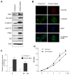
Overexpression of Wnt3 activated Wnt/β-catenin signaling in FOCUS HCC cells. (A) Western blot analysis in FOCUS-Wnt3 or FOCUS-C cells. Wnt3 was overexpressed in FOCUS-Wnt3 as demonstrated by anti-Wnt3 and anti-myc tag mAbs. Expression levels of Cyclin D1, GS, and c-Myc proteins were also increased. Note that the anti-c-Myc mAb does not recognize the myc-tag, but rather the c-Myc protein. Actin was used as a loading control. (B) Control (FOCUS-C) or FOCUS-Wnt3 cells were double-immunostained with anti-myc tag (red color) and anti-β-catenin (green color) Abs; the nucleus was counterstained with the DAPI (blue color). The bottom panel reveals the merged images indicating nuclear localization of β-catenin. (C) The TCF transcriptional activity was increased by 3 fold in FOCUS-Wnt3 cells compared with control (*P < 0.01). (D) Wnt3 expression increased the FOCUS cell proliferation rate. The results are expressed as the mean ± SE of triplicate assays. *P < 0.05 versus control
Fig. 4
Fig. 4
Inhibition of Wnt/β-catenin signaling by Wnt3 siRNA. (A) FOCUS cells were transfected with either siRNA GLO (a control for transfection efficiency and silencing), Wnt3 siRNA, or β-catenin siRNA and immunblotted with either anti-Wnt3 or anti-β-catenin mAbs (left panel). Expression levels of Wnt3 and β-catenin were plotted as a ratio to actin (right panel). (B) Effects of Wnt3 siRNA on the TCF transcriptional activities in FOCUS HCC cell lines. Wnt3 siRNA, control siRNA (siRNA GLO), or β-catenin siRNA were co-transfected in the presence of a TCF reporter gene. The TCF transcriptional activity was decreased in FOCUS HCC cells (30% for Wnt3 siRNA, and 43% for β-catenin siRNA). (C) FOCUS cell proliferation was reduced by Wnt3 siRNA compared to control. The results are expressed as the mean ± SE of triplicate assays. *P < 0.05 versus control.
Fig. 5
Fig. 5
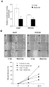
Anti-Wnt3 mAb treatment inhibits Wnt/β-catenin signaling. (A) Effects of anti-Wnt3 mAb on TCF transcriptional activities in HCC cell lines. The TCF transcriptional activities were decreased in Huh7 (60%), and FOCUS (40%) cells by the anti-Wnt3 mAb. *P < 0.001 versus control. (B) Delayed wound healing exhibited by the anti-Wnt3 mAb. Both Huh7 and FOCUS cells treated with anti-Wnt3 mAb showed delayed wound closure. At 24 h, most of the wound was closed with migrating FOCUS cells treated with the control IgG, while it remained open in those treated with anti-Wnt3 mAb. Graph of the wound closure (percent) plotted against time (bottom panel).
Fig. 6
Fig. 6
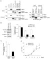
Activation of Wnt/β-catenin signaling is mediated by a Wnt3 and FZD7 interaction. (A) Co-immunoprecipitation assay demonstrating the interaction between Wnt3 and FZD7. Huh7 and FOCUS cells were transfected with either Wnt3-myc and FZD7-EE or FZD7-ΔCRD-EE plasmids. Cell lysates were first immunopurified using monoclonal anti-EE antibody immobilized onto a Sepharose Fast Flow matrix, and immunoblotted with anti-EE or anti-myc antibody. (Left panel) Demonstration of Wnt3, FZD7, and FZD7-ΔCRD protein expression in Huh7 and FOCUS cells after transfection. (Right panel) Immunoblotting with anti-EE or anti-myc antibodies after immunoprecipitation. Note that Wnt3 was detected only in an immunoprecipitation derived from cells co-transfected with Wnt3-myc and FZD7-EE but not from cells transfected with Wnt3-myc and FZD7-ΔCRD-EE, which lacks the putative ligand-binding domain. (B) Western blot analysis to evaluate the knockdown of FZD7 expression by FZD7 siRNA. Transfection of FZD7 siRNA in FOCUS-Wnt3 reduced FZD7 expression (by 65%) as well as β-catenin accumulation (by 80%) (left panel). Expression levels of Wnt3, FZD7 and β-catenin were plotted as a ratio to actin (right panel). (C) FZD7 siRNA abolished the high TCF transcriptional activity in FOCUS-Wnt3 mediated by Wnt3 overexpression. (D) FOCUS-Wnt3 cell proliferation was reduced by FZD7 siRNA compared to control. The results are expressed as the mean ± SE of triplicate assays. *P < 0.05 versus control.

Similar articles

Cited by

References

    1. Logan CY, Nusse R. The Wnt signaling pathway in development and disease. Annu Rev Cell Dev Biol. 2004;20:781–810. - PubMed
    1. Caricasole AFT, Iacovelli L, Barletta E, Caruso A, Melchiorri D, Terstappen GC, et al. Functional characterization of Wnt7a signaling in PC12 cells: interaction with A FZD5 x LRP6 receptor complex and modulation by Dickkopf proteins. J Biol Chem. 2003;278:37024. - PubMed
    1. Winn RA, Marek L, Han SY, Rodriguez K, Rodriguez N, Hammond M, et al. Restoration of Wnt-7a expression reverses non-small cell lung cancer cellular transformation through frizzled-9-mediated growth inhibition and promotion of cell differentiation. J Biol Chem. 2005;280:19625–19634. - PubMed
    1. Wang Z, Shu W, Lu MM, Morrisey EE. Wnt7b activates canonical signaling in epithelial and vascular smooth muscle cells through interactions with Fzd1, Fzd10, and LRP5. Mol Cell Biol. 2005;25:5022. - PMC - PubMed
    1. Gazit A, Yaniv A, Bafico A, Pramila T, Igarashi M, Kitajewski J, et al. Human frizzled 1 interacts with transforming Wnts transduce a TCF dependent transcriptional response. Oncogene. 1999;18:5959–5966. - PubMed

Publication types

MeSH terms