. 2008 Apr 18;283(16):10753-63.
doi: 10.1074/jbc.M801263200. Epub 2008 Feb 25.

Protein phosphatase 2A is a negative regulator of transforming growth factor-beta1-induced TAK1 activation in mesangial cells

Affiliations

Protein phosphatase 2A is a negative regulator of transforming growth factor-beta1-induced TAK1 activation in mesangial cells

Sung Il Kim et al. J Biol Chem. .

Abstract

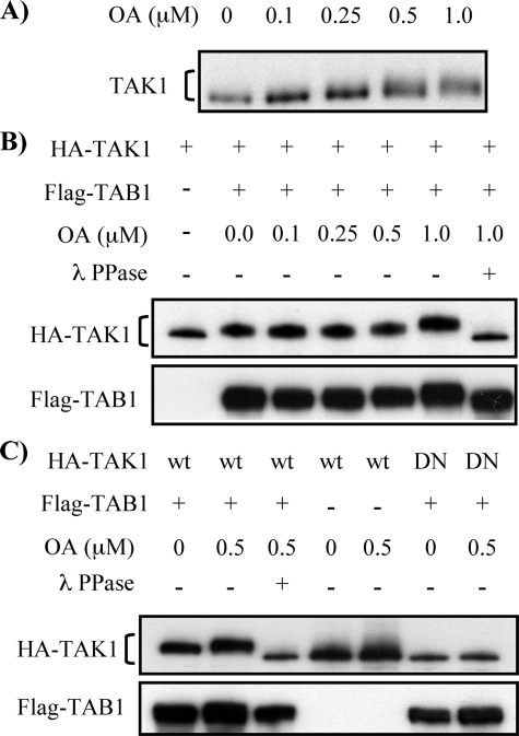
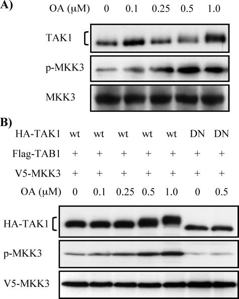
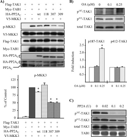
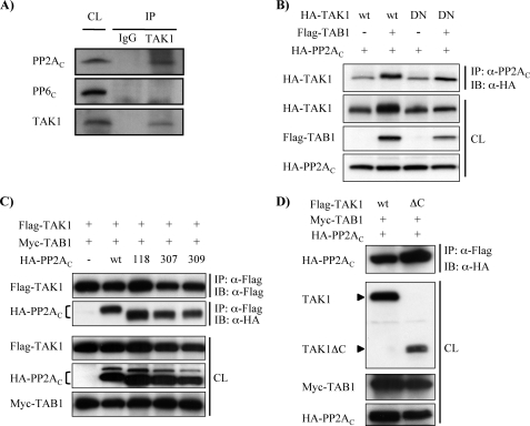
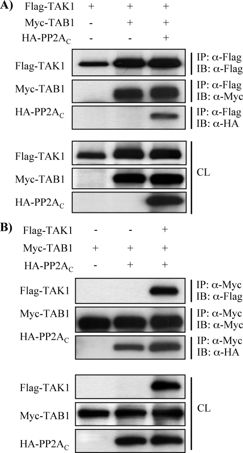
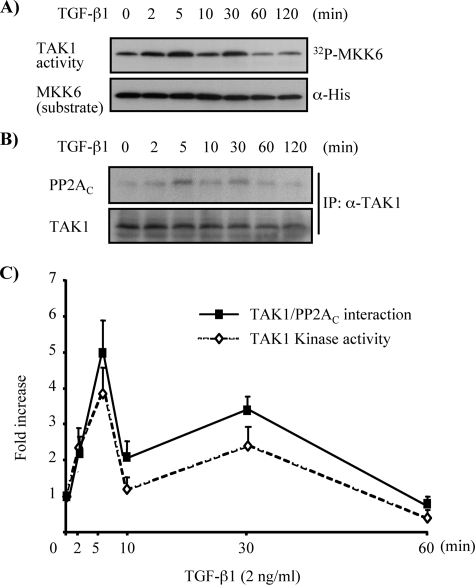
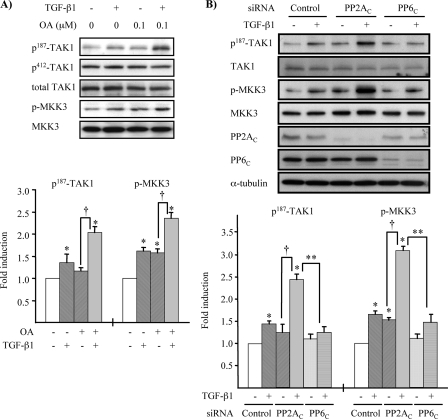
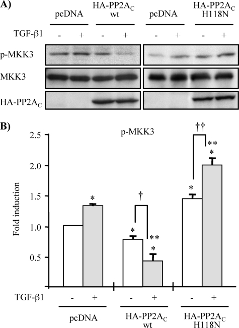
TAK1 (transforming growth factor (TGF)-beta-activated kinase 1) is a serine/threonine kinase that is rapidly activated by TGF-beta1 and plays a vital function in its signal transduction. Once TAK1 is activated, efficient down-regulation of TAK1 activity is important to prevent excessive TGF-beta1 responses. The regulatory mechanism of TAK1 inactivation following TGF-beta1 stimulation has not been elucidated. Here we demonstrate that protein phosphatase 2A (PP2A) plays a pivotal role as a negative regulator of TAK1 activation in response to TGF-beta1 in mesangial cells. Treatment with okadaic acid (OA) induces autophosphorylation of Thr-187 in the activation loop of TAK1. In vitro dephosphorylation assay suggests that Thr-187 in TAK1 is a major dephosphorylation target of PP2A. TGF-beta1 stimulation rapidly activates TAK1 in a biphasic manner, indicating that TGF-beta1-induced TAK1 activation is tightly regulated. The association of PP2A(C) with TAK1 is enhanced in response to TGF-beta1 stimulation and closely parallels TGF-beta1-induced TAK1 activity. Attenuation of PP2A activity by OA treatment or targeted knockdown of PP2A(C) with small interfering RNA enhances TGF-beta1-induced phosphorylation of TAK1 at Thr-187 and MKK3 (MAPK kinase 3). Endogenous TAK1 co-precipitates with PP2A(C) but not PP6(C), another OA-sensitive protein phosphatase, and knockdown of PP6(C) by small interfering RNA does not affect TGF-beta1-induced phosphorylation of TAK1 at Thr-187 and MKK3. Moreover, ectopic expression of phosphatase-deficient PP2A(C) enhances TAK1-mediated MKK3 phosphorylation by TGF-beta1 stimulation, whereas the expression of wild-type PP2A(C) suppresses the MKK3 phosphorylation. Taken together, our data indicate that PP2A functions as a negative regulator in TGF-beta1-induced TAK1 activation.


Figures

FIGURE 1.
FIGURE 1.
Inhibition of Ser/Thr protein phosphatases by OA induces TAK1 phosphorylation. A, OA treatment increases mobility shift in endogenous TAK1. MMC grown to subconfluence were treated with increasing concentrations of OA for 6 h as indicated. Cell lysates were subjected to Western blot analysis with anti-TAK1 antibody. B, OA treatment enhances TAK1 phosphorylation induced by coexpression of TAK1 and TAB1. MMC transfected with expression vector encoding HA-TAK1 alone or together with FLAG-TAB1 were treated with increasing concentrations of OA for 6 h, as indicated. Expression of HA-TAK1 and FLAG-TAB1 was determined by Western blot analysis with anti-HA and anti-FLAG antibodies, respectively. Four hundred units of λ protein phosphatase (λ PPase) were added to cell lysates and incubated for 30 min at 30 °C to dephosphorylate TAK1. C, TAK1 kinase activity is required for its phosphorylation. Wild-type HA-TAK1 (wt) or kinase-deficient mutant of HA-TAK1 (DN) was coexpressed with FLAG-TAB1 in MMC, as indicated, and treated with 0.5 μm OA for 6 h. Western blot analysis and treatment with λ PPase were performed as described in B.
FIGURE 2.
FIGURE 2.
OA-induced TAK1 phosphorylation mediates MKK3 phosphorylation. A, OA treatment increases endogenous MKK3 phosphorylation. MMC grown to subconfluence were treated with increasing concentrations of OA for 6 h as indicated. Cell lysates were subjected to Western blot analysis with anti-TAK1, anti-p-MKK3/6, and anti-MKK3 antibodies, respectively. B, OA-induced MKK3 phosphorylation is dependent on TAK1 activation. Wild-type HA-TAK1 (wt) or kinase-deficient mutant of HA-TAK1 (DN) was coexpressed with FLAG-TAB1 and V5-MKK3 in MMC and treated with increasing concentrations of OA for 6 h. Cell lysates were subjected to Western blot analysis with anti-HA, anti-p-MKK3/6, and anti-V5 antibodies, respectively.
FIGURE 3.
FIGURE 3.
PP2A down-regulates phosphorylation of TAK1 and MKK3. A, ectopic expression of wild type or mutants of PP2AC changes TAK1 activity. MMC were transfected as indicated with expression vectors for V5-MKK3, FLAG-TAK1, Myc-TAB1, and wild-type HA-PP2AC (wt) or mutants of PP2AC (H118N, Y307F, or L309Q). TAK1 activity was assessed by MKK3 phosphorylation. Expression of each transfected gene was confirmed by Western blot analysis with anti-V5, anti-FLAG, anti-Myc, and anti-HA antibodies, respectively. Expression levels of endogenous and exogenous PP2AC were evaluated by Western blot with anti-PP2AC antibody (bottom panel). Densitometry data are expressed as the percentage of p-MKK3 signals, quantified as the ratio to total V5-MKK3, compared with control transfection cells with FLAG-TAK1, Myc-TAB1, and V5-MKK3, but without HA-PP2AC (lane 2); the results represent mean ± S.E. of three independent experiments (*, p < 0.05 versus control transfection). B, OA treatment induces TAK1 phosphorylation at Thr-187. MMC grown to subconfluence were treated with OA for 30 min with the indicated concentrations. Cell lysates were subjected to Western blot analysis with anti-phospho-Thr-187-TAK1 (p187-TAK1), anti-phospho-Ser-412-TAK1 (p412-TAK1), and anti-TAK1 (total TAK1), respectively. Densitometry data are presented as -fold increase in p187-TAK1 or p412-TAK1 signals, quantified as the ratio to total TAK1, compared with control untreated cells; the results represent mean ± S.E. of three independent experiments (*, p < 0.05 versus untreated cells). C, dephosphorylation of TAK1 by PP2A primarily targets p-Thr-187 in the TAK1 activation loop. Phosphorylated HA-TAK1 was obtained by coexpression with FLAG-TAB1 in MMC followed by immunoprecipitation with anti-HA antibodies. The immunoprecipitates were incubated with 0.02, 0.1, and 0.2 units of purified PP2A for 20 min at 30 °C. Following the reaction, dephosphorylation of TAK1 was evaluated by Western blotting with anti-phospho-Thr-187-TAK1 and anti-phospho-Ser-412-TAK1 antibodies. A relative equivalent amount of HA-TAK1 in the immunoprecipitated samples was verified by Western blotting with anti-HA antibody. Expression of FLAG-TAB1 was confirmed by subjecting cell lysates to Western blot analysis with anti-FLAG antibody.
FIGURE 4.
FIGURE 4.
PP2A associates with TAK1. A, endogenous PP2AC is stably associated with TAK1. Cell lysates from MMC grown to subconfluence were immunoprecipitated with rabbit anti-TAK1 antibody. Nonimmunized rabbit IgG was used for control. Immunoprecipitates (IP) and cell lysates (CL) were subjected to immunoblotting with anti-PP2AC, anti-PP6C, and anti-TAK1 antibodies. B, phosphorylation status of TAK1 is not essential for the association of TAK1 with PP2AC. Wild-type HA-TAK1 (wt) or kinase-deficient mutant of HA-TAK1 (DN) was coexpressed with FLAG-TAB1 and HA-PP2AC, as indicated, in MMC. Cell lysates were subjected to immunoprecipitation with anti-PP2AC antibody, followed by immunoblotting (IB) with anti-HA antibody. Expression of the transfected genes was confirmed by subjecting cell lysates (CL) to Western blotting with corresponding anti-HA and anti-FLAG antibodies, as indicated. C, phosphatase activity of PP2AC does not affect the association of PP2AC with TAK1. Wild-type HA-PP2AC (wt) or mutants of PP2AC (H118N, Y307F, or L309Q) were coexpressed with FLAG-TAK1 and Myc-TAB1, as indicated, in MMC. Cell lysates were subjected to immunoprecipitation with anti-FLAG antibody for TAK1, followed by immunoblotting with either anti-FLAG or anti-HA antibodies. Expression of the transfected genes was confirmed by subjecting cell lysates to Western blotting with corresponding anti-FLAG, anti-HA, and anti-Myc antibodies. D, PP2AC interacts with the N-terminal region of TAK1. MMC were transfected with expression constructs encoding either full-length FLAG-TAK1 (wt) or C-terminally truncated TAK1 (ΔC) together with Myc-TAB1 and HA-PP2AC, as indicated. Cell lysates were subjected to immunoprecipitation with anti-FLAG antibody, followed by immunoblotting with anti-HA antibody. Expression of the transfected genes was confirmed in cell lysates (CL) subjected to Western blotting with corresponding anti-TAK1, anti-Myc, and anti-HA antibodies.
FIGURE 5.
FIGURE 5.
PP2AC associates with TAB1 independent of TAK1. A, FLAG-TAK1 was coexpressed with Myc-TAB1 and HA-PP2AC as indicated in MMC. Cell lysates were subjected to immunoprecipitation (IP) with anti-FLAG antibody, followed by immunoblotting (IB) with anti-FLAG, anti-Myc, and anti-HA antibodies, respectively. Expression of the transfected genes was confirmed in cell lysates (CL) subjected to Western blotting with corresponding anti-FLAG, anti-Myc, and anti-HA antibodies. B, Myc-TAB1 was coexpressed with FLAG-TAK1 and HA-PP2AC as indicated in MMC. Cell lysates were subjected to immunoprecipitation with anti-Myc antibody followed by immunoblotting with anti-FLAG, anti-Myc, and anti-HA antibodies, respectively. Expression of the transfected genes was confirmed in cell lysates subjected to Western blotting with corresponding anti-FLAG, anti-Myc, and anti-HA antibodies.
FIGURE 6.
FIGURE 6.
Association of PP2AC with TAK1 parallels activation and inactivation of endogenous TAK1 by TGF-β1 stimulation. MMC grown to subconfluence were rendered quiescent in medium supplemented with 0.5% FBS for 16 h and then stimulated with TGF-β1 (2 ng/ml) for the indicated times. Endogenous TAK1 in cell lysates was immunoprecipitated with anti-TAK1 antibody. One-half of the immunoprecipitate was subjected to immune complex kinase assay to assess endogenous TAK1 activity using His-MKK6 as a substrate and phosphorylation of MKK6 by TAK1 was visualized by autoradiography (A, top). The other half of the immunoprecipitate (IP) was subjected to immunoblotting with anti-PP2AC antibody to evaluate the association of endogenous TAK1 with PP2AC (B, top). Immunoblotting with anti-His and anti-TAK1 antibodies served as loading controls (bottom). C, densitometry data are presented as -fold increase in [32P]MKK6 and coprecipitated PP2AC, quantified as the ratio to total MKK6 substrate or total immunoprecipitated TAK1, compared with control untreated cells; the results represent mean ± S.E. of three independent experiments (⋄, 32P-labeled His-MKK6 versus His-MKK6; ▪, PP2AC versus TAK1 after immunoprecipitation).
FIGURE 7.
FIGURE 7.
Down-regulation of PP2A activity enhances TGF-β1-induced TAK1 and MKK3 phosphorylation. A, low dose OA increases TGF-β1-induced phosphorylation of Thr-187 in TAK1 and MKK3. MMC grown to subconfluence were rendered quiescent in medium supplemented with 0.5% FBS for 16 h and pretreated with 0.1 μm OA for 30 min, followed by treatment with TGF-β1 (2 ng/ml) for 5 min. Endogenous TAK1 and MKK3 phosphorylation were determined by Western blot analysis with anti-phospho-Thr-187-TAK1 (p187-TAK1), anti-phospho-Ser-412-TAK1 (p412-TAK1), and anti-phospho-MKK3/6 antibodies. Reblotting with anti-TAK1 (total TAK1) and anti-MKK3 antibodies was performed for loading controls. Quantitative analysis of levels of phospho-Thr-187-TAK1 and p-MKK3 was determined by densitometry, and data are presented as the mean ± S.E. band density quantified as the ratio to total TAK1 and MKK3 and normalized to nontreated conditions (*, p < 0.05 versus nontreated conditions; †, p < 0.05 versus OA pretreatment only). B, knockdown of PP2AC with siRNA enhances TGF-β1-induced phosphorylation of Thr-187 in TAK1 activation loop and MKK3. MMC transfected with siRNA targeted against PP2AC, PP6C, or control nontargeting siRNA (Control) were incubated in medium containing 15% FBS for 24 h and then rendered quiescent in medium supplemented with 0.5% FBS for 16 h prior to treatment with TGF-β1 (2 ng/ml) for 5 min. Endogenous TAK1 and MKK3 phosphorylation were determined by Western blot analysis with anti-phospho-Thr-187-TAK1 (p187-TAK1) and anti-p-MKK3/6 antibodies. Reblotting with anti-TAK1, anti-MKK3, and anti-α-tubulin antibodies served as loading controls. Reduction of PP2AC and PP6C protein expression was examined by Western blotting with anti-PP2AC and anti-PP6C antibodies. Densitometry data are presented as -fold increase in phospho-Thr-187-TAK1 or p-MKK3, quantified as the ratio to total TAK1 or MKK3, compared with control siRNA transfected cells without TGF-β1 treatment; the results represent mean ± S.E. of three independent experiments (*, p < 0.05 versus control siRNA-transfected cells without TGF-β1 treatment; **, p < 0.05 versus PP2AC siRNA-transfected cells with TGF-β1 treatment; †, p < 0.05 versus PP2AC siRNA-transfected cells without TGF-β1 treatment).
FIGURE 8.
FIGURE 8.
Ectopic expression of wild-type and phosphatase-deficient mutant of PP2AC affects TGF-β1-induced MKK3 phosphorylation. A, MMC were transiently transfected with expression vector encoding wild-type HA-PP2AC (wt) or phosphatase-deficient mutant of HA-PP2AC (H118N), as indicated. As controls, MMC were transfected with empty vector pcDNA3.1 (lanes 1 and 2 of each panel). Following transfection, cells were incubated in medium containing 15% FBS for 18 h and then rendered quiescent in medium supplemented with 0.5% FBS for 16 h, prior to treatment with TGF-β1 (2 ng/ml) for 5 min. Endogenous TAK1 activation was assessed by alteration in MKK3 phosphorylation determined by Western blot analysis with anti-p-MKK3/6 antibody. Immunoblotting with anti-MKK3 and anti-HA antibodies were performed for loading controls. B, densitometry data are presented as -fold increase in p-MKK3, quantified as the ratio to total MKK3, compared with empty vector pcDNA3.1 controls without TGF-β1 treatment; empty vector pcDNA3.1 controls without TGF-β1 treatment (*, p < 0.05 versus empty vector controls without TGF-β1 treatment; **, p < 0.05 versus empty vector controls with TGF-β1 treatment; †, p < 0.05 versus wild-type HA-PP2Ac-transfected cells without TGF-β1 treatment; ††, p < 0.05 versus mutant HA-PP2Ac (H118N)-transfected cells without TGF-β1 treatment).

Similar articles

Cited by

References

    1. Heldin, C. H., Miyazono, K., and ten Dijke, P. (1997) Nature 390 465-471 - PubMed
    1. Massague, J. (1998) Annu. Rev. Biochem. 67 753-791 - PubMed
    1. Border, W. A., and Noble, N. A. (1994) N. Engl. J. Med. 331 1286-1292 - PubMed
    1. Cheng, J., and Grande, J. P. (2002) Exp. Biol. Med. 227 943-956 - PubMed
    1. Massague, J., Blain, S. W., and Lo, R. S. (2000) Cell 103 295-309 - PubMed

Publication types