

Review
doi: 10.1016/j.cell.2008.01.015.
Stem cells, the molecular circuitry of pluripotency and nuclear reprogramming
Affiliations
- PMID: 18295576
- PMCID: PMC4142810
- DOI: 10.1016/j.cell.2008.01.015
Item in Clipboard
Review
Stem cells, the molecular circuitry of pluripotency and nuclear reprogramming
Cell.
.
Abstract
Reprogramming of somatic cells to a pluripotent embryonic stem cell-like state has been achieved by nuclear transplantation of a somatic nucleus into an enucleated egg and most recently by introducing defined transcription factors into somatic cells. Nuclear reprogramming is of great medical interest, as it has the potential to generate a source of patient-specific cells. Here, we review strategies to reprogram somatic cells to a pluripotent embryonic state and discuss our understanding of the molecular mechanisms of reprogramming based on recent insights into the regulatory circuitry of the pluripotent state.
Figures
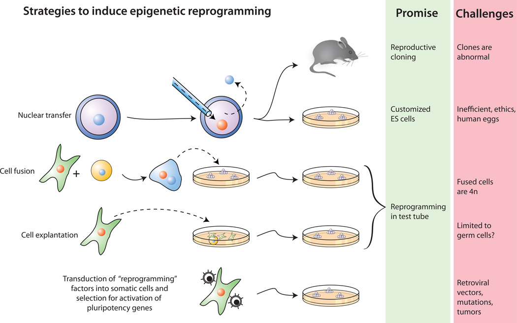
(i) Nuclear transfer involves the injection of a somatic nucleus into an enucleated oocyte which, upon transfer into a surrogate mother, can give rise to a clone (“reproductive cloning”), or, upon explantation in culture, can give rise to genetically matched embryonic stem (ES) cells (“somatic cell nuclear transfer”, SCNT). (ii) Cell fusion of somatic cells with ES cells results in the generation of hybrids that show all features of pluripotent ES cells. (iii) Explantation of somatic cells in culture selects for immortal cell lines that may be pluripotent or multipotent. At present, spermatogonial stem cells are the only source of pluripotent cells that can be derived from postnatal animals. (iv) Transduction of somatic cells with defined factors can initiate reprogramming to a pluripotent state.
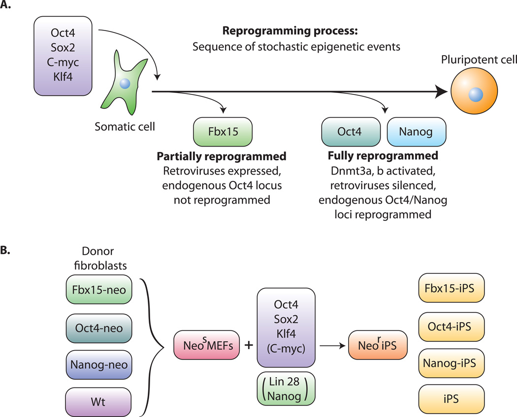
A. Transduction of the four transcription factors Oct4, Sox2, c-myc and Klf4 into fibroblasts initiates the conversion to partially reprogrammed cells that express Fbx15 or to fully reprogrammed iPS cells that express Oct4 or Nanog. The process involves a sequence of stochastic epigenetic events. B. Selection schemes. Cells carrying a drug resistance marker in the Fbx15, the Oct4 or the Nanog gene are transduced with the four factors and selected for drug resistance.
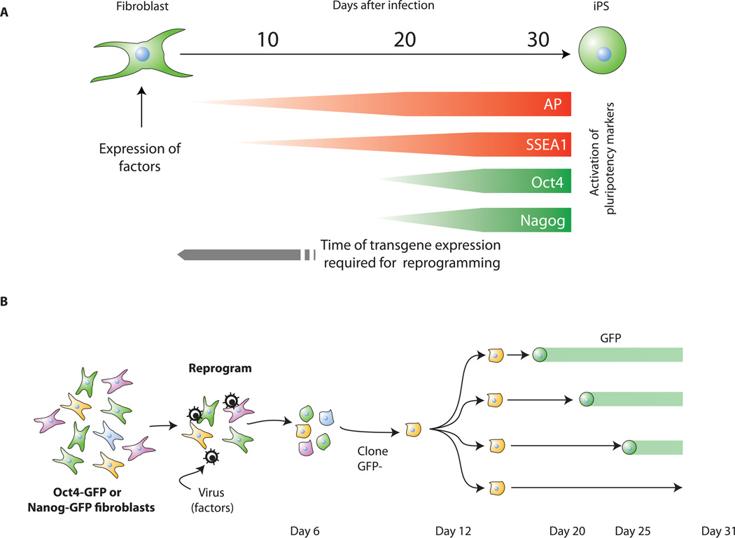
A. Kinetics of pluripotency-marker appearance. Alkaline phosphatase (AP) and SSEA1 positive cells are already detected 3 and 9 days, respectively, after factor transduction whereas GFP expressed from the endogenous Oct4 or Nanog loci first appear only after 2 weeks. The virally transduced factors need to be expressed for about 2 weeks to initiate the reprogramming process (Brambrink et al., 2008). B. Oct4-GFP or Nanog-GFP fibroblasts were transduced with the four factors. Colonies displaying a transformed phenotype, were GFP negative and were cloned a few days after infection. Further cloning yielded subclones that activated GFP at different times (Meissner et al., 2007). Because the subclones were derived from the same infected cell, stochastic epigenetic events must be important for reprogramming.
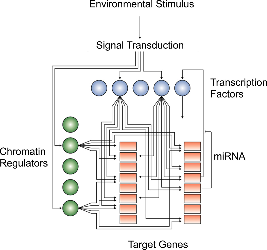
Cartoon showing hypothetic connections between signal transduction pathways, transcription factors (blue balls), chromatin regulators (green balls) and their target genes (orange squares) to form an image of transcriptional regulatory circuitry. Some target genes produce miRNAs, which function at post-transcriptional levels.
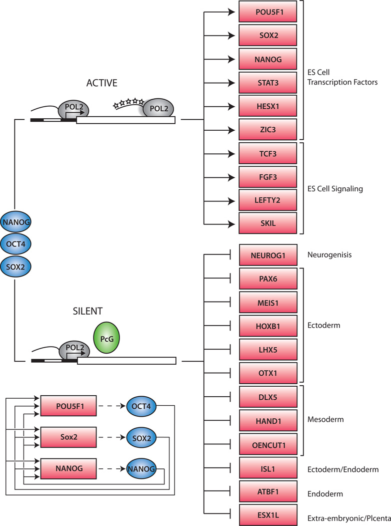
The Oct4, Sox2 and Nanog transcription factors (blue) occupy actively transcribed genes, including transcription factors and signaling components necessary to maintain the ES cell state. The three regulators also occupy silent genes encoding transcription factors that, if expressed, would promote other more differentiated cell states. At this latter set of genes, RNA polymerase II initiates transcription but does not produce complete transcripts due to the repressive action of PcG proteins. The PcG proteins prevent RNA polymerase from transitioning into a fully modified transcription elongation apparatus (represented by phosphorylated “stars” on the tail of the POL2 enzyme at the top of the figure). The interconnected autoregulatory loop, where Oct4, Nanog and Sox2 bind together at each of their own promoters, is shown in the bottom left of the figure.
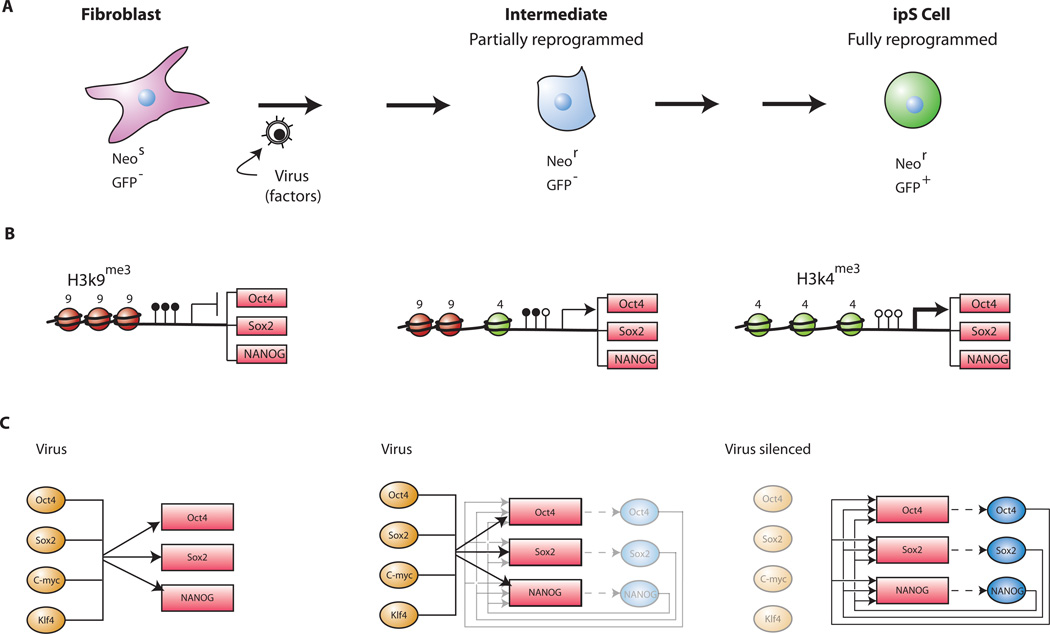
A. Sequential changes of phenotype and activation of Oct4, Nanog and Sox2. Fibroblasts do not express Oct4 or Nanog. After transduction with the four transcription factors Oct4, Sox2, c-myc and Klf4 the infected fibroblasts assume a transformed phenotype. The endogenous Oct4 or Nanog genes become transcribed at a low level that is sufficient for drug resistance in cells carrying the neo gene in either locus (Maherali et al., 2007; Okita et al., 2007; Wernig et al., 2007) but not sufficient to produce GFP expression in Oct4-GFP or Nanog-GFP cells. In a stochastic sequence of epigenetic events the endogenous Oct4 and Nanog genes become fully activated only after 2 to 3 weeks as indicated by the appearance of GFP+ iPS cells in Oct4-GFP or Nanog-GFP fibroblasts (Brambrink et al., 2008; Meissner et al., 2007) (compare Figure 3b). B. Sequential changes of histone and DNA modification. In fibroblasts the promoters of Oct4, Nanog and Sox2 are methylated (filled lollipops) and histone H3 is methylated at K9. During the reprogramming process the repressive H3K9me3 histone marks may be gradually replaced by the transcriptionally active H3K4me3 histone marks and the CpG sites become gradually demethylated (open lollipops). This transient epigenetic state may permit an expression level that is sufficient to give drug resistance (driving the inserted neo gene) but not sufficient to give GFP expression. Additional epigenetic changes alter the histone modification and the DNA methylation conformation to one that is fully derepressed allowing normal factor expression and resulting in GFP+ iPS cells. C. Molecular circuitry during reprogramming. The transduced factors may interact with the endogenous pluripotency genes encoding Oct4, Nanog and Sox2 and gradually activate the autoregulatory loop that sustains normal factor expression. During the reprogramming process the de novo methyltransferases Dnmt3a and Dnmt3b become activated and in turn de novo methylate and silence the virally transduced factors. The pluripotent state is now maintained by the autoregulatory expression of the three transcription factors Oct4, Nanog and Sox2.
Similar articles
-
Stem cells, pluripotency and nuclear reprogramming.J Thromb Haemost. 2009 Jul;7 Suppl 1:21-3. doi: 10.1111/j.1538-7836.2009.03418.x. J Thromb Haemost. 2009. PMID: 19630760 Review.
-
Reprogramming somatic cells towards pluripotency by cellular fusion.Curr Opin Genet Dev. 2012 Oct;22(5):459-65. doi: 10.1016/j.gde.2012.07.005. Epub 2012 Aug 3. Curr Opin Genet Dev. 2012. PMID: 22868176 Review.
-
Faithful reprogramming to pluripotency in mammals - what does nuclear transfer teach us?Int J Dev Biol. 2010;54(11-12):1609-21. doi: 10.1387/ijdb.103195jm. Int J Dev Biol. 2010. PMID: 21404182 Review.
-
Reprogramming of two somatic nuclei in the same ooplasm leads to pluripotent embryonic stem cells.Stem Cells. 2013 Nov;31(11):2343-53. doi: 10.1002/stem.1497. Stem Cells. 2013. PMID: 23922292
-
The role of the reprogramming method and pluripotency state in gamete differentiation from patient-specific human pluripotent stem cells.Mol Hum Reprod. 2018 Apr 1;24(4):173-184. doi: 10.1093/molehr/gay007. Mol Hum Reprod. 2018. PMID: 29471503
Cited by
-
OCT4/SOX2-independent Nanog autorepression modulates heterogeneous Nanog gene expression in mouse ES cells.EMBO J. 2012 Dec 12;31(24):4547-62. doi: 10.1038/emboj.2012.321. Epub 2012 Nov 23. EMBO J. 2012. PMID: 23178592 Free PMC article.
-
Simultaneous and quantitative monitoring transcription factors in human embryonic stem cell differentiation using mass spectrometry-based targeted proteomics.Anal Bioanal Chem. 2021 Mar;413(8):2081-2089. doi: 10.1007/s00216-021-03160-7. Epub 2021 Mar 2. Anal Bioanal Chem. 2021. PMID: 33655347
-
Ectopic expression of OCT4B1 Decreases Fertility Rate and Changes Sperm Parameters in Transgenic Mice.Iran J Biotechnol. 2022 Jul 1;20(3):e3019. doi: 10.30498/ijb.2022.278266.3019. eCollection 2022 Jul. Iran J Biotechnol. 2022. PMID: 36381279 Free PMC article.
-
SNF5 is an essential executor of epigenetic regulation during differentiation.PLoS Genet. 2013 Apr;9(4):e1003459. doi: 10.1371/journal.pgen.1003459. Epub 2013 Apr 25. PLoS Genet. 2013. PMID: 23637628 Free PMC article.
-
Induced neuronal cells: how to make and define a neuron.Cell Stem Cell. 2011 Dec 2;9(6):517-25. doi: 10.1016/j.stem.2011.11.015. Cell Stem Cell. 2011. PMID: 22136927 Free PMC article. Review.
References
-
- Alon U. Network motifs: theory and experimental approaches. Nat Rev Genet. 2007;8:450–461. - PubMed
-
- Alvarez-Dolado M, Pardal R, Garcia-Verdugo JM, Fike JR, Lee HO, Pfeffer K, Lois C, Morrison SJ, Alvarez-Buylla A. Fusion of bone-marrow-derived cells with Purkinje neurons, cardiomyocytes and hepatocytes. Nature. 2003;425:968–973. - PubMed
-
- Azuara V, Perry P, Sauer S, Spivakov M, Jorgensen HF, John RM, Gouti M, Casanova M, Warnes G, Merkenschlager M, et al. Chromatin signatures of pluripotent cell lines. Nat Cell Biol. 2006;8:532–538. - PubMed
-
- Balsam LB, Wagers AJ, Christensen JL, Kofidis T, Weissman IL, Robbins RC. Haematopoietic stem cells adopt mature haematopoietic fates in ischaemic myocardium. Nature. 2004;428:668–673. - PubMed
Publication types
MeSH terms
Substances
Grants and funding
LinkOut - more resources
Full Text Sources
Other Literature Sources
