. 2008 Feb 15;22(4):543-57.
doi: 10.1101/gad.1614408.

BMP signaling in dermal papilla cells is required for their hair follicle-inductive properties

Affiliations

BMP signaling in dermal papilla cells is required for their hair follicle-inductive properties

Michael Rendl et al. Genes Dev. .

Abstract

Hair follicle (HF) formation is initiated when epithelial stem cells receive cues from specialized mesenchymal dermal papilla (DP) cells. In culture, DP cells lose their HF-inducing properties, but during hair growth in vivo, they reside within the HF bulb and instruct surrounding epithelial progenitors to orchestrate the complex hair differentiation program. To gain insights into the molecular program that maintains DP cell fate, we previously purified DP cells and four neighboring populations and defined their cell-type-specific molecular signatures. Here, we exploit this information to show that the bulb microenvironment is rich in bone morphogenetic proteins (BMPs) that act on DP cells to maintain key signature features in vitro and hair-inducing activity in vivo. By employing a novel in vitro/in vivo hybrid knockout assay, we ablate BMP receptor 1a in purified DP cells. When DPs cannot receive BMP signals, they lose signature characteristics in vitro and fail to generate HFs when engrafted with epithelial stem cells in vivo. These results reveal that BMP signaling, in addition to its key role in epithelial stem cell maintenance and progenitor cell differentiation, is essential for DP cell function, and suggest that it is a critical feature of the complex epithelial-mesenchymal cross-talk necessary to make hair.


Figures
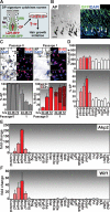
Figure 1.
Figure 1.
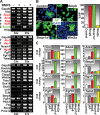
Growth factor screen and identification of BMPs as regulators of DP signature genes. (A) Schematic of screen. DP cells were isolated by FACSorting from K14-H2B-GFP/Lef1-RFP transgenic mice, placed in culture where they were treated with DP niche signature growth factors, and subsequently screened for their ability to maintain DP signature gene expression in vitro and hair induction in vivo. (B) Sections of P4 K14-H2B-GFP back skins after exposure to substrate solution for AP, a highly expressed DP signature gene and a classical DP marker of early anagen. Arrows point to AP-positive DPs (left) and corresponding HFs in the fluorescent channels (right). Bar, 100 μm. (C) Progressive loss of AP activity by DP cells in culture. BrdU was administered to monitor proliferation, AP activity was assayed with substrate, and DAPI was used to detect nuclei. Shown are representative images of cultures harvested at the times indicated at the top, and as quantified for AP(+) (left graph) and BrdU(+) and BrdU(+)/AP(+) (right graph). Bar, 50 μm. Error bars indicate SD. (D) BMP6-mediated maintenance of AP activity in cultured DP cells. DP cells were cultured in the presence of a subset of growth factors/cytokines expressed by the DP niche and for which the DP possesses corresponding surface receptors. The relative effects on cell proliferation and maintenance of AP activity were quantified as in C. Error bars indicate SD of triplicate experiments. (E) Large-scale screen for DP niche growth factors/cytokines affecting AP gene expression (Akp2) in DP cells. Note that of the 27 factors tested, only BMPs maintained Akp2 at the mRNA level as detected by real-time PCR. Error bars indicate SD of duplicate experiments. (F) Screen results of Wif1 expression. A similar pattern of BMP-mediated maintenance was found for the DP signature gene Wif1 by real-time PCR. Error bars indicate SD of duplicate experiments.
Figure 2.
Figure 2.
Maintenance of molecular signature features of cultured DP cells by BMP signaling. (A) FACSorted DP cells from P4 K14-H2B-GFP/Lef1-RFP mice were cultured in medium ± 400 ng/mL BMP6. Semiquantitative RT–PCRs were conducted on a battery of DP signature genes, and data for the two most relevant PCR cycle numbers are shown. DP signature genes that are either enhanced (red, up arrow) or diminished (green, down arrow) upon BMP6 treatment are highlighted. Note that Nog (Noggin) is up and only Bmp6 is decreased, suggesting a feedback circuit. (B,C) Testing for AP activity and DP signature gene maintenance by retroviral expression of Bmp6, Bmpr1a*, and Wnt3a. DP cells were infected with expression constructs for Bmp6, the constitutively active form of Bmpr1a* or Wnt3a. (B) AP activity (green; counterstain is DAPI in blue) was detected (left) and quantified (right). Bar, 50 μm. (C) Real-time PCR of DP signature genes are presented as fold change relative to control. Vim (Vimentin) expression served as control. Error bars indicate SD of duplicate experiments.
Figure 3.
Figure 3.
Evidence of BMP signaling in the DP in vivo and in vitro. (A) Bmp6 mRNA is specifically expressed in the DP as judged by RT–PCR of FACS-purified DP niche cell populations and skin fractions (top) and by in situ hybridization (bottom; shown is hair bulb region; lines demarcate the DP area). (HF) Back skin follicles; (Wh) whisker HFs; (Mx) matrix; (ORS) outer root sheath; (DF) dermal fraction; (Mc) melanocytes; (Der) dermis; (Epi) epidermis. (B) Real-time PCR detects Bmpr1a mRNAs in all isolated hair bulb cell fractions. (C,D) Immunofluorescence of pSMAD1 of back skin hair bulb regions (C) and cultured DP cells ± 400 ng/mL BMP6 (D). Antibodies are shown, with color-coding according to the secondary antibodies. (Blue) DAPI. β4-Integrin or white lines denote demarcation between DP and follicle. KIT was used to distinguish Mc. Graph at right presents quantification of the culture data. (E) Dose-dependent effects on four BMP-sensitive DP signature genes, and demonstration that the effects can be blocked by the BMP inhibitor Noggin (NOG). FACS-sorted DP cells were cultured in medium supplemented as shown. Real-time PCR was performed and normalized relative to untreated control (Co). Error bars indicate SD of duplicate experiments.
Figure 4.
Figure 4.
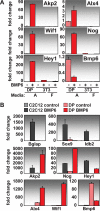
BMP6-mediated DP signature gene regulation is DP-specific and not recapitulated in dermal fibroblasts or differentiating osteogenic cells. (A) FACS-purified DP cells were cultured in AmnioMAX medium (A) ± 400 ng/mL BMP6 for 6 d. 3T3 dermal fibroblasts were grown in both A and F media for comparisons and were treated in parallel. Cells were harvested, and isolated mRNAs were subjected to real-time PCR. Shown are representative data for six genes. All fold changes were normalized relative to untreated DP control. Note that DP signature gene regulation by BMP6 was observed only with DP and not 3T3 cells. (B) Multipotent C2C12 cells (gray), known to differentiate into osteoblasts when treated with BMPs, and DP cells (red) were cultured and tested as above for expression of known BMP-induced osteoblast genes (top and middle rows), or DP signature genes (middle and bottom rows). All fold changes were normalized relative to the untreated C2C12 control sample. Error bars indicate SD of duplicate experiments.
Figure 5.
Figure 5.
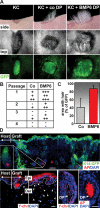
BMP6 treatment of DP cultures enhances their inductive ability in vivo. (A) K14-GFPactin-expressing KC prepared from newborn male back skins were grafted onto back skins of female Nude mice alone (left panel) or in the presence of FACS-purified male DP cells (passage 1) that were cultured ± 400 ng/mL BMP6 (middle and right panel). After 3–4 wk, grafts were photographed in bright-field or GFP epifluorescence. (B) Summary of hair growth in 10 independent experiments. The amount of hair growth was classified as follows: no growth (−); few hairs formed (<50) (+); medium hair growth (50–150) (++); full dense hair growth (>150) (+++). Each line represents an individual experiment. Note that control DP cells usually entered senescence in the fourth through sixth passages. (C) Quantification of area of hair growth. The graft area that was covered by hair growth was measured and expressed as percentage of the total graft area as determined by GFP expression. (D) Skin sections from grafts of KC + BMP6-treated DP cells. (Top) The host/graft boundary was identified by fluorescence microscopy. AP activity (red) marks the DP cells. Bar, 100 μm. (Bottom) FISH with a Y-chromosome probe (Y-chr) identifies the engrafted cells. Note that neither the male dermal fibroblasts (arrowheads) nor the male DP cells (arrows) were seen when male KCs were grafted alone (not shown). Bars, 25 μm.
Figure 6.
Figure 6.
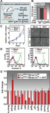
DP-specific targeting of the Bmpr1a gene and effects on DP cells in vitro. (A) Schematic of DP-specific knockout strategy. FACS-purified male DP cells from K14-H2B-GFP/Lef1-RFP/Rosa-CreERtam/Bmpr1aflfl mice were treated with OHT to activate Cre-mediated gene ablation in vitro and were grafted together with female K14-H2B-GFP KC onto female Nude mice in in vivo hair growth chamber-graft assays. (B) Real-time PCR verification of quantitative Bmpr1a exon 2 ablation within 3 d after culture initiation and OHT treatment. (C,D) Normal proliferation, morphology, and apoptosis levels after Bmpr1a ablation in pure DP cells. Apoptosis assays were performed by Annexin V and propidium iodide (PI) stainings followed by FACS. TNFα treatments were conducted as positive controls. (E) Real-time PCR analysis of DP signature genes upon Bmpr1a ablation. RNAs were isolated just prior to chamber grafting (see Fig. 7); i.e., after 7–12 d at passage 1, when DP signature genes are still expressed. Note the selective effects on expression of BMP6-sensitive DP signature genes in Bmpr1a-deficient DP cultures. Bars are the means of three independent experiments.
Figure 7.
Figure 7.
DP-specific ablation of Bmpr1a diminishes their hair induction capacity. DP cells from male Bmpr1a knockout (OHT) and control (Mock) mice were grafted together with female K14-H2B-GFP KC onto the backs of female Nude mice. (A) Side view of the graft area after 3 wk. Shown is a representative example of four separate experiments. (Right frames) GFP epifluorescence served as control for comparable graft performance. (B,C) Serial sections of grafts stained with hematoxylin/eosin (H+E) (B) and examined by GFP fluorescence to ensure fair comparisons of graft samples (C). Arrowheads denote aberrant follicle-like downgrowths in grafts containing Bmpr1a-null DP cells. (D,E) Y-chromosome (Y-chr) FISH (red). DAPI counterstaining in blue. Frames in E are magnified images of boxed areas from D to show integration of both wild-type and Bmpr1a-null DP-derived male cells within the dermis of the grafts. Note that wild-type but not Bmpr1a-null DP cultures generated HFs with DP (arrows) and dermal sheaths. Note presence of additional DP-derived male cells within the dermis (arrowheads). Asterisks denote autofluorescence within the cornified layer of the epidermis and the hair shafts. White lines highlight the BM around the DP (Mock) and at the epidermal/dermal border (OHT). Bars, 100 μm. (F) Summary of hair growth in eight experiments. The amount of hair growth was classified as follows: no growth (−); few hairs formed (<50) (+); medium hair growth (50–150) (++); full dense hair growth (>150) (+++). Each line represents an individual experiment. (G) Quantification of area of hair growth. The graft area that was covered by hair growth was measured and is expressed as percentage of the total graft area as determined by GFP expression.
Figure 8.
Figure 8.
In vivo alterations of DP signature gene expression in grafted dermal cells derived from Bmpr1a-null DP cells. (A,B) Serial sections of grafts were stained for AP activity (A) or WIF1 (B) and subjected to fluorescence microscopy. Boxed areas (labeled iiii) were magnified and are shown in C. Arrows point to expected sites of expression, while arrowheads indicate erroneous expression. (D) pSMAD1 immunofluorescence of serial sections of grafts. Shown are HFs corresponding to the ones in panels i and ii in C. Note that aberrant AP/WIF1-positive cells were pSMAD1-deficient. (EG) Immunofluorescence of graft sections stained for DP markers HOXA9, HHIP, and NCAM. Note aberrant NCAM in dermal cells lining the follicle-like downgrowths. Bars, 100 μm.

Similar articles

Cited by

References

    1. Akiyama S., Katagiri T., Namiki M., Yamaji N., Yamamoto N., Miyama K., Shibuya H., Ueno N., Wozney J.M., Suda T. Constitutively active BMP type I receptors transduce BMP-2 signals without the ligand in C2C12 myoblasts. Exp. Cell Res. 1997;235:362–369. - PubMed
    1. Alonso L., Fuchs E. The hair cycle. J. Cell Sci. 2006;119:391–393. - PubMed
    1. Andl T., Ahn K., Kairo A., Chu E.Y., Wine-Lee L., Reddy S.T., Croft N.J., Cebra-Thomas J.A., Metzger D., Chambon P., et al. Epithelial Bmpr1a regulates differentiation and proliferation in postnatal hair follicles and is essential for tooth development. Development. 2004;131:2257–2268. - PubMed
    1. Badea T.C., Wang Y., Nathans J. A noninvasive genetic/pharmacologic strategy for visualizing cell morphology and clonal relationships in the mouse. J. Neurosci. 2003;23:2314–2322. - PMC - PubMed
    1. Blanpain C., Lowry W.E., Geoghegan A., Polak L., Fuchs E. Self-renewal, multipotency, and the existence of two cell populations within an epithelial stem cell niche. Cell. 2004;118:635–648. - PubMed

Publication types

MeSH terms