. 2008 Mar;118(3):1110-22.
doi: 10.1172/JCI32376.

Protein disulfide isomerase acts as an injury response signal that enhances fibrin generation via tissue factor activation

Affiliations

Protein disulfide isomerase acts as an injury response signal that enhances fibrin generation via tissue factor activation

Christoph Reinhardt et al. J Clin Invest. 2008 Mar.

Abstract

The activation of initiator protein tissue factor (TF) is likely to be a crucial step in the blood coagulation process, which leads to fibrin formation. The stimuli responsible for inducing TF activation are largely undefined. Here we show that the oxidoreductase protein disulfide isomerase (PDI) directly promotes TF-dependent fibrin production during thrombus formation in vivo. After endothelial denudation of mouse carotid arteries, PDI was released at the injury site from adherent platelets and disrupted vessel wall cells. Inhibition of PDI decreased TF-triggered fibrin formation in different in vivo murine models of thrombus formation, as determined by intravital fluorescence microscopy. PDI infusion increased - and, under conditions of decreased platelet adhesion, PDI inhibition reduced - fibrin generation at the injury site, indicating that PDI can directly initiate blood coagulation. In vitro, human platelet-secreted PDI contributed to the activation of cryptic TF on microvesicles (microparticles). Mass spectrometry analyses indicated that part of the extracellular cysteine 209 of TF was constitutively glutathionylated. Mixed disulfide formation contributed to maintaining TF in a state of low functionality. We propose that reduced PDI activates TF by isomerization of a mixed disulfide and a free thiol to an intramolecular disulfide. Our findings suggest that disulfide isomerases can act as injury response signals that trigger the activation of fibrin formation following vessel injury.


Figures
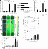
Figure 1
Figure 1. TF-dependent luminal fibrin generation in vivo is controlled by PDI.
(A) Platelet-secreted disulfide isomerases activate microparticle TF. Left: Monocyte microparticles (MP; 106) were incubated with collagen-activated platelets (P; 5 × 10⁷). Microparticles (α-TF/MP) or activated platelets (α-TF/P) were selectively preincubated with anti-TF antibody (10 μg/ml). Unbound antibody was removed by centrifugation. *P < 0.05. Right: Microparticles were incubated with activated platelets for 10 minutes, then bacitracin, anti-PDI antibody, or control antibody were added to bypass inhibition of platelet stimulation. TF activity was determined by 2-stage assay. *P < 0.05 versus MP/P. Means ± SD; n = 5. (B) Platelet-released PDI stimulates TF activity. Supernatants (P-SN) recovered from activated platelets (109) were incubated with microparticles (106) plus anti-PDI antibody or control antibody (15 minutes). The procoagulant activity (2-stage assay) was suppressed by anti-TF antibody. *P < 0.05 versus control antibody. n = 4. (C) Microparticles accelerate fibrin production in mice. Microparticles (107; in 250 μl) were infused into WT mice after carotid ligation injury. MP (α-TF), microparticles precoated with α-TF. Top: Fibrin (pseudocolored) was visualized by intravital videofluorescence microscopy using an Alexa Fluor 488–labeled anti-fibrin antibody. Measurements were performed before (pre injury) or 5 and 30 minutes after injury. Scale bar: 50 μm. Bottom: Quantitative analyses of the data. Means from 2 experiments, 30-minute values. (D) PDI contributes to microparticle-driven initiation of coagulation. Anti-PDI antibody (also recognizing murine PDI, 800 μg/animal) or control IgG were infused into WT mice (15 minutes after injury), then microparticles (107) were added (arrows). Fibrin formation at the injury site was monitored. *P < 0.05 versus isotype IgG. n = 4–6. (E) Microparticle recruitment is unchanged by PDI suppression. Anti-PDI antibody was infused 15 minutes after carotid injury. Then DCF-tagged microparticles were infused. Labeled microparticles at lesion site were measured by intravital microscopy. n = 3–4.
Figure 3
Figure 3. PDI contributes to trigger TF-dependent fibrin formation under conditions of suppressed platelet adhesion.
(A) TF-dependent initiation of coagulation in living animals. Left: Fibrin formation was assessed 15 minutes after carotid artery ligation by intravital microscopy in hTF mice and low-hTF mice. Scale bar: 50 μm. Right: Quantitation of fibrin formation at site of vessel injury. Effect of anti-PDI antibody (or control antibody; infused before implementing the vessel injury) on fibrin formation in hTF animals. *P < 0.05 versus isotype control. n = 4. (B) PDI contributes to fibrin formation in mice with normal platelet counts. Fibrin generation was assessed before or after vascular injury (ligation) in WT (SV129S1) mice pretreated with the indicated antibodies prior to induction of vessel injury. *P < 0.05 versus isotype control. n = 4. (C) GPVI inhibition decreases platelet adhesion. Firm platelet adhesion of rhodamine-labeled platelets to the injured artery (ligation) was visualized 15 minutes after injury in the presence of control antibody or neutralizing anti-GPVI antibody (hTF mice). *P < 0.05. n = 8. (D) PDI regulates fibrin formation under conditions of decreased platelet adhesion. Platelet adhesion was inhibited by infusion of anti-GPVI antibody (hTF mice). Thrombocytopenia was induced by injection of anti-GPIbα antibody (platelet-depleted WT mice). Both groups were then treated intravenously with anti-PDI antibody or control antibody, vessels were injured, and fibrin formation was determined after 15 minutes. *P < 0.05 versus isotype control. n = 7–10. (E) PDI increases fibrin formation after wire-induced vessel injury. Fibrin formation after wire-induced injury of the murine carotid artery was assessed by western blotting in the presence of α-PDI or control IgG. The anti-fibrin antibody specifically detects fibrin but not fibrinogen. Thrombi were excised 15 minutes after vascular injury.
Figure 5
Figure 5. PDI decreases TF free thiols.
(A) PDI lowers TF free thiols. Top panels: sTF (1.7 μg/ml) was exposed for 15 minutes to platelet releasates from collagen-activated platelets (5 × 10⁸), TRX (10 μM), or PDI (1.8 μM). In all cases, sTF was then incubated with biotin-MPB. One aliquot was then pulled down with streptavidin agarose, and subsequently the biotinylated proteins were detected by immunoblotting with anti-TF antibody (MPB-IB). Another aliquot of the sTF previously incubated with MPB was directly subjected to western blotting. Bottom panels: Densitometric analyses of the effects of TRX and PDI on sTF free thiols. *P < 0.05. n = 4. (B) PDI causes disulfide formation of cell TF. TF expressing monocytes (106) were incubated for 15 minutes with PDI or vehicle. The cells were subsequently washed and then incubated with biotin-MPB. The biotinylated proteins were selected with streptavidin-agarose. Biotinylated TF species were detected by immunoblotting with anti-TF antibody. Western blots were performed under identical conditions.
Figure 6
Figure 6. Constitutive glutathionylation of TF.
(A) Basal glutathionylation of cell TF. Solubilized monocytes were immunoprecipitated with anti-GSH antibody or with isotype control antibody, as described in Methods. This was followed by western blotting with control antibody or anti-TF antibody. (B) Structure of C209 containing peptide STDSPVEC*MGQEK as identified by MS/MS fragmentation. The spectrum was generated by collision-induced dissociation (CID) of the doubly charged 858.33-m/z ion. CID of doubly charged precursor ions predominately forms singly charged product ions that are indicative of the sequence of a peptide. The product ions thus generated are b-ions from the N-terminal site and y-ions from the C-terminal site. Peptide fragments observed in the mass spectrometer are shown in bold. The mass difference between b7 and b6 ions shows that the cysteine contains a modification equal to a glutathionylation from the N-terminal site. This is confirmed by the same mass difference between the y5 and y6 ion from the C-terminal site. (C) Glutathionylation attenuates TF activation. Cell lysis increases procoagulant activity, relative to intact cells, which is completely inhibited by anti-TF antibody. Increasing concentrations of DTT suppress TF activity. Glutathionylation in lysed cells was induced by preincubation with DTT (0.1 mM), followed by incubation with GSH (0.1 mM plus 0.1 mM diamide; 15 minutes). This increased the density of the band representing glutathionylated TF in immunoblots of lysed monocytes by 5.5-fold. Procoagulant activity of the lysates was determined by coagulation factor concentrate. *P < 0.05 versus control. n = 3–9.
Figure 7
Figure 7. Deglutathionylation and TF disulfide bond formation by reduced PDI.
(A) Incorporation of glutathione into sTF is reverted by GRX and DTT. sTF (1.7 μg/ml) incorporates biotin-labeled glutathione (0.1 mM, in the presence of 0.1 mM diamide; 15 minutes). Where indicated, DTT (1 mM) and GRX (4 μM; in the presence of 1 mM GSH) were added together with biotin-labeled glutathione. The biotinylated proteins were captured by streptavidin-agarose and immunoblotted with anti-TF antibody (GSH-IB). In parallel, sTF was analyzed by western blotting. (B) Deglutathionylation of TF by PDI. Left: sTF was labeled with biotin-labeled glutathione in the absence (control) or presence of native PDI, fully oxidized PDI (PDIox), or fully reduced PDI (PDIred; all at 1.8 μM, 15 minutes). Biotinylated proteins selected by streptavidin-agarose were immunoblotted with anti-TF antibody, and in parallel, sTF was analyzed by western blotting. Right: Quantification of labeling of sTF with biotin-glutathione by densitometry. *P < 0.05 (versus control). n = 4. (C) Reduced forms of PDI promote disulfide bond formation of TF. Left: sTF was exposed to vehicle (control) and native PDI, PDIox, or PDIred (all at 1.8 μM, 15 minutes, room temperature). Then, the samples were incubated with biotin-MPB. The biotinylated proteins were selected with streptavidin-agarose, followed by detection of biotinylated sTF species by immunoblotting with anti-TF antibody. In parallel, western blots were performed. Right: Quantification of free thiols of sTF by densitometry. *P < 0.05 versus control.
Figure 8
Figure 8. TF activation requires reduced PDI.
(A) TF activation by PDI. TF-expressing monocytes were incubated with native PDI (1.8 μM), TRX (10 μM), or GRX (4 μM; in the presence of 1 mM GSH) for 15 minutes at room temperature. Then, the procoagulant activity was determined by 2-stage assay. Apart from anti-TF antibody (10 μg/ml), pretreatment of monocytes with the free thiol–selective reagent DTNB also inhibited stimulation of TF activity by PDI (data not shown). *P < 0.05 (versus control); n = 4. (B) Relevance of redox-active cysteines for TF activation by PDI. Bottom: TF-expressing monocytes were incubated with native PDI and a PDI mutant in which the redox-active cysteines were substituted by serine (PDIΔCys1,2). Additionally, the cells were incubated with PDIred and PDIox (all at 1.8 μM). Then, the procoagulant activity was determined (2-stage assay). n = 4. Top: In contrast to native PDI, PDIΔCys1,2 did not deglutathionylate sTF. *P < 0.05 versus control; #P < 0.05 versus native PDI.
Figure 9
Figure 9. Model for the activation of TF-dependent coagulation start by PDI.
Top: PDI exposure at the vessel injury site contributes to enable the initial step of the extrinsic pathway of blood coagulation, conversion of TF from the functionally inactive to the active form. After vessel injury, PDI can be secreted by activated platelets (P) adhering at the injury site and released from cells of the damaged vessel wall. The exposed PDI might interact with TF expressed by vessel wall cells and TF carrying blood components such as microparticles rapidly recruited to the vessel lesion. In parallel, the exposed PDI can directly contribute to amplify platelet activation. Bottom: Hypothetical molecular mechanism of TF activation by PDI. TF-dependent fibrin formation is low when the Cys186/Cys209 pair of TF is glutathionylated and/or in the free thiol form. Mixed disulfides with glutathione (and other types of linkages) at Cys209 are a constitutive redox form of the protein. PDI can cleave the mixed disulfides of TF when its redox-active cysteines are in the reduced form. This could potentially result in the formation of an unstable PDI-TF intermediate. Through nucleophilic attack by the vicinal free thiol, the intermediate might be disintegrated and be converted into a stable disulfide. The isomerization reaction proposed is thus in principle similar to the PDI-catalyzed isomerization suggested to mediate the correct positioning of protein disulfides in the ER (21). After completing the redox exchange, the same PDI molecule can potentially stimulate additional TF molecules, which would amplify TF activation.
Figure 2
Figure 2. Specific exposure of PDI in the wounded area of the vessel wall.
(A) Vessel injury causes appearance of intraluminal PDI in association with platelets. Fifteen minutes after ligation, vessel segments containing the injured area were excised and subjected to immunohistochemistry using an Alexa Fluor 594–labeled anti-PDI antibody. Platelets were stained with Alexa Fluor 488–labeled anti-CD41 antibody, and nuclei were visualized with DAPI. Arrows indicate PDI located in the vessel wall. Arrowheads show platelet-associated PDI. In parallel, the vessel segments were exposed to labeled isotype control antibodies. (B) PDI exposure at vessel injury site. Alexa Fluor 488–labeled anti-PDI antibody (or Alexa Fluor 488–labeled control antibody) was infused into WT and thrombocytopenic (platelet-depleted) mice (22) before and 15 and 25 minutes after vessel injury. The distribution of the labels at the injury location was visualized by intravital microscopy. Values obtained with anti-PDI antibody were normalized to values obtained with control antibody. #P < 0.05 compared with pre-injury; *P < 0.05 compared with control. n = 6.
Figure 4
Figure 4. TF disulfide exchange supports fibrin production in vivo.
(A) Prevention of TF disulfide switching inhibits fibrin generation. Monocyte microparticles were preincubated for 30 minutes ex vivo with DTNB (1 mM; DTNB-MP) or vehicle (control-MP). Then they were infused into the murine blood. Fibrin generation at the lesion site (ligation injury) was determined 15 minutes after infusion of microparticles. *P < 0.05. n = 4. (B) PDI inhibition requires functioning disulfide exchange in vivo. The anti-PDI antibody (or control antibody) was infused following incorporation of DTNB-pretreated microparticles into the murine blood circulation. Fibrin generation at the lesion site (ligation injury) was determined 15 minutes after infusion of microparticles. n = 4. (C) TF-driven fibrin formation is enhanced by PDI. PDI (200 μg/animal) was included in the blood circulation of hTF mice, and fibrin generation was registered 10 minutes after vessel injury at the lesion site (ligation injury). Scale bar: 50 μm. *P < 0.05. n = 6.

Similar articles

Cited by

References

    1. Dahlback B. Blood coagulation. Lancet. 2000;355:1627–1632. - PubMed
    1. Schenone M., Furie B.C., Furie B. The blood coagulation cascade. Curr. Opin. Hematol. 2004;11:272–277. - PubMed
    1. Gomez K., McVey K.H. Tissue factor initiated blood coagulation. Front. Biosci. 2006;11:1349–1359. - PubMed
    1. Mackman N., Tilley R.E., Key N.S. Role of the extrinsic pathway of blood coagulation in hemostasis and thrombosis. Arterioscler. Thromb. Vasc. Biol. . 2007;27:1687–1693. - PubMed
    1. Steffel J., Lüscher T.F., Tanner F.C. Tissue factor in cardiovascular diseases: molecular mechanisms and clinical implications. Circulation. . 2006;113:722–731. - PubMed

Publication types