

High-resolution CT imaging of carotid artery atherosclerotic plaques
- PMID: 18272562
- PMCID: PMC8128558
- DOI: 10.3174/ajnr.A0950
High-resolution CT imaging of carotid artery atherosclerotic plaques
Abstract
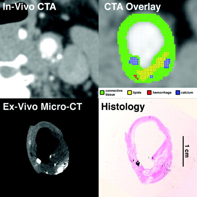
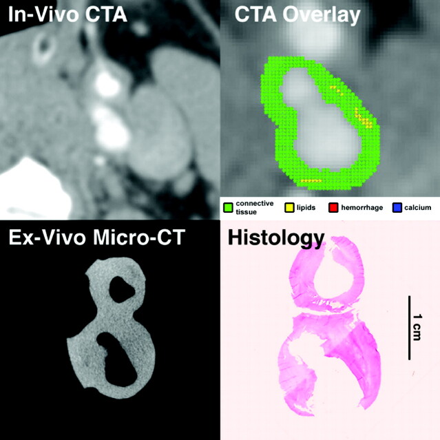
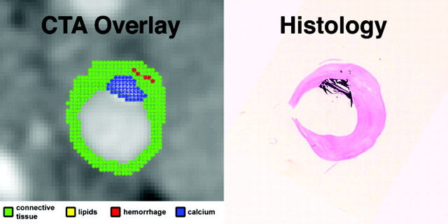
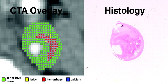
Background and purpose: Plaque morphologic features have been suggested as a complement to luminal narrowing measurements for assessing the risk of stroke associated with carotid atherosclerotic disease, giving rise to the concept of "vulnerable plaque." The purpose of this study was to evaluate the ability of multidetector-row CT angiography (CTA) to assess the composition and characteristics of carotid artery atherosclerotic plaques with use of histologic examination as the gold standard.
Materials and methods: Eight patients with transient ischemic attacks who underwent carotid CTA and "en bloc" endarterectomy were enrolled in a prospective study. An ex vivo micro-CT study of each endarterectomy specimen was obtained, followed by histologic examination. A systematic comparison of CTA images with histologic sections and micro-CT images was performed to determine the CT attenuation associated with each component of the atherosclerotic plaques. A computer algorithm was subsequently developed that automatically identifies the components of the carotid atherosclerotic plaques, based on the density of each pixel. A neuroradiologist's reading of this computer analysis was compared with the interpretation of the histologic slides by a pathologist with respect to the types and characteristics of the carotid plaques.
Results: There was a 72.6% agreement between CTA and histologic examination in carotid plaque characterization. CTA showed perfect concordance for calcifications. A significant overlap between densities associated with lipid-rich necrotic core, connective tissue, and hemorrhage limited the reliability of individual pixel readings to identify these components. However, CTA showed good correlation with histologic examination for large lipid cores (kappa = 0.796; P < .001) and large hemorrhages (kappa = 0.712; P = .102). CTA performed well in detecting ulcerations (kappa = 0.855) and in measuring the fibrous cap thickness (R(2) = 0.77; P < .001).
Conclusion: The composition of carotid atherosclerotic plaques determined by CTA reflects plaque composition defined by histologic examination.
Figures
Similar articles
-
Vulnerable carotid plaque imaging and histopathology without a dedicated MRI receiver coil.Neuroradiol J. 2017 Apr;30(2):120-128. doi: 10.1177/1971400916678244. Epub 2017 Jan 10. Neuroradiol J. 2017. PMID: 28071288 Free PMC article.
-
Contemporary carotid imaging: from degree of stenosis to plaque vulnerability.J Neurosurg. 2016 Jan;124(1):27-42. doi: 10.3171/2015.1.JNS142452. Epub 2015 Jul 31. J Neurosurg. 2016. PMID: 26230478 Review.
-
Utility of Dual-Layer Spectral Detector CTA to Characterize Carotid Atherosclerotic Plaque Components: An Imaging-Histopathology Comparison in Patients Undergoing Endarterectomy.AJR Am J Roentgenol. 2022 Mar;218(3):517-525. doi: 10.2214/AJR.21.26540. Epub 2021 Sep 22. AJR Am J Roentgenol. 2022. PMID: 34549604
-
Correlation between fissured fibrous cap and contrast enhancement: preliminary results with the use of CTA and histologic validation.AJNR Am J Neuroradiol. 2014 Apr;35(4):754-9. doi: 10.3174/ajnr.A3759. Epub 2013 Oct 24. AJNR Am J Neuroradiol. 2014. PMID: 24157737 Free PMC article. Clinical Trial.
-
[Dual source computed tomography in analysis of significance and morphology carotid plaques].Przegl Lek. 2013;70(3):118-22. Przegl Lek. 2013. PMID: 24003664 Review. Polish.
Cited by
-
Association between carotid artery plaque type and cerebral microbleeds.AJNR Am J Neuroradiol. 2012 Dec;33(11):2144-50. doi: 10.3174/ajnr.A3133. Epub 2012 May 24. AJNR Am J Neuroradiol. 2012. PMID: 22627799 Free PMC article.
-
The Accuracy of Noninvasive Imaging Techniques in Diagnosis of Carotid Plaque Morphology.Open Access Maced J Med Sci. 2015 Jun 15;3(2):224-30. doi: 10.3889/oamjms.2015.039. Epub 2015 Mar 27. Open Access Maced J Med Sci. 2015. PMID: 27275225 Free PMC article.
-
Carotid plaque imaging profiling in subjects with risk factors (diabetes and hypertension).Cardiovasc Diagn Ther. 2020 Aug;10(4):1005-1018. doi: 10.21037/cdt.2020.01.13. Cardiovasc Diagn Ther. 2020. PMID: 32968657 Free PMC article. Review.
-
Association between carotid plaque enhancement shown by multidetector CT angiography and histologically validated microvessel density.Eur Radiol. 2012 Oct;22(10):2237-45. doi: 10.1007/s00330-012-2467-5. Epub 2012 May 10. Eur Radiol. 2012. PMID: 22572988
-
Evaluation of the effect of tofogliflozin on the tissue characteristics of the carotid wall-a sub-analysis of the UTOPIA trial.Cardiovasc Diabetol. 2022 Feb 5;21(1):19. doi: 10.1186/s12933-022-01451-6. Cardiovasc Diabetol. 2022. PMID: 35123483 Free PMC article. Clinical Trial.
References
-
- Beneficial effect of carotid endarterectomy in symptomatic patients with high-grade carotid stenosis. North American Symptomatic Carotid Endarterectomy Trial Collaborators. N Engl J Med 1991;325:445–53 - PubMed
-
- Barnett HJ, Taylor DW, Eliasziw M, et al. Benefit of carotid endarterectomy in patients with symptomatic moderate or severe stenosis. North American Symptomatic Carotid Endarterectomy Trial Collaborators. N Engl J Med 1998;339:1415–25 - PubMed
-
- Randomised trial of endarterectomy for recently symptomatic carotid stenosis: final results of the MRC European Carotid Surgery Trial (ECST). Lancet 1998;351:1379–87 - PubMed
-
- Mayberg MR, Wilson SE, Yatsu F, et al. Carotid endarterectomy and prevention of cerebral ischemia in symptomatic carotid stenosis. Veterans Affairs Cooperative Studies Program 309 Trialist Group. JAMA 1991;266:3289–94 - PubMed
-
- Ebrahim S, Papacosta O, Whincup P, et al. Carotid plaque, intima media thickness, cardiovascular risk factors, and prevalent cardiovascular disease in men and women: the British Regional Heart Study. Stroke 1999;30:841–50 - PubMed
Publication types
MeSH terms
Grants and funding
LinkOut - more resources
Full Text Sources
Other Literature Sources
Medical
Research Materials
Miscellaneous