. 2008 Mar;40(3):290-8.
doi: 10.1038/ng.82. Epub 2008 Feb 10.

MicroRNA Mirn140 modulates Pdgf signaling during palatogenesis

Affiliations

MicroRNA Mirn140 modulates Pdgf signaling during palatogenesis

Johann K Eberhart et al. Nat Genet. 2008 Mar.

Abstract

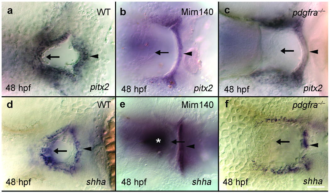
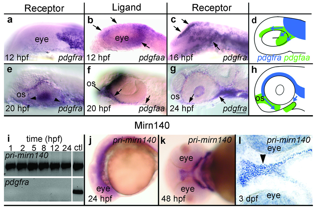
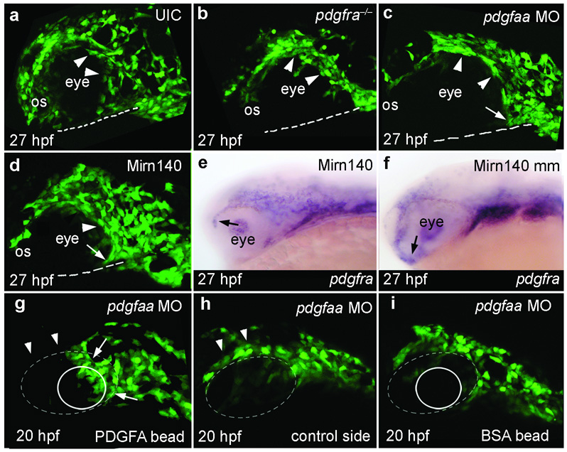
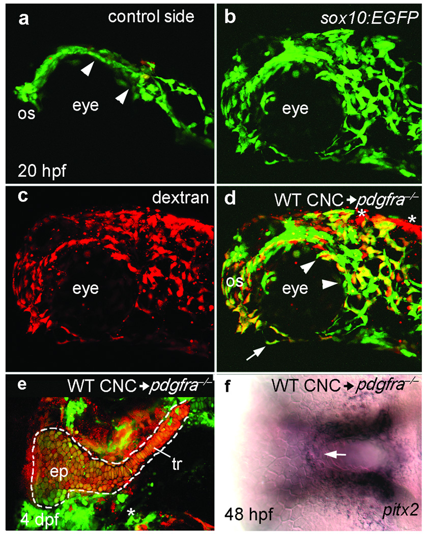
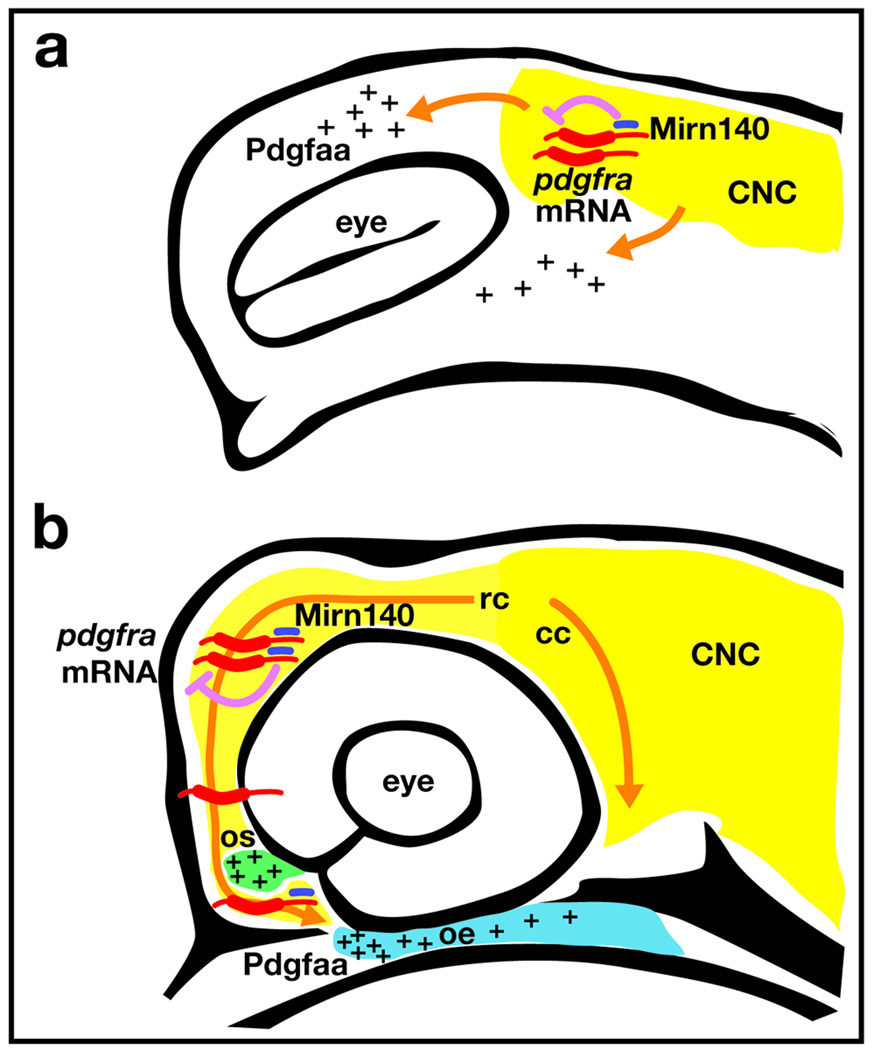
Disruption of signaling pathways such as those mediated by sonic hedgehog (Shh) or platelet-derived growth factor (Pdgf) causes craniofacial abnormalities, including cleft palate. The role that microRNAs play in modulating palatogenesis, however, is completely unknown. We show that, in zebrafish, the microRNA Mirn140 negatively regulates Pdgf signaling during palatal development, and we provide a mechanism for how disruption of Pdgf signaling causes palatal clefting. The pdgf receptor alpha (pdgfra) 3' UTR contained a Mirn140 binding site functioning in the negative regulation of Pdgfra protein levels in vivo. pdgfra mutants and Mirn140-injected embryos shared a range of facial defects, including clefting of the crest-derived cartilages that develop in the roof of the larval mouth. Concomitantly, the oral ectoderm beneath where these cartilages develop lost pitx2 and shha expression. Mirn140 modulated Pdgf-mediated attraction of cranial neural crest cells to the oral ectoderm, where crest-derived signals were necessary for oral ectodermal gene expression. Mirn140 loss of function elevated Pdgfra protein levels, altered palatal shape and caused neural crest cells to accumulate around the optic stalk, a source of the ligand Pdgfaa. These results suggest that the conserved regulatory interactions of mirn140 and pdgfra define an ancient mechanism of palatogenesis, and they provide candidate genes for cleft palate.


Figures

Figure 1
Figure 1
Overexpression of Mirn140 phenocopies pdgfra mutants. (a-c) Animals injected with Mirn140 duplex had cranial hemorrhaging (arrows) at 48 hpf (b), mimicking the phenotype of zebrafish (c) and mouse pdgfra mutants,. (d–f) Frontal views of 6 dpf larvae show that, compared to uninjected controls (UIC) (d), Mirn140 duplex injected (e) and pdgfra mutant (f) animals develop hypoplastic upper lips (arrowheads). (g–l) In Alcian/Alizarin-stained palates of 6 dpf uninjected controls (UIC) (g) or Mirn140 mis-match control injected embryos (j) trabeculae (tr) fuse at the trabecular communis in the midline and extent anteriorly forming the ethmoid plate (ep), but Mirn140 duplex injected (h,k) and pdgfra mutants (i,l) show both mild (h,i) and severe (k,l) phenotypes that include complete clefting of the palatal skeleton. (m) The b1059 allele is a mutation of pdgfrab. 1059 was genetically mapped to linkage group 20 (LG20) between the polymorphic markers z20582 and z14542, with 9 cross-overs and 7 cross-overs, respectively, out of 434 meioses. Sequence analysis of wild-type and b1059 mutant embryos revealed a missense mutation in the second tyrosine kinase (TK) domain of pdgfra. Protein sequence alignment of this region of the second tyrosine kinase domain (amino acids 841–870) of Pdgfra and related receptors shows the non-conservative I855N missense mutation (asterisk) in the second tyrosine kinase domain. This domain is highly conserved in Pdgfra across species as well as across related receptors such as Pdgfrb and Kit. n=notochord.
Figure 2
Figure 2
The oral ectoderm is similarly disrupted in Mirn140 duplex injected embryos and pdgfra mutants. Ventral views, anterior to the left, of oral ectoderm labeled with pitx2 (a–c) and shha (d–f) riboprobe in wild-type (a,d), Mirn140 duplex injected embryos (b,e) and pdgfra mutants (c,f). The roof of the oral ectoderm (arrows), adjacent to the normal location of palatal precursors, expressed neither gene in Mirn140 duplex injected embryos or pdgfra mutants. Loss of gene expression was specific to the roof of the oral ectoderm as the floor of the oral ectoderm expressed both pitx2 and shha (arrowheads). The embryo in e is severely affected and shha staining in the ventral brain is evident (asterisk).
Figure 3
Figure 3
Mirn140 regulates Pdgfra levels. (a) Compared to uninjected controls (UIC), Mirn140 duplex injection reduces and mirn140 morpholino injection elevates the level of the endogenous Pdgfra protein, respectively, detected by anti-human PDGFRA antibody. Anti-mouse Actin antibody is used as a loading control. (b) Schematic of the GFP-pdgfra mRNA injected to test for interaction of Mirn140 with the pdgfra 3’UTR, which bears a predicted Mirn140 binding site. (c–f) 27 hpf embryos injected with GFP-pdgfra alone (c) or with GFP-pdgfra and Mirn140-mismatch (Mirn140mm) duplex (d) fluoresce more strongly than animals injected with GFP-pdgfra and Mirn140 duplex (e). Morpholino knockdown of Mirn140 with Dicer-inhibitor MO increased GFP fluorescence above controls (f). (g) Pixel density analysis of GFP fluorescence confirmed results of c–f; Error bars indicate standard deviations. (h–k) Synthetic pdgfra mRNA truncated to remove the Mirn140 binding site (pdgfra*) rescued the cleft palate phenotype of Mirn140 over-expression, in three independent trials. (h) Distribution of palatal phenotypes after injection of Mirn140 duplex alone (n=54) or co-injection of Mirn140 and pdgfra* mRNA (n=117), in percent of animals. Mildly affected fish had near normal trabeculae and lateral ethmoid plate, but lacked the medial ethmoid plate like Fig. 1m; and severely affected fish lacked both the ethmoid plate and trabeculae like Fig. 1n. (i–k) Alizarin/Alcian-stained palates of 6 dpf uninjected control (UIC) (i) and co-injected embryos (j, k).
Figure 4
Figure 4
mirn140 overlaps with pdgfra during crest cell migration. (a–h) pdgfaa expression predicts the migratory pathway of pdgfra-expressing crest cells. (a–c) Lateral views of pdgfra (a,c) or pdgfaa (b) expression in 12 hpf (a,b) and 16 hpf (c) embryos. Premigratory crest expresses pdgfra (a) while the midbrain rudiment expresses pdgfaa (b). By 16 hpf, the position of pdgfra-expressing crest cells (c) mirrors the 12 hpf distribution of pdgfaa (compare arrows in b,c). (d) Schematic of early crest cell migration (blue arrows) relative to 12 hpf pdgfaa expression (green). (e-g) Lateral views of 20 hpf (e,f) and 24 hpf (g) embryos stained with pdgfra (e,g) or pdgfaa (f) riboprobe. When pdgfra-expressing crest cells have migrated to the optic stalk (os) and are near the oral ectoderm (e, arrowheads) the optic stalk, oral ectoderm, and cells between these two tissues express pdgfaa (f, arrows). By 24 hpf, neural crest cells are condensing on the oral ectoderm (g, arrows). (h) Schematic depicting crest cell migration (blue, arrows) to the oral ectoderm relative to pdgfaa expression (green). (i-l) mirn140 expression overlaps with pdgfra. (i) RT-PCR detects mirn140 from one hpf onward. Controls for genomic contamination utilized primers targeted to intronic sequence and failed to yield PCR product except in PCRs utilizing genomic DNA (ctl) (j-l) mirn140 transcripts detected by pri-mirn140 riboprobe are broadly distributed during crest cell migration (j, lateral view) and become restricted to post-migratory crest cells (k, ventral view), including the palatal skeleton (l, arrowhead, horizontal section).
Figure 5
Figure 5
Pdgf signaling, modulated by Mirn140, guides palatal skeleton precursors to the oral ectoderm. (a–d) Lateral views of 27 hpf sox10:EGFP transgenic embryos. (a) Neural crest cells disperse and migrate around the eye and optic stalk (os) to reach the oral ectoderm (dashed line) in uninjected control (UIC) embryos. (b–d) In contrast, most palatal precursor cells do not disperse (arrowheads) or migrate to the oral ectoderm in pdgfra mutants (b), pdgfaa morpholino injected embryos (c), or Mirn140 duplex injected embryos (d). In all three circumstances rostrally migrating crest cells stop migrating at the optic stalk. Caudally migrating crest is less severely effected in pdgfaa morpholino injected embryos and Mirn140 duplex injected embryos than in pdgfra mutants (arrows, c,d, see Supplementary Figure 5). (e–f) Lateral views of 27 hpf embryos stained with pdgfra riboprobe. pdgfra mRNA is similarly present in Mirn duplex (e) and mismatch control (f) injected embryos demonstrating that Mirn140’s effect on migration is not through regulation of pdgfra transcripts. (g–i) Lateral views of pdgfaa morpholino injected (depleting endogenous Pdgfaa), sox10:GFP transgenic embryos implanted with rat recombinant PDGFA (g,h) or BSA (i) loaded beads (circles) just medial to the eye (dashed oval). (g) Crest cells accumulated adjacent to PDGFA beads (arrows, n=8) and fewer crest cells were present above the eye (arrowheads), compared to the control side of the same embryo (h) suggesting that crest cells were rerouted to the Pdgfa bead. (i) Control BSA beads did not attract crest cells.
Figure 6
Figure 6
Pdgfra is required in neural crest for neural crest cell migration, palatal skeleton development and proper oral ectoderm specification. (a–d) Lateral views of the control (a) and experimental side (b–d) of a 20 hpf pdgfra mutant embryo that received a neural crest cell transplantation from a pdgfra+;sox10:EGFP embryo (WT CNC). (a) Mutant neural crest cells (green) are not dispersed (arrowheads) and failed to migrate beyond the optic stalk (os) on the side of the embryo that did not receive transplanted crest cells. (b–d) Donor pdgfra+;sox10:EGFP transgenic cells, labeled with Alexa dextran 568 (c), have dispersed, migrated around the optic stalk, and reached the oral ectoderm (arrow in d) in a pdgfra‒/‒ environment (n=21). (d) Merged imaged of b,c. Asterisks mark the location of two small patches of non-neural crest cells in the transplant. (e) Flat mounted palatal skeleton of another pdgfra mutant that received a neural crest cell transplant from a pdgfra+;fli1:EGFP transgenic donor (n=5). pdgfra+ crest-derived cartilage was present unilaterally in the ethmoid plate (ep) and trabeculae (tr) of the palatal skeleton (outlined). The trabecula and a portion of the ethmoid plate on the control side of the mutant embryo host were missing (asterisk). (f) 48 hpf ventral view of the embryo in a-d showing pdgfra+ crest rescues pitx2 expression in the roof of the pdgfra‒/‒ oral ectoderm (arrow, n=8). The contaminant cells, shown in d, are distant from the oral ectoderm and unlikely to influence oral ectodermal gene expression.
Figure 7
Figure 7
Loss of Mirn140 function alters palatal skeleton morphology and neural crest cell migration. (a–e) At 6dpf the length-to-width ratio (as shown in b–e) was calculated in injected and control embryos. Compared to uninjected controls (UIC, n=9) and mirn140 morpholino (MO) + Mirn140 duplex co-injected embryos (n=13), ratios were significantly larger and smaller in mirn140 morpholino injected embryos (n=8) and Mirn140 duplex injected embryos (n=11), respectively. Levels not connected by the same letter are significantly different at the 0.05% level (Tukey-Kramer HSD; one-way ANOVA: F1,57=186.7, p<0.0001). Error bars indicate standard deviation. (f-h) Compared to controls (f, n=8) fewer rostrally migrating neural crest cells had migrated from the optic stalk (os) to the oral ectoderm (dashed line) in sox10:EGFP transgenic embryos injected with mirn140 morpholino (g, n=10) or pdgfra* mRNA (h, n=10). Neural crest cells did encircle the optic stalk in these embryos, unlike the effects of Pdgf loss-of-function (see Fig. 5). (i–k) The resultant palatal phenotypes in the same embryos imaged in f–h. Compared to controls (i) Palatal morphology was altered in mirn140 morpholino injected embryos (j) but not pdgfa* injected embryos (k).
Figure 8
Figure 8
Model of how Mirn140 modulates Pdgf signaling during palatogenesis. (a) Pdgfra signaling is required for neural crest cell dispersion. Neural crest cells (yellow) express pdgfra (red) and mirn140 (blue) as they disperse into regions of pdgfaa expression (pluses). Mirn140 inhibits Pdgfra production (pink arrow), but the overall level of Pdgf signaling is sufficient to promote crest dispersion along Pdgfaa-positive pathways. (b) When rostrally migrating crest cells reach the optic stalk, stoichiometric differences in pdgfra and Mirn140 levels regulate their final migration. To envelope the optic stalk (green), crest cells require relatively higher levels of Pdgfra. Crest cells that migrate on towards the oral ectoderm (blue) must first decrease the levels of Pdgfra via Mirn140, in order to leave the optic stalk.

Comment in

  • MicroRNAs in facial development.
    Clouthier DE. Clouthier DE. Nat Genet. 2008 Mar;40(3):268-9. doi: 10.1038/ng0308-268. Nat Genet. 2008. PMID: 18305475 No abstract available.

Similar articles

Cited by

References

    1. Osumi-Yamashita N, Ninomiya Y, Doi H, Eto K. The contribution of both forebrain and midbrain crest cells to the mesenchyme in the frontonasal mass of mouse embryos. Developmental Biology. 1994;164:409–419. - PubMed
    1. Trainor PA, Melton KR, Manzanares M. Origins and plasticity of neural crest cells and their roles in jaw and craniofacial evolution. International Journal of Developmental Biology. 2003;47:541–553. - PubMed
    1. Wada N, et al. Hedgehog signaling is required for cranial neural crest morphogenesis and chondrogenesis at the midline in the zebrafish skull. Development. 2005;132:3977–3988. - PubMed
    1. Eberhart JK, Swartz ME, Crump JG, Kimmel CB. Early Hedgehog signaling from neural to oral epithelium organizes anterior craniofacial development. Development. 2006;133:1069–1077. - PubMed
    1. Trainor PA, Krumlauf R. Hox genes, neural crest cells and branchial arch patterning. Current Opinion in Cell Biology. 2001;13:698–705. - PubMed

Publication types

MeSH terms

Substances