. 2008 Mar;44(3):527-38.
doi: 10.1016/j.yjmcc.2007.12.006. Epub 2008 Jan 3.

TGF-beta1-induced plasminogen activator inhibitor-1 expression in vascular smooth muscle cells requires pp60(c-src)/EGFR(Y845) and Rho/ROCK signaling

Affiliations

TGF-beta1-induced plasminogen activator inhibitor-1 expression in vascular smooth muscle cells requires pp60(c-src)/EGFR(Y845) and Rho/ROCK signaling

Rohan Samarakoon et al. J Mol Cell Cardiol. 2008 Mar.

Abstract

TGF-beta1 and its target gene encoding plasminogen activator inhibitor-1 (PAI-1) are major causative factors in the pathology of tissue fibrosis and vascular disease. The increasing complexity of TGF-beta1 action in the cardiovascular system requires analysis of specific TGF-beta1-initiated signaling events that impact PAI-1 transcriptional regulation in a physiologically-relevant cell system. TGF-beta1-induced PAI-1 expression in both primary cultures and in an established line (R22) of vascular smooth muscle cells (VSMC) was completely blocked by inhibition of epidermal growth factor receptor (EGFR) activity or adenoviral delivery of a kinase-dead EGFR(K721A) construct. TGF-beta1-stimulated PAI-1 expression, moreover, was preceded by EGFR phosphorylation on Y845 (a src kinase target residue) and required pp60(c-src) activity. Infection of VSMC with an adenovirus encoding the EGFR(Y845F) mutant or transfection with a dominant-negative pp60(c-src) (DN-Src) expression vector effectively decreased TGF-beta1-stimulated, but not PDGF-induced, PAI-1 expression implicating the pp60(c-src) phosphorylation site EGFR(Y845) in the inductive response. Consistent with these findings, TGF-beta1 failed to induce PAI-1 synthesis in src kinase-deficient (SYF(-/-/-)) fibroblasts and reexpression of a wild-type pp60(c-src) construct in SYF(-/-/-) cells rescued the PAI-1 response to TGF-beta1. TGF-beta1-induced EGFR activation, but not SMAD2 activation, moreover, was virtually undetectable in SYK(-/-/-) fibroblasts in comparison to wild type (SYK(+/+/+)) counterparts, confirming an upstream signaling role of src family kinases in EGFR(Y845) phosphorylation. Genetic EGFR deficiency or infection of VSMCs with EGFR(K721A) virtually ablated TGF-beta1-stimulated ERK1/2 activation as well as PAI-1 expression but not SMAD2 phosphorylation. Transient transfection of a dominant-negative RhoA (DN-RhoA) expression construct or pretreatment of VSMC with C3 transferase (a Rho inhibitor) or Y-27632 (an inhibitor of p160ROCK, a downstream effector of Rho) also dramatically attenuated the TGF-beta1-initiated PAI-1 inductive response. In contrast to EGFR pathway blockade, interference with Rho/ROCK signaling effectively inhibited TGF-betaR-mediated SMAD2 phosphorylation and nuclear accumulation. TGF-beta1-stimulated SMAD2 activation, moreover, was not sufficient to induce PAI-1 expression in the absence of EGFR signaling both in VSMC and mouse embryonic fibroblasts. Thus, two distinct pathways involving the EGFR/pp60(c-src)/MEK-ERK pathway and Rho/ROCK-dependent SMAD2 activation are required for TGF-beta1-induced PAI-1 expression in VSMC. The identification of such novel interactions between two TGF-beta1-activated signaling networks that specifically impact PAI-1 transcription in VSMC may provide therapeutically-relevant targets to manage the pathophysiology of PAI-1-associated cardiovascular/fibrotic diseases.


Figures

Fig. 1
Fig. 1
EGFR signaling is required for TGF-β1-stimulated PAI-1 expression. Serum-deprived primary VSMC (A) and R22 cells [52] (B) were maintained under quiescence conditions or stimulated with TGF-β1 (1 ng/ml; for 1–4 h) with or without pretreatment with the EGFR inhibitor AG1478. AG1478 decreased basal PAI-1 levels in primary VSMC in a dose-dependent manner and completely blocked PAI-1 induction in response to TGF-β1 in primary cells as well as in R22 VSMC at a final concentration of 2.5 μM (A,B). A molecular genetic approach was utilized to confirm involvement of the EGFR in TGF-β1-initiated signaling. TGF-β1-induced PAI-1 expression was effectively suppressed by adenoviral delivery of a kinase-dead (EGFRKD) EGFRK721A-GFP mutant receptor but not with a control-GFP viral construct (C). Use of an antibody specific for EGFR1 confirmed over-expression of the EGFRKD in infected R22 cells which migrated above endogenous EGFR due to the presence of the GFP tag. A summary of three independent Western blot experiments (mean±SD) with statistical analysis appears in (D). Similarly, TGF-β1 (0.05 to 0.2 ng/ml) stimulated PAI-1 synthesis and ERK1/2 phosphorylation in EGFR+/+ MEFs but not in EGFR−/− fibroblasts (E,F). Confluent EGFR+/+ and EGFR−/− MEFs were serum-deprived for one day prior to addition of TGF-β1 (at concentrations indicated in E or at 0.1 ng/ml in F). After 4 h, cells were extracted, lysate proteins separated by electrophoresis and membrane transfers probed with antibodies to EGFR (to confirm EGFR status), PAI-1, ERK2, pERK1/2 and pSMAD2 (E). TGF-β1 effectively increased pSMAD2 levels in both EGFR+/+ and EGFR−/− MEFs indicating that TGF-β1 signaling (at least to SMAD2) is intact in wild-type and mutant fibroblasts. Data plotted (in F) is the mean±SD of 3 independent experiments. Actin, EGFR and ERK2 provided loading controls.
Fig. 2
Fig. 2
TGF-β1-stimulated PAI-1 expression requires src kinase signaling. src phosphorylation at the Y416 residue in the activation loop of the kinase domain was evident 15 min after TGF-β1 stimulation (A). Transient transfection of a dominant-negative pp60c-src (DN-Src) construct ablated TGF-β1-initiated PAI-1 expression (B). Comparison of wild-type (SYF+/+/+) and triple (c-src, c-yes, c-fyn) src family kinase-deficient (SYF−/−/−) mouse embryonic fibroblasts (MEFs) indicated that PAI-1 was inducible by TGF-β1 only in wild-type cells confirming src kinase involvement in the inductive response (C,D). Data plotted (D) is the mean±SD of triplicate independent assessments. Reexpression of pp60c-src in SYF−/−/− MEFs (SYF−/−/− c-src+) restores PAI-1 inducibility in response to TGF-β1 (E). Total cellular pp60c-src or ERK2 served as a loading controls for Western analyses.
Fig. 3
Fig. 3
EGFRY845 phosphorylation is required for TGF-β1-induced PAI-1 expression. EGFR phosphorylation (at the Y845 site) was evident within 7 min (by immunocytochemistry; A) and 10 min (by Western blotting; B) after TGF-β1 (0.1 ng/ml) addition to serum-deprived EGFR+/+ MEFs (A,B). VSMC exhibited similar EGFR activation kinetics. Serum-deprived R22 cells were incubated with TGF-β1 (1 ng/ml) and EGFRY845 phosphorylation determined by Western analysis with pY845-specific antibodies (C). Phosphorylation of the Y845 site in R22 cells occurred between 5 and 15 min post-TGF-β1 stimulation. TGF-β1-induced PAI-1 expression was effectively attenuated by adenoviral delivery of a EGFRY845F-GFP mutant receptor but not with a control-GFP viral construct (D). PDGF-dependent PAI-1 induction, in contrast, was not affected by expression of the EGFRY845F mutant. Visual examination of R22 cultures infected with the EGFRY845-GFP or GFP-only adenoviruses confirmed expression of the adenoviral constructs (top panels in D). The histogram (in E) is a summary of three independent Western blot experiments (mean±SD), at a similar multiplicity of infection (MOI=50–100), for both the GFP and EGFRY845F-GFP viruses; insert is representative blot of the triplicate Western analysis. Stripped membranes were probed with antibodies to ERK2 or the EGFR to assess protein loading levels.
Fig. 4
Fig. 4
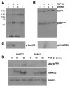
EGFR activation in response to TGF-β1 stimulation requires src tyrosine kinase activity. TGF-β1 induced tyrosine phosphorylation of the EGFR and the pp60c-src non-receptor tyrosine kinase (as detected with the phospho-tyrosine-specific antibody 4G10) within 30 min of growth factor addition (1 ng/ml) (A). The identity of the phospho-proteins was determined by blotting with antibodies to pp60c-src and the EGFR (B). Inhibition of src kinase activity with the specific src family inhibitor SU6656 (1 μM) dramatically deceased both basal and TGF-β1-induced Src Y416 phosphorylation (C) without altering total pp60c-src levels (B). TGF-β1-stimulated phosphorylation of the EGFR (A), particularly at the Y845 site (C), is completely blocked by pretreatment with SU6656 suggesting an upstream signaling role of src family members in EGFR activation. Consistent with these findings (in A–C), EGFR phosphorylation in response to TGF-β1 was evident in SYF+/+/+ but not SYF−/−/− MEFs whereas SMAD2 activation (pSMAD2) was similar in both cell types (D). Total SMAD2 levels provided a loading control.
Fig. 5
Fig. 5
SMAD2 phosphorylation in TGF-β1-treated VSMC is independent of EGFR/MEK-ERK signaling. TGF-β1-stimulates a sustained phosphorylation (for at least 4 h) of SMAD2 and ERK1/2 activation upon addition to quiescent R22 cells (A). Pretreatment with AG1478 for 30 min prior to a 2 h incubation with TGF-β1 did not affect SMAD2 phosphorylation but effectively blocked both TGF-β1-mediated PAI-1 induction (see Fig. 1A) and ERK1/2 phosphorylation (B). Similarly, the MEK inhibitors U0126 (10 μM) and PD98059 (10–20 μM) dramatically attenuated TGF-β1-stimulated PAI-1 expression in both R22 (C) and primary (D) VSMC. U0126 blocked the TGF-β1-stimulated increase in pERK1/2 levels but did not impact SMAD2 activation in R22 cells (E). Each experimental group is presented in triplicate (in B,E). As anticipated from data in (B,E), SMAD2 was rapidly phosphorylated in both EGFR−/− and wild-type MEFs in response to TGF-β1 (F). Increased ERK1/2 phosphorylation was evident only in EGFR+/+ fibroblasts, initially at 15 min (F) and more significantly at 2 h (G) after growth factor addition, confirming participation of the EGFR in ERK activation by TGF-β1. The low basal pERK1/2 in EGFR−/− cells suggests a role for EGFR signaling in ERK phosphorylation even in quiescent fibroblasts. ERK2 (A,C), the EGFR (B,E), actin (D), SMAD2/3 (F), ERK1/2 (G) provide loading controls.
Fig. 6
Fig. 6
RhoA signaling is required for TGF-β1-mediated PAI-1 expression. TGF-β1 stimulation of serum-deprived R22 cells significantly increased cellular levels of the GTP-bound form of Rho declining to the basal state by 24 h (A). Pretreatment with the Rho inhibitor C3 transferase (for 12 h) induced actin cytoskeletal modifications and cell shape changes, consistent with role of Rho proteins in actin remodeling and confirming the activity of C3 transferase in the VSMC cell system (B). TGF-β1-induced PAI-1 expression, was effectively suppressed by pretreatment of R22 cells (C) and primary VSMC (D) with C3 transferase as well as by transient transfection of R22 VSMC with a dominant-negative RhoAN17 (DN-RhoA) construct (2.5 μg) (E). Data plotted in (F) is a summary of three independent Western blot experiments (mean+SD) using R22 cells. C3=C3 transferase; numbers in parentheses in (F) on the x-axis is the concentration (in μg/ml) of C3 transferase. Total Rho (A), ERK2 (C,E) EGFR (D) levels provided loading controls.
Fig. 7
Fig. 7
Crucial role of ROCK in PAI-1 induction. To determine the potential involvement of ROCK as a downstream Rho effector of PAI-1 expression, quiescent VSMC were treated with the ROCK inhibitor Y-27632 (20 μM). Visualization of Texas Red phalloidin-stained R22 cells clearly indicated that actin organization and cell shape were dramatically altered by addition of Y-27632 to R22 cells (A). Y-27632 incubation prior to a 4-h stimulation with TGF-β1 (1 ng/ml) completely blocked PAI-1 induction even at the lowest concentration (5 μM) in both primary VSMC (B) and R22 cells (C). Data plotted (D) is the mean+SD of three independent Western blot experiments for R22 VSMC. Y=Y-27632 in (D); numbers in parentheses indicate drug concentration (μM). Actin, EGFR (B) and ERK2 (C,D) served as loading controls.
Fig. 8
Fig. 8
TGF-β1-induced SMAD2/3 activation and nuclear accumulation is suppressed by inhibition of Rho/ROCK signaling. Pretreatment of quiescent R22 cells with C3 transferase (C3) or Y-27632 effectively inhibited SMAD2 phosphorylation in response to TGF-β1 (A–C). Data plotted (C) is the mean+SD of three independent Western blot experiments for R22 VSMC. Y=Y-27632 in (D); numbers in parentheses indicate drug concentration (μM). TGF-β1-stimulated pSMAD2 (depicted in green) nuclear accumulation (D) is similarly suppressed by pretreatment with Y-27632 (at all concentrations between 5–20 μM, a range that effectively blocked PAI-1 induction [Figs. 7B–D]) indicting that interference with either member of the Rho pathway (Rho, ROCK) had similar consequences on both SMAD2 and SMAD3 activation. Nuclei were visualized by DAPI staining (D). Exposure to TGF-β1 was for 4 h in each case. Membranes were reprobed with antibodies to Rho (A) and SMAD2/3 (B) to confirm loading levels. For the time course study, quiescent R22 cells were treated with TGF-β1 for times indicated in the text with or without Y-27632 (20 μM) pretreatment. Equal amounts of lysates from each time point were probed with pSMAD2, pSMAD3, total SMAD2/3, TGF-βRI, PAI-1 or Rho antibodies (E).
Fig. 9
Fig. 9
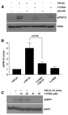
Inhibition of ROCK signaling with Y-27632 attenuates ERK1/2 but not EGFRY845 phosphorylation in TGF-β1-stimulated VSMC. To determine the effect of ROCK signaling in ERK1/2 activation, R22 cells were stimulated with TGF-β1 for 2 h with or without pretreatment with Y-27632 (20 μM) or AG1478 (2.5 μM which was used as a control since interference with EGFR signaling blocks TGF-β1-induced ERK activation, Fig. 5B); lysates were probed for pERK1/2 and total ERK1/2 levels. Y-27632 effectively attenuated the increase in ERK1/2 phosphorylation in response to TGF-β1 (A,B). Histogram in (B) illustrates the mean+SD of three independent Western blot experiments. Although inhibition of ROCK signaling significantly decreased TGF-β1-stimulated pERK1/2 levels (A,B), Y-27632 did not affect TGF-β1-induced EGFR phosphorylation (170 kDa pEGFR as detected with the 4G10 monoclonal antibody; e.g., Fig. 4A) (C). Blots were reprobed with antibodies to ERK2 (A,B) or total EGFR (C) for loading assessments.
Fig. 10
Fig. 10
Model for TGF-β1-induced PAI-1 expression in VSMC. The available data indicates that TGF-β1 activates two distinct signaling pathways that initiate transcription of PAI-1 in VSMC. Rho/ROCK are required for SMAD phosphorylation as well as ERK activation (through yet to be defined mechanisms) while the pp60c-src-activated EGFR (at the Y845 site) signals to MEK-ERK initiating likely ERK/USF interactions resulting in USF phosphorylation and a subtype (USF-1→USF-2) switch [e.g., 45,46] at the PAI-1 PE1/PE2 E box sites. Collectively, these two promoter-level events stimulate high levels of PAI-1 in response to TGF-βR occupancy. The actual mechanism underlying EGFR activation in response to TGF-β1 is currently under investigation but may involve direct recruitment of src kinases to the EGFR or the processing and release of a membrane-anchored EGFR ligand (e.g., HB-EGF). Similarly, events associated with TGF-β1 stimulation of the RhoA/ROCK pathway are presently unclear.

Similar articles

Cited by

References

    1. Kutz SM, Hordines J, McKeown-Longo PJ, Higgins PJ. TGF-β1-induced PAI-1 gene expression requires MEK activity and cell-to-substrate adhesion. J Cell Sci. 2001;114:3905–14. - PubMed
    1. Yue J, Mulder KM. Transforming growth factor-beta signal transduction in epithelial cells. Pharmacol Ther. 2001;91:1–34. - PubMed
    1. Bottinger EP, Bitzer M. TGF-β signaling in renal disease. J Am Soc Nephrol. 2002;13:2600–10. - PubMed
    1. Bhowmick NA, Ghiassi M, Aakre M, Brown K, Singh V, Moses HL. TGF-β-induced RhoA and p160ROCK activation is involved in the inhibition of Cdc25A with resultant cell-cycle arrest. Proc Natl Acad Sci U S A. 2003;100:15548–53. - PMC - PubMed
    1. Derynck R, Zhang Y. Smad-dependent and Smad-independent pathways in TGF-β family signalling. Nature. 2003;425:577–84. - PubMed

Publication types

MeSH terms