DNA damage-induced ubiquitylation of RFC2 subunit of replication factor C complex
- PMID: 18245774
- PMCID: PMC2431014
- DOI: 10.1074/jbc.M709835200
DNA damage-induced ubiquitylation of RFC2 subunit of replication factor C complex
Abstract
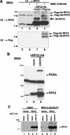
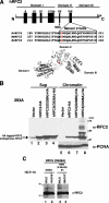
Many proteins involved in DNA replication and repair undergo post-translational modifications such as phosphorylation and ubiquitylation. Proliferating cell nuclear antigen (PCNA; a homotrimeric protein that encircles double-stranded DNA to function as a sliding clamp for DNA polymerases) is monoubiquitylated by the RAD6-RAD18 complex and further polyubiquitylated by the RAD5-MMS2-UBC13 complex in response to various DNA-damaging agents. PCNA mono- and polyubiquitylation activate an error-prone translesion synthesis pathway and an error-free pathway of damage avoidance, respectively. Here we show that replication factor C (RFC; a heteropentameric protein complex that loads PCNA onto DNA) was also ubiquitylated in a RAD18-dependent manner in cells treated with alkylating agents or H(2)O(2). A mutant form of RFC2 with a D228A substitution (corresponding to a yeast Rfc4 mutation that reduces an interaction with replication protein A (RPA), a single-stranded DNA-binding protein) was heavily ubiquitylated in cells even in the absence of DNA damage. Furthermore RFC2 was ubiquitylated by the RAD6-RAD18 complex in vitro, and its modification was inhibited in the presence of RPA. The inhibitory effect of RPA on RFC2 ubiquitylation was relatively specific because RAD6-RAD18-mediated ubiquitylation of PCNA was RPA-insensitive. Our findings suggest that RPA plays a regulatory role in DNA damage responses via repression of RFC2 ubiquitylation in human cells.
Figures


Similar articles
-
Regulation of DNA damage tolerance in mammalian cells by post-translational modifications of PCNA.Mutat Res. 2017 Oct;803-805:82-88. doi: 10.1016/j.mrfmmm.2017.06.004. Epub 2017 Jun 21. Mutat Res. 2017. PMID: 28666590 Review.
-
Replication protein A dynamically regulates monoubiquitination of proliferating cell nuclear antigen.J Biol Chem. 2019 Mar 29;294(13):5157-5168. doi: 10.1074/jbc.RA118.005297. Epub 2019 Jan 30. J Biol Chem. 2019. PMID: 30700555 Free PMC article.
-
PCNA Monoubiquitination Is Regulated by Diffusion of Rad6/Rad18 Complexes along RPA Filaments.Biochemistry. 2020 Dec 15;59(49):4694-4702. doi: 10.1021/acs.biochem.0c00849. Epub 2020 Nov 27. Biochemistry. 2020. PMID: 33242956 Free PMC article.
-
Regulation of HLTF-mediated PCNA polyubiquitination by RFC and PCNA monoubiquitination levels determines choice of damage tolerance pathway.Nucleic Acids Res. 2018 Nov 30;46(21):11340-11356. doi: 10.1093/nar/gky943. Nucleic Acids Res. 2018. PMID: 30335157 Free PMC article.
-
Regulation of Rad6/Rad18 Activity During DNA Damage Tolerance.Annu Rev Biophys. 2015;44:207-28. doi: 10.1146/annurev-biophys-060414-033841. Annu Rev Biophys. 2015. PMID: 26098514 Free PMC article. Review.
Cited by
-
En bloc transfer of polyubiquitin chains to PCNA in vitro is mediated by two different human E2-E3 pairs.Nucleic Acids Res. 2012 Nov 1;40(20):10394-407. doi: 10.1093/nar/gks763. Epub 2012 Aug 16. Nucleic Acids Res. 2012. PMID: 22904075 Free PMC article.
-
Copy number variants at Williams-Beuren syndrome 7q11.23 region.Hum Genet. 2010 Jul;128(1):3-26. doi: 10.1007/s00439-010-0827-2. Epub 2010 May 1. Hum Genet. 2010. PMID: 20437059 Review.
-
Rad18 E3 ubiquitin ligase activity mediates Fanconi anemia pathway activation and cell survival following DNA Topoisomerase 1 inhibition.Cell Cycle. 2011 May 15;10(10):1625-38. doi: 10.4161/cc.10.10.15617. Epub 2011 May 15. Cell Cycle. 2011. PMID: 21478670 Free PMC article.
-
Investigating RFC1 expansions in sporadic amyotrophic lateral sclerosis.J Neurol Sci. 2021 Nov 15;430:118061. doi: 10.1016/j.jns.2021.118061. Epub 2021 Aug 31. J Neurol Sci. 2021. PMID: 34537679 Free PMC article.
-
RFC2: a prognosis biomarker correlated with the immune signature in diffuse lower-grade gliomas.Sci Rep. 2022 Feb 24;12(1):3122. doi: 10.1038/s41598-022-06197-5. Sci Rep. 2022. PMID: 35210438 Free PMC article.
References
-
- Hartwell, L. H., and Weinert, T. A. (1989) Science 246 629-634 - PubMed
-
- Carr, A. M. (2002) DNA Repair (Amst.) 1 983-994 - PubMed
-
- Kastan, M. B., and Bartek, J. (2004) Nature 432 316-323 - PubMed
-
- Sancar, A., Lindsey-Boltz, L. A., Unsal-Kacmaz, K., and Linn, S. (2004) Annu. Rev. Biochem. 73 39-85 - PubMed
Publication types
MeSH terms
Substances
Grants and funding
LinkOut - more resources
Full Text Sources
Research Materials
Miscellaneous

