

Fragments of extracellular matrix as mediators of inflammation
- PMID: 18243041
- PMCID: PMC2478752
- DOI: 10.1016/j.biocel.2007.12.005
Fragments of extracellular matrix as mediators of inflammation
Abstract
Classically, the extracellular matrix (ECM) was viewed as a supporting structure for stabilizing the location of cells in tissues and for preserving the architecture of tissues. This conception has changed dramatically over the past few decades with discoveries that ECM has profound influences on the structure, viability, and functions of cells. Much of the data supporting this new paradigm has been obtained from studies of normal and pathological structural cells such as fibroblasts, smooth muscle cells, and malignant cells, as, for example, breast cancer epithelial cells. However, there has also been recognition that effects of ECM on cells extend to inflammatory cells. In this context, attention has been drawn to fragments of ECM components. In this review, we present information supporting the concept that proteolytic fragments of ECM affect multiple functions and properties of inflammatory and immune cells. Our focus is particularly upon neutrophils, monocytes, and macrophages and fragments derived from collagens, elastin, and laminins. Hyaluronan fragments, although they are not products of proteolysis, are also discussed, as they are a notable example of ECM fragments that exhibit important effects on inflammatory cells. Further, we summarize some exciting recent developments in this field as a result of mouse models in which defined ECM fragments and their receptors are clearly implicated in inflammation in vivo. Thus, this review underscores the idea that proteolysis of ECM may well have implications that go beyond modifying the structural environment of cells and tissues.
Figures
Similar articles
-
The extracellular matrix in IBD: a dynamic mediator of inflammation.Curr Opin Gastroenterol. 2017 Jul;33(4):234-238. doi: 10.1097/MOG.0000000000000368. Curr Opin Gastroenterol. 2017. PMID: 28562487 Free PMC article. Review.
-
Extracellular matrix proteins and leukocyte function.J Leukoc Biol. 1995 May;57(5):699-702. doi: 10.1002/jlb.57.5.699a. J Leukoc Biol. 1995. PMID: 7759947 Review.
-
Role of extracellular matrix and its regulators in human airway smooth muscle biology.Cell Biochem Biophys. 2006;44(1):139-46. doi: 10.1385/CBB:44:1:139. Cell Biochem Biophys. 2006. PMID: 16456242 Review.
-
Extracellular matrix in development: insights from mechanisms conserved between invertebrates and vertebrates.Cold Spring Harb Perspect Biol. 2011 Dec 1;3(12):a005082. doi: 10.1101/cshperspect.a005082. Cold Spring Harb Perspect Biol. 2011. PMID: 21917993 Free PMC article. Review.
-
The extracellular matrix as a multitasking player in disease.FEBS J. 2019 Aug;286(15):2830-2869. doi: 10.1111/febs.14818. Epub 2019 Apr 11. FEBS J. 2019. PMID: 30908868 Review.
Cited by
-
Extra Cellular Matrix Remodeling: An Adjunctive Target for Spinal Cord Injury and Intervertebral Disc Degeneration.Neurospine. 2022 Sep;19(3):632-645. doi: 10.14245/ns.2244366.183. Epub 2022 Sep 30. Neurospine. 2022. PMID: 36203290 Free PMC article.
-
Anti-HMG-CoA Reductase, Antioxidant, and Anti-Inflammatory Activities of Amaranthus viridis Leaf Extract as a Potential Treatment for Hypercholesterolemia.Evid Based Complement Alternat Med. 2016;2016:8090841. doi: 10.1155/2016/8090841. Epub 2016 Mar 9. Evid Based Complement Alternat Med. 2016. PMID: 27051453 Free PMC article.
-
Xenogenic esophagus scaffolds fixed with several agents: comparative in vivo study of rejection and inflammation.J Biomed Biotechnol. 2012;2012:948320. doi: 10.1155/2012/948320. Epub 2012 Mar 8. J Biomed Biotechnol. 2012. PMID: 22505820 Free PMC article.
-
Extracellular matrix and fibroblast communication following myocardial infarction.J Cardiovasc Transl Res. 2012 Dec;5(6):848-57. doi: 10.1007/s12265-012-9398-z. Epub 2012 Aug 28. J Cardiovasc Transl Res. 2012. PMID: 22926488 Free PMC article. Review.
-
New insights into arterial stiffening: does sex matter?Am J Physiol Heart Circ Physiol. 2018 Nov 1;315(5):H1073-H1087. doi: 10.1152/ajpheart.00132.2018. Epub 2018 Jul 20. Am J Physiol Heart Circ Physiol. 2018. PMID: 30028199 Free PMC article. Review.
References
-
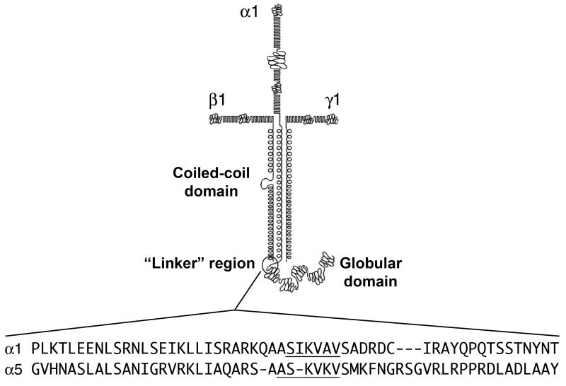
- Adair-Kirk TL, Atkinson JJ, Broekelmann TJ, Doi M, Tryggvason K, Miner JH, Mecham RP, Senior RM. A site on laminin alpha 5, AQARSAASKVKVSMKF, induces inflammatory cell production of matrix metalloproteinase-9 and chemotaxis. J Immunol. 2003;171:398–406. - PubMed
-
- Adair-Kirk TL, Atkinson JJ, Kelley DG, Arch RH, Miner JH, Senior RM. A chemotactic peptide from laminin alpha 5 functions as a regulator of inflammatory immune responses via TNF alpha-mediated signaling. J Immunol. 2005;174:1621–1629. - PubMed
-
- Alon R, Cahalon L, Hershkoviz R, Elbaz D, Reizis B, Wallach D, Akiyama SK, Yamada KM, Lider O. TNF-alpha binds to the N-terminal domain of fibronectin and augments the beta 1-integrin-mediated adhesion of CD4+ T lymphocytes to the glycoprotein. J Immunol. 1994;152:1304–1313. - PubMed
-
- Andersson E, Rydengard V, Sonesson A, Morgelin M, Bjorck L, Schmidtchen A. Antimicrobial activities of heparin-binding peptides. Eur J Biochem. 2004;271:1219–1226. - PubMed
-
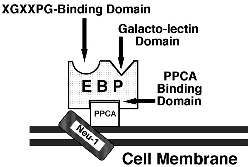
- Baranek T, Debret R, Antonicelli F, Lamkhioued B, Belaaouaj A, Hornebeck W, Bernard P, Guenounou M, Le Naour R. Elastin receptor (spliced galactosidase) occupancy by elastin peptides counteracts proinflammatory cytokine expression in lipopolysaccharide-stimulated human monocytes through NF-kappaB down-regulation. J Immunol. 2007;179:6184–6192. - PubMed
Publication types
MeSH terms
Substances
Grants and funding
LinkOut - more resources
Full Text Sources
Other Literature Sources
Miscellaneous
