. 2008 Feb 20;27(4):629-41.
doi: 10.1038/emboj.2008.5. Epub 2008 Jan 31.

Inflammatory cardiac valvulitis in TAX1BP1-deficient mice through selective NF-kappaB activation

Affiliations

Inflammatory cardiac valvulitis in TAX1BP1-deficient mice through selective NF-kappaB activation

Hidekatsu Iha et al. EMBO J. .

Abstract

Nuclear factor kappa B (NF-kappaB) is a key mediator of inflammation. Unchecked NF-kappaB signalling can engender autoimmune pathologies and cancers. Here, we show that Tax1-binding protein 1 (TAX1BP1) is a negative regulator of TNF-alpha- and IL-1beta-induced NF-kappaB activation and that binding to mono- and polyubiquitin by a ubiquitin-binding Zn finger domain in TAX1BP1 is needed for TRAF6 association and NF-kappaB inhibition. Mice genetically knocked out for TAX1BP1 are born normal, but develop age-dependent inflammatory cardiac valvulitis, die prematurely, and are hypersensitive to low doses of TNF-alpha and IL-1beta. TAX1BP1-/- cells are more highly activated for NF-kappaB than control cells when stimulated with TNF-alpha or IL-1beta. Mechanistically, TAX1BP1 acts in NF-kappaB signalling as an essential adaptor between A20 and its targets.


Figures
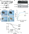
Figure 1
Figure 1
Generation of TAX1BP1−/− mice. (A) Targeted disruption of the mouse TAX1BP1 gene. The targeting vector pMm151(NEO)KO, the thymidine kinase cassette (TK) located at the 5′ end of the targeting vector (indicated by a shaded box), and the Neo cassette (indicated by a hatched box) are indicated. Arrows show the sense direction for each ORF. Homologous recombination at the TAX1BP1 locus was confirmed by PCR by using specific primers indicated by small arrows. H3: HindIII; Cl: ClaI; Hp: HpaI; Sa: SalI; Sp: SpeI; B: BamHI. (B) TAX1BP1 KO was confirmed by PCR (upper panel) or by blotting using a polyclonal antibody against TAX1BP1 (middle panel). Proteins were normalized using anti-tubulin (lower panel). (C) Left: gross appearance of representative 45- and 75-day TAX1BP1+/+ and TAX1BP1−/− mice. Right: growth curves of TAX1BP1+/+ and TAX1BP1−/− mice. TAX1BP1−/− and WT controls were weighed at 2-week intervals for 75 days and again at 240 days. Ten animals were used per group. Data represent mean weight±s.e. (P<0.0001). (D) Death of mice over an 18 months follow-up. The Kaplan–Meier curves show that TAX1BP1−/− mice have lower survival than WT or TAX1BP1+/− mice (log-rank P<0.001).
Figure 2
Figure 2
TAX1BP1−/− mice show severe cardiac valvulitis and skin dermatitis with inflammatory cellular infiltrates. (A) Valvulitis in haematoxylin and eosin (H&E)-stained valves (Va) from 21-day, 6-month-, and 12-month-old TAX1BP1−/− mice. In panels 5 and 6, valve leaflets from TAX1BP1−/− mice are grossly hypertrophic with cellular infiltration and collagen invasion. (B) Immunostaining of valve leaflets from TAX1BP1−/− mice using anti-F4/80 (F4/80 is a macrophage-restricted cell surface glycoprotein) and anti-CD3, revealing the presence of macrophages (panel 1) and T cells (panel 2) (arrowheads). H&E (panel 3) and anti-myeloperoxidase (panel 4) stainings show mononuclear cells, neutrophils (arrowheads), and cellular debris near small capillary (Ca); magnification × 400. Arteritis and periarteritis in TAX1BP1−/− mice are shown. The lower magnification (panel 5) shows heart blood vessel (Ve) with surrounding infiltration of mononuclear cells and neutrophils; magnification × 200. The enlarged panel 6 shows severe inflammation characterized by the infiltration of mononuclear cells and neutrophils near a small capillary; magnification × 400. (C) Skin inflammation in TAX1BP1 KO mice. Inflammation observed in H&E-stained skin from TAX1BP1−/− mice (panels 1 and 2). Sections of TAX1BP1 KO skin were stained with H&E (panel 3) and for T cells (CD3) (panel 4), macrophages (F4/80) (panel 5), and neutrophils (myeloperoxidase) (panel 6); magnification × 100. Enlargement of panels 4–6 shows the presence of T cells (anti-CD3 staining), neutrophils (anti-myeloperoxidase staining), and macrophages (anti-F4/80 staining); magnification × 400.
Figure 3
Figure 3
Increased hypersensitivity of TAX1BP1−/− mice to inflammatory challenges. (A) Kaplan–Meier curves of TAX1BP1 mice after sublethal TNF-α or IL-1β challenges. TAX1BP1−/− mice succumbed to low doses of TNF-α and IL-1β. Twelve-week-old TAX1BP1+/+, TAX1BP1+/−, and TAX1BP1−/− littermates were challenged with proinflammatory cytokine TNF-α (0.5 mg/kg) (TAX1BP1+/+, closed circle; TAX1BP1+/−, closed triangle; TAX1BP1−/−, closed square) or IL-1β (0.5 mg/kg) (TAX1BP1+/+, open circle; TAX1BP1+/−, open triangle; TAX1BP1−/−, open square). Survival was monitored every 2 h, and the experiment was terminated after 24 h. The number of mice in each experimental group was n=14. No control mice (+PBS n=14) died (data not shown); the experiments were performed twice. (B) Kaplan–Meier curves of TAX1BP1 bone marrow chimaera mice after TNF-α challenge. Five- to 6-week-old female TAX1BP1−/− or TAX1BP1+/+ recipient mice were first sublethally irradiated and then transplanted with the indicated donor bone marrow. Eight weeks after transplantation, bone marrow chimaera mice were challenged with TNF-α (0.5 mg/kg body weight, intraperitoneal injection). Survival was monitored every 2 h and the experiment was terminated after 24 h. The number of mice in each experimental group was n=12; the experiments were performed twice. Statistical analysis was performed using a log-rank test (P<0.001).
Figure 4
Figure 4
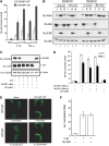
TAX1BP1 inhibits TNF-α- and IL-1β-triggered NF-κB activation. (A) TAX1BP1 KO MEFs show higher NF-κB responses to TNF-α and IL-1β than WT MEFs. MEFs were transfected with an NF-κB-luc reporter and an RSV-β-gal plasmid as an internal control. At 16 h after transfection, increasing concentrations of IL-1β (10–50 ng/ml) and TNF-α (10–50 ng/ml) were added for 8 h and the cells were harvested for luciferase assay. (B) Macrophages from TAX1BP1 KO mice show a higher NF-κB response to TNF-α and IL-1β than WT macrophages. Peritoneal macrophages from WT and KO mice were stimulated for 30 and 90 min at 37°C with IL-1β (100 ng/ml) and TNF-α (100 ng/ml) and cell lysates were examined by immunoblotting (IB) using anti-phospho (P) Ser 32/36 IκBα, anti-IκBα, anti-phospho (P) T183 JNK1, or anti-JNK1, as indicated. (C) 293T cells were co-transfected with 2 μg TAX1BP1-specific siRNA (lanes 3–5) or control GFP siRNA (lane 6) in a DNA transfection mix of 100 ng of GFP plasmid, 50 ng of NF-κB-luc, and 50 ng of RSV-β-gal and adjusted to equal concentrations with pCDNA3. TAX1BP1, GFP, and tubulin expressions were verified by IB. (D) To analyse NF-κB activation, cells transfected with or without the indicated siRNA were treated with either IL-1β (50 ng/ml) or TNF-α (50 ng/ml) and NF-κB-luciferase reporter expression was assayed 8 h later. (E) TAX1BP1 blocks IL-1-triggered nuclear translocation of GFP-p65. 293-IL1R cells were transfected with 250 ng of GFP-p65 and then with either 500 ng of control pcDNA3 (mock) or TAX1BP1 (TAX1BP1 WT). At 24 h after transfection, cells were treated with IL-1β (100 ng/ml) for 40 min. Green fluorescence (GFP-p65) was monitored for 40 min with a Leica laser-scanning microscope. The top row shows untreated cells at the beginning; the bottom row shows cells after 40 min of IL-1β treatment. (F) TAX1BP1 has no effect on IL-1β-induced AP-1 activation. 293T cells were transfected with 1.6 μg of total plasmid DNA containing a mixture of AP-1-luciferase reporter and RSV-galactosidase plasmid, without (−) or with (+) TAX1BP1 plasmid. At 40 h after transfection, cells were treated with IL-1β (10 ng/ml) for 8 h. Cells were then harvested for luciferase assay.
Figure 5
Figure 5
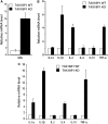
Increased expression of proinflammatory cytokines TNF-α and IL-1β in TAX1BP1−/− tissue. (A) Real-time PCR analysis of IκBα mRNA expression in heart tissue isolated from four control and five TAX1BP1 KO mice. (B) Real-time PCR analysis of proinflammatory cytokines in heart tissue isolated from four control and five TAX1BP1 KO mice. (C) Real-time PCR analysis of proinflammatory cytokines in skin isolated from four control and five KO mice. All the results are the mean values relative to GADPH; error bars indicate SE of triplicate samples.
Figure 6
Figure 6
The second Zn finger of TAX1BP1 contains a novel ubiquitin-binding domain. (A) Schematic representations of TAX1BP1 WT, which contains three coiled-coil (CC) and two ubiquitin-binding domains (open rectangles), and four TAX1BP1 ubiquitin-binding domain mutants (UBZ1 mutant: F737A; UBZ2 mutant: F764A; UBZ1+2 mutant: F737A+F764A; UBZ* mutant: C757A+C760A), which are mutated (X) in the indicated first (UBZ1) or second (UBZ2) Zn finger(s). (B) Binding of TAX1BP1 to ubiquitin. HA-TAX1BP1 was expressed in 293T cells and cell lysates were prepared 40 h later. Total cell lysates were incubated for 8 h at 4°C with either GST-ubiquitin (Ub) (lane 3) or GST-ubiquitin Ile44 mutant (UBI44A) (lane 4) immobilized on glutathione Sepharose. The binding of TAX1BP1 was visualized by immunoblotting (IB) with anti-HA. (C) Mutation of the UBZ2 domain disrupts the ubiquitin-binding potential of TAX1BP1. FLAG-TAX1BP1, FLAG-TAX1BP1 UBZ1, FLAG-TAX1BP1 UBZ2, or FLAG-TAX1BP1 UBZ* was transfected into 293T cells. Binding to immobilized GST (cont) or GST-Ub (Ub) was analysed by IB with anti-FLAG. (D) The UBZ2 domain is sufficient to bind K48- and K63-linked ubiquitin. Purified polyubiquitin chains (K48- or K63-linked) were incubated with immobilized intact individual UBZ domains fused to GST (GST-UBZ2, GST-UBZ1) or both UBZ domains fused to GST (GST-UBZ1+2) for 2 h at 4°C. Binding of polyubiquitin chains to GST fusion proteins was visualized by IB using anti-Ub. Ponseau S stained the GST fusion proteins used in the binding assays.
Figure 7
Figure 7
TAX1BP1's ubiquitin-binding domain is required for inhibition of IL-1β triggered NF-κB activation. (A) UBZ2 mutation disrupts the binding of TAX1BP1 to TRAF6. 293T cells were co-transfected with HA-tagged TRAF6 and the indicated FLAG-tagged TAX1BP1 UBZ mutants. 24 h after transfection, cell lysates were subjected to immunoprecipitation (IP) with 1 μg of mouse monoclonal anti-FLAG antibody (α-FLAG), followed by immunoblotting (IB) with mouse monoclonal anti-FLAG antibody coupled to HRP (α-FLAG-HRP), or mouse monoclonal anti-HA (α-HA). Transfection efficiency was verified by IB with anti-FLAG-HRP and anti-HA. (B) TAX1BP1 UBZ* exerts a dominant-negative effect on TAX1BP1–TRAF6 binding. Cells were transfected with FLAG-TRAF6 and/or Myc-TAX1BP1 or HA-TAX1BP1 UBZ*. After 48 h, cell lysates were immunoprecipitated with anti-FLAG, followed by IB with anti-Myc or anti-FLAG (upper panels). Protein amounts were verified by IB with anti-tubulin (lower panel). * indicates a background band. (C) The UBZ2 domain is essential for TAX1BP1's NF-κB inhibitory potential. 293T cells were transfected with 2 μg of total plasmid DNA containing a mixture of NF-κB luciferase reporter with pActβ-galactosidase plasmid and the indicated WT or TAX1BP1 UBZ mutant plasmids. After 24 h, cells were stimulated for 6 h with 20 ng/ml IL-1β and assayed for luciferase. Values shown are normalized against β-galactosidase activity.
Figure 8
Figure 8
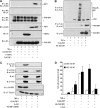
Expression of TAX1BP1 reduces ubiquitination of TRAF6. (A) TAX1BP1 overexpression inhibits ubiquitination of ectopically expressed TRAF6. 293T cells were transfected with FLAG-TRAF6, HA-TAX1BP1 (WT; lanes 4–6), or HA-TAX1BP1 UBZ* (UBZ*; lanes 7 and 8) with Myc-Ub. At 40 h after transfection, cells were stimulated with IL-1β for 8 h. A 300 μg portion of cell lysate was subjected to immunoprecipitation (IP) with 40 μl of anti-FLAG-conjugated agarose beads, followed by immunoblotting (IB) with either rabbit polyclonal anti-Myc (top panel) or rabbit polyclonal anti-FLAG (second panel). HA-TAX1BP1 in the cell lysates was assessed by IB using anti-HA, and protein loadings were normalized by IB with anti-actin (lower panels). (B) TAX1BP1 overexpression inhibits IL-1β-induced ubiquitination of cell endogenous TRAF6. 293T cells were transfected with Myc-Ub (lanes 1 and 2) or co-transfected with HA-TAX1BP1 (WT) (lanes 3 and 4) and Myc-Ub. At 40 h after transfection, cells were stimulated as indicated with IL-1β (50 ng/ml) for 8 h (lanes 2 and 4). A 300 μg portion of cell lysate was subjected to IP with 40 μl of anti-TRAF6-conjugated agarose beads, followed by IB with either rabbit polyclonal anti-TRAF6 (middle panel) or rabbit polyclonal anti-Myc (upper panel). HA-TAX1BP1 in the cell lysates was assessed by IB using anti-HA, and protein loadings were normalized by IB with anti-actin (lower panel). (C) Reduced ability of A20 to moderate IL-1β's induction of NF-κB in TAX1BP1−/− MEF cells. WT and TAX1BP1−/− MEFs were transfected with NF-κB-luc and RSV-β-gal, with or without increasing amounts of A20 plasmid. At 40 h after transfection, cells were treated with IL-1β (50 ng/ml) for 8 h and then harvested for luciferase analysis. (D) TAX1BP1 fails to moderate IL-1β's induction of NF-κB in A20−/− MEFs. WT and A20−/− MEFs were transfected with NF-κB-luc and RSV-β-gal, with or without increasing amounts of TAX1BP1 or A20 plasmids. At 40 h after transfection, cells were treated with IL-1β (50 ng/ml) for 8 h and then harvested for luciferase analysis. (E) IL-1β induces the binding of A20 to TRAF6, which is abolished by overexpression of TAX1BP1 UBZ*. 293T cells were transfected with FLAG-TRAF6, E-tagged A20, and HA-tagged TAX1BP1 UBZ*, as indicated. Cells were stimulated with IL-1β (50 ng/ml) for 8 h. Binding of TRAF6 to A20 was analysed by co-immunoprecipitation. Total expression of FLAG-TRAF6, HA-TAX1BP1 UBZ*, and E-A20 in the cell lysate was assessed by IB using anti-FLAG, anti-HA, and anti-E. (F) TRAF6 and A20 do not bind each other in TAX1BP1 KO cells. TAX1BP1+/+ and TAX1BP1−/− MEFs were transfected with E-A20 and FLAG-TRAF6. Binding of A20 to TRAF6 was analysed as described in (E).
Figure 9
Figure 9
TAX1BP1 binds RIP1 and inhibits TNF-α-induced ubiquitination of RIP1. (A) TNF-α induces the binding of TAX1BP1 to RIP1. 293T cells were transfected with FLAG-RIP1, HA-TAX1BP1, and E-TRAF2. At 40 h after transfection, cells were stimulated with TNF-α for 8 h. Total expression of transfected proteins was assessed by immunoblotting (IB) using anti-FLAG, anti-HA, and anti-E respectively. A 300 μg portion of cell lysate was subjected to immunoprecipitation (IP) with 40 μl of anti-HA-conjugated agarose beads, followed by IB with rabbit polyclonal anti-FLAG, anti-HA, or anti-E as indicated. * indicates a background band. (B) Overexpression of TAX1BP1 inhibits TNF-α-induced RIP1 ubiquitination. 293T cells were co-transfected with HA-TAX1BP1 and FLAG-RIP1. At 48 h after transfection, cells were stimulated with TNF-α (50 ng/ml) for 30 min (lanes 2, 4, and 6). RIP1 was immunoprecipitated with anti-FLAG. Binding of RIP1 and TAX1BP1 was revealed by IB with anti-HA (middle panel) and RIP1 ubiquitination was revealed by IB with anti-FLAG (top panel). HA-TAX1BP1 in the cell lysates was assessed by IB using anti-HA. (C) TAX1BP1 is needed for A20–RIP1 complex formation. 293T cells were transfected with control or TAX1BP1-siRNA. Cells were stimulated for 30 min at 37°C with TNF-α (50 ng/ml). A 300 μg portion of cell lysates was immunoprecipitated with 40 μl of anti-FLAG-conjugated agarose beads, followed by IB with either rabbit polyclonal anti-FLAG or anti-E. TAX1BP1 in the cell lysates was assessed by IB using anti-TAX1BP1, and protein loadings were normalized by IB with anti-tubulin (lower panel). (D) TAX1BP1 fails to moderate TNF-α-induced NF-κB activation in A20−/− MEFs. WT and A20−/− MEFs were transfected with an NF-κB-luc plasmid and an RSV-β-gal plasmid, with FLAG-RIP1, HA-TAX1BP1, or E-A20 plasmids. At 40 h after transfection, cells were treated with TNF-α (10 ng/ml) for 8 h and then harvested for luciferase assay.

Similar articles

Cited by

References

    1. Beg AA, Sha WC, Bronson RT, Baltimore D (1995) Constitutive NF-kappa B activation, enhanced granulopoiesis, and neonatal lethality in I kappa B alpha-deficient mice. Genes Dev 9: 2736–2746 - PubMed
    1. Bienko M, Green CM, Crosetto N, Rudolf F, Zapart G, Coull B, Kannouche P, Wider G, Peter M, Lehmann AR, Hofmann K, Dikic I (2005) Ubiquitin-binding domains in Y-family polymerases regulate translesion synthesis. Science 310: 1821–1824 - PubMed
    1. Boone DL (2004) The ubiquitin-modifying enzyme A20 is required for termination of Toll-like receptor responses. Nat Immunol 5: 1052–1060 - PubMed
    1. Cao Z, Henzel WJ, Gao X (1996) IRAK: a kinase associated with the interleukin-1 receptor. Science 271: 1128–1131 - PubMed
    1. Chen ZJ (2005) Ubiquitin signalling in the NF-[kappa]B pathway. Nat Cell Biol 7: 758–765 - PMC - PubMed

Publication types

MeSH terms