Comparative Study
. 2008 Jan 30;28(5):1099-108.
doi: 10.1523/JNEUROSCI.4906-07.2008.

Netrin-1 is a novel myelin-associated inhibitor to axon growth

Affiliations
Comparative Study

Netrin-1 is a novel myelin-associated inhibitor to axon growth

Karin Löw et al. J Neurosci. .

Abstract

We investigated the influence of the bifunctional guidance molecule netrin-1 on axonal growth in the injured adult spinal cord. In the adult, netrin-1 is expressed on mature oligodendrocytes, cells of the central canal, and the meninges. Netrin-1 protein in white matter is selectively enriched adjacent to paranodal loops of myelin in nodes of Ranvier. The repulsion-mediating netrin-1 uncoordinated-5 (UNC5) receptors are expressed by neurons of the corticospinal and rubrospinal projections, and by intrinsic neurons of the spinal cord, both before and after spinal cord injury. Neutralization of netrin-1 in myelin prepared from adult rat spinal cord using UNC5 receptor bodies increases neurite outgrowth from UNC5-expressing spinal motor neurons in vitro. Furthermore, axon regeneration is inhibited in a netrin-1-enriched zone, devoid of other myelin-associated inhibitors, within spinal cord lesion sites in vivo. We conclude that netrin-1 is a novel oligodendrocyte-associated inhibitor that can contribute to axonal growth failure after adult spinal cord injury.


Figures

Figure 1.
Figure 1.
Netrin-1 expression in vivo. a–i, In situ hybridization was performed on rat spinal cord sections using rat netrin-1 antisense (Net-1 anti; a, c, d, e, f, g, h, i) or sense (b) α-35S-UTP labeled probes. a, b, Darkfield images of coronal sections through the adult intact cervical spinal cord. Note the netrin-1 signal in the central canal and throughout gray and white matter, with highest white matter expression levels in the dorsolateral fasciculus (white arrows). c, Autoradiography of sagittal section of adult spinal cord. Netrin-1 is expressed by meninges (m) surrounding the spinal cord and central canal (cc; arrowhead). d, Brightfield image of horizontal section of adult intact cervical spinal cord (Nissl counterstained). Netrin-1 is expressed by cells constituting the central canal. Inset, Coronal section of central canal. e, Coronal section of rat E15 spinal cord (darkfield). Netrin-1 is expressed in floor plate (FP) and by cells constituting the central canal as expected. f, Horizontal section through dorsolateral white matter of adult rat spinal cord. Combination of netrin-1 in situ hybridization with APC (mature oligodendrocyte marker) immunocytochemistry demonstrates netrin-1 expression by mature oligodendrocytes. Inset, Coronal section through dorsolateral white matter. g, h, Netrin-1 does not colocalize with GFAP (g, circled cells) or the neuronal marker NeuN (h). i, Detection of netrin-1 expression by in situ hybridization in the thoracic (T6) spinal cord adjacent to a T7 transection site, 2 weeks after lesion. k, RT-PCR of netrin-1 from spinal cord 1, 2, and 4 weeks after T7 transection, normalized to the housekeeping gene Rplp1. l, Immunolabeling for netrin-1 identifies its presence and enrichment in perinodal segments of adult white matter (black reaction product), using a counterstain for the perinodal marker Caspr (brown). m, n, Netrin-1 labeling in intact white matter of the dorsolateral funiculus (m), and persistent presence 3 months after spinal cord injury in the dorsolateral funiculus (n) located 500 μm rostral to injury site.
Figure 2.
Figure 2.
UNC5 receptor expression in corticospinal and rubrospinal neurons. a1–d1, UNC5A in situ signal in plane of photo emulsion (brightfield). a2–d2, Same section under UV excitation to visualize FG retrogradely labeled neurons. a3–d3, Overlay of columns 1 and 2 with color rendering. a1–b3, Motor cortex, layer V, corticospinal neurons. Intact (a1–a3) and lesioned (1 week; b1–b3) corticospinal neurons express UNC5A. c1–d3, Intact (c1–c3) and lesioned (1 week; d1–d3) rubrospinal neurons also express UNC5A.
Figure 3.
Figure 3.
UNC5 expression in spinal cord. a, b, f, g, l, m, Coronal sections through adult intact cervical spinal cord (darkfield). c, d, h, i, n, o, Coronal sections through adult thoracic spinal cord (T6) after a sham lesion (c, h, n) and 2 weeks after a T7 full transection lesion (d, i, o). Expression of the netrin-receptors UNC5A (c, d), UNC5C (h, i) and DCC (n, o) is restricted to the spinal cord gray matter, and expression patterns are maintained 2 weeks after spinal cord transection. Compared with sham-lesioned animals, levels of UNC5A (e), UNC5C (k) and DCC (p) by real-time PCR are reduced to 42.8 ± 9.3% (SEM), 58.5 ± 8.6% (SEM) and 80.3 ± 0.85% (SEM) in the adjacent rostral spinal cord and to 29.8 ± 5.9% (SEM), 44.8 ± 6.3% (SEM) and 65.8 ± 5.7% (SEM) in the adjacent caudal spinal cord segments, respectively, 4 weeks after T7 complete transection (ANOVA, *p < 0.05, **p < 0.01, ***p < 0.001).
Figure 4.
Figure 4.
Netrin-1 neutralization enhances neurite outgrowth in vitro. a, b, UNC5A (a) and UNC5C (b) are expressed in motor neuron pools (mn) in ventral E15 rat spinal cord as expected, by in situ hybridization; thus, these neurons were used as a test system in vitro for effects of netrin-1 neutralization on axonal outgrowth. c, Detection of MAG in myelin purified from adult rat spinal cord by Western blot, indicating that the myelin isolate is of expected quality for in vitro studies. d, Netrin-1 ELISA demonstrating binding of UNC5B receptor body in a dose dependent, saturatable manner. Recombinant netrin-1 was coated at 20 μg/ml and incubated with increasing concentrations (1–128 μg/ml) of UNC5B receptor body. e, f, neurite outgrowth from UNC5-expressing E15 spinal motor neurons on adult rat spinal cord myelin is significantly enhanced when netrin-1 is neutralized by UNC5 receptor body (e), compared with control cultures containing IgG (f) (for details, see Materials and Methods). g, The length of the longest neurite of spinal motor neurons after 48 h on myelin was increased by 81% after netrin-1 neutralization with UNC5 receptor bodies (***p < 0.001). Thus, netrin-1 is present in adult rat spinal cord myelin in sufficient quantities to inhibit axonal outgrowth from UNC5-expressing neurons in vitro. h, Immunocytochemical verification of UNC5A expression by E15 spinal motor neurons. i, secondary antibody only control.
Figure 5.
Figure 5.
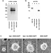
In vitro characterization of engineered netrin-1-overexpressing fibroblasts. a, Northern blot to detect bicistronic netrin-1 (Net-1) IRES EGFP (5490 bp, lane 2) or netrin-1 HA IRES EGFP RNA (5530 bp, lane 3) in fibroblasts transduced with the respective netrin-1 IRES EGFP or netrin-1 HA IRES EGFP MLVs, using the 1494 bp HincII-ScaI fragment of the mouse netrin-1 cDNA as a probe. b, Confirmation of netrin-1 secretion from transduced fibroblasts by Western blot. A ∼85 kDa protein was specifically detected in high salt cell surface extracts of fibroblasts transduced with netrin-1 HA IRES EGFP MLV (lane 4) using a polyclonal antibody directed against the HA epitope (Y11; Santa Cruz Biotechnology; 200 ng/ml). HA-tagged trkB served as a positive control (lane 1). c, Axon outgrowth from E13 rat dorsal spinal cord explants was elicited in coculture with netrin-1 IRES EGFP MLV (A) and netrin-1 HA IRES EGFP MLV (B) transduced fibroblasts but not control fibroblasts infected with EGFP MLV (C) after 18 h in coculture.
Figure 6.
Figure 6.
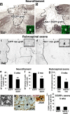
Netrin-1 inhibits axonal regeneration in vivo. Rats underwent cervical spinal cord lesions and were grafted with either netrin-1 secreting or control fibroblasts. a–d, Expression of netrin-1 within fibroblast grafts was monitored by expression of EGFP reporter (insets, a, b) or by in situ hybridization with netrin-1 antisense probe (insets, c, d). a, b, The density of neurofilament-labeled axons was 51% greater in control fibroblast grafts (a) than in netrin-1-secreting fibroblast grafts (b) 5 weeks after lesions and grafting (e; p < 0.01), and 57% greater by 3 months after lesion and grafting (f; p < 0.01). Thus, netrin-1 inhibits the growth of a general spinal cord population of axons. Shown in a and b are grafts 5 weeks after lesion. Similarly, the growth of UNC5A-expressing rubrospinal axons is 83% greater in (c) control fibroblast grafts than in (d) netrin-1-secreting grafts, 5 weeks after lesion (g; p < 0.05). h, i, UNC5A expression in DRGs is confined to large caliber neurons, which do not express CGRP (black arrows). j, CGRP-labeled sensory axons, which do not express UNC5 receptors, are not repelled from netrin-1-expressing fibroblast grafts. g, Graft; r, rostral; c, caudal; d, dorsal; ri, right.

Similar articles

Cited by

References

    1. Azanchi R, Bernal G, Gupta R, Keirstead HS. Combined demyelination plus Schwann cell transplantation therapy increases spread of cells and axonal regeneration following contusion injury. J Neurotrauma. 2004;21:775–788. - PubMed
    1. Benson MD, Romero MI, Lush ME, Lu QR, Henkemeyer M, Parada LF. Ephrin-B3 is a myelin-based inhibitor of neurite outgrowth. Proc Natl Acad Sci USA. 2005;102:10694–10699. - PMC - PubMed
    1. Blesch A, Tuszynski MH. GDNF gene delivery to injured adult CNS motor neurons promotes axonal growth, expression of the trophic neuropeptide CGRP, and cellular protection. J Comp Neurol. 2001;436:399–410. - PubMed
    1. Blesch A, Tuszynski MH. Cellular GDNF delivery promotes growth of motor and dorsal column sensory axons after partial and complete spinal cord transections and induces remyelination. J Comp Neurol. 2003;467:403–417. - PubMed
    1. Bradbury EJ, Moon LD, Popat RJ, King VR, Bennett GS, Patel PN, Fawcett JW, McMahon SB. Chondroitinase ABC promotes functional recovery after spinal cord injury. Nature. 2002;416:636–640. - PubMed

Publication types

MeSH terms

LinkOut - more resources