. 2008 Apr 25;374(1):82-99.
doi: 10.1016/j.virol.2007.11.036. Epub 2008 Jan 24.

Ectromelia virus BTB/kelch proteins, EVM150 and EVM167, interact with cullin-3-based ubiquitin ligases

Affiliations

Ectromelia virus BTB/kelch proteins, EVM150 and EVM167, interact with cullin-3-based ubiquitin ligases

Brianne A Wilton et al. Virology. .

Abstract

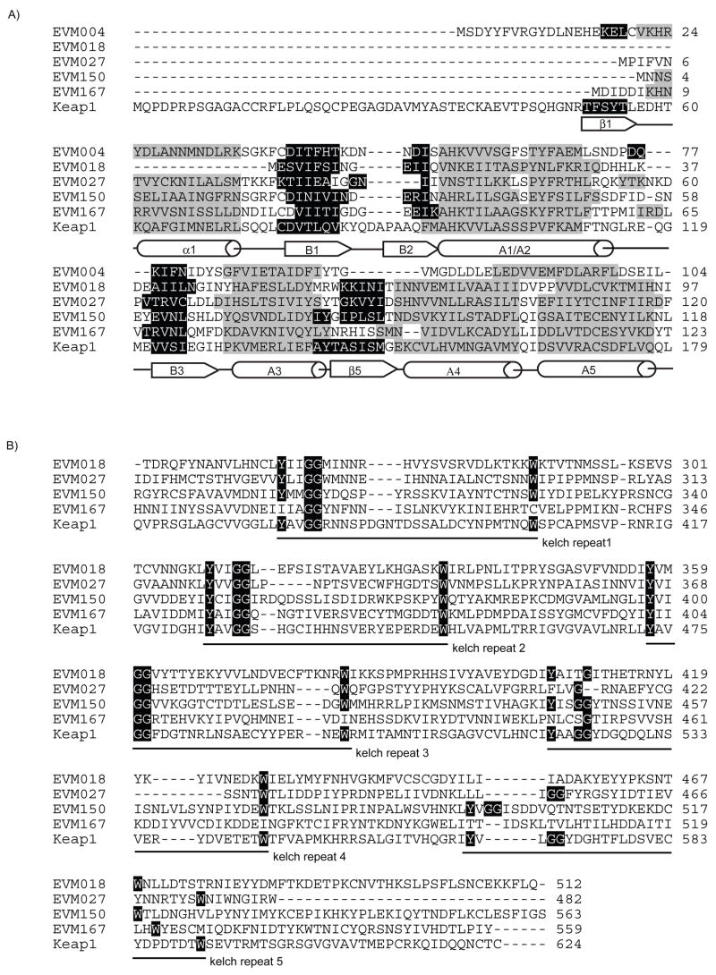
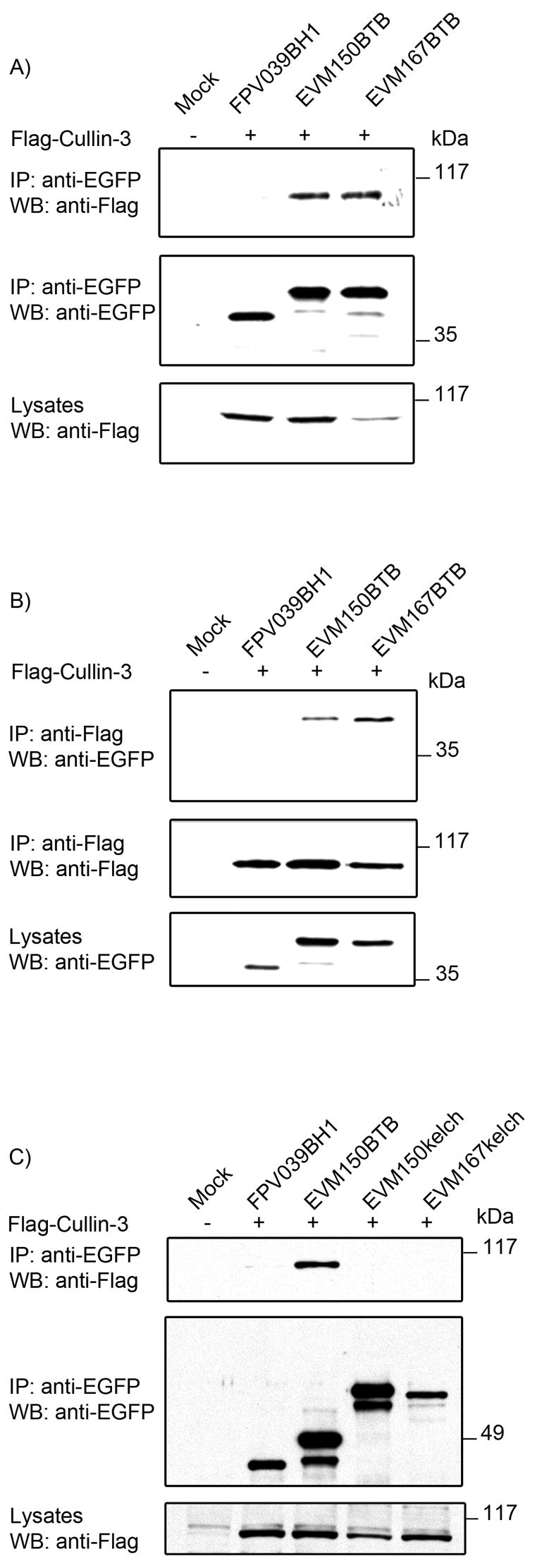
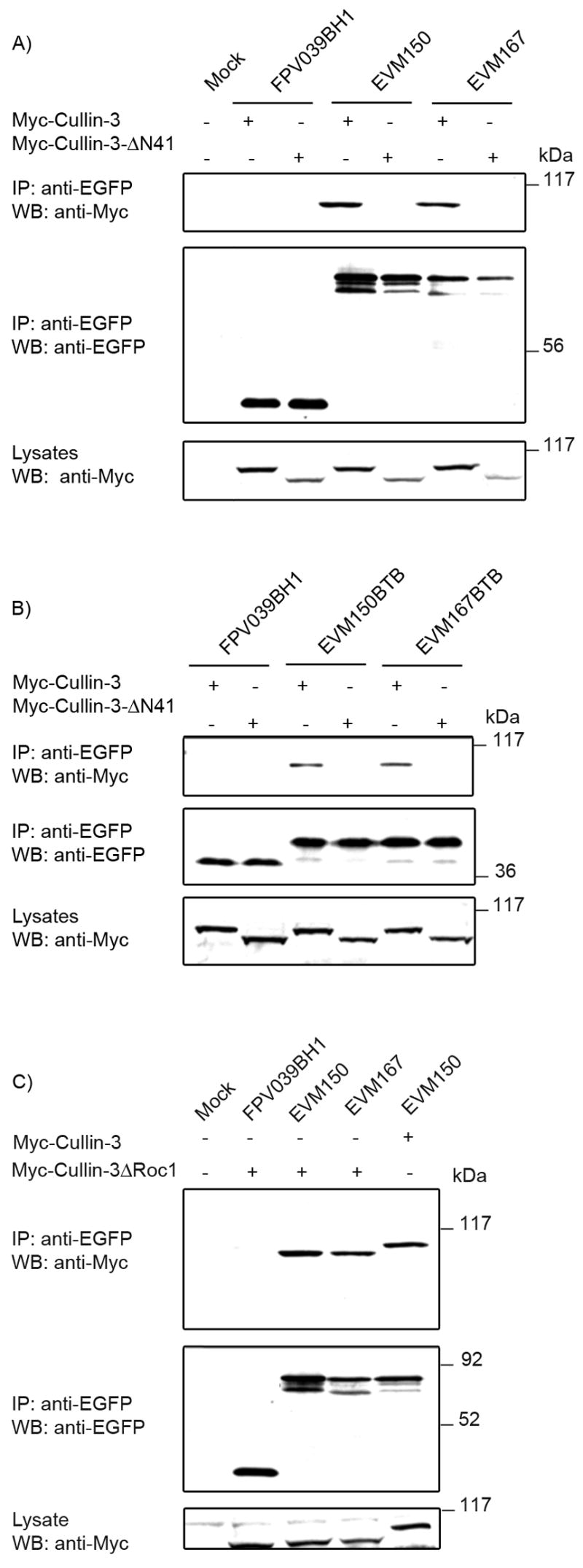
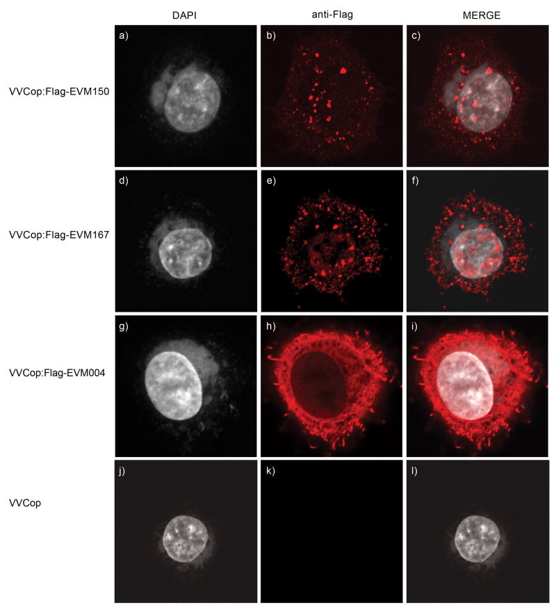
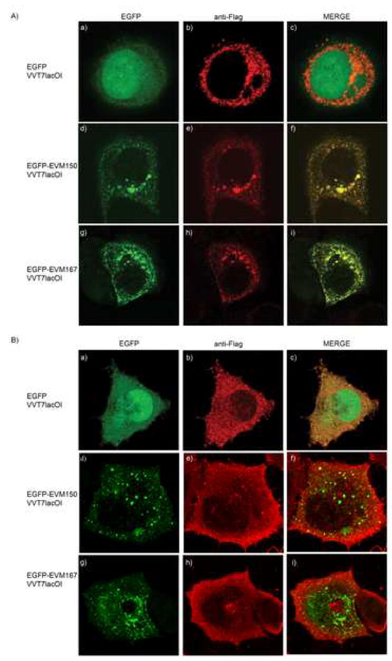
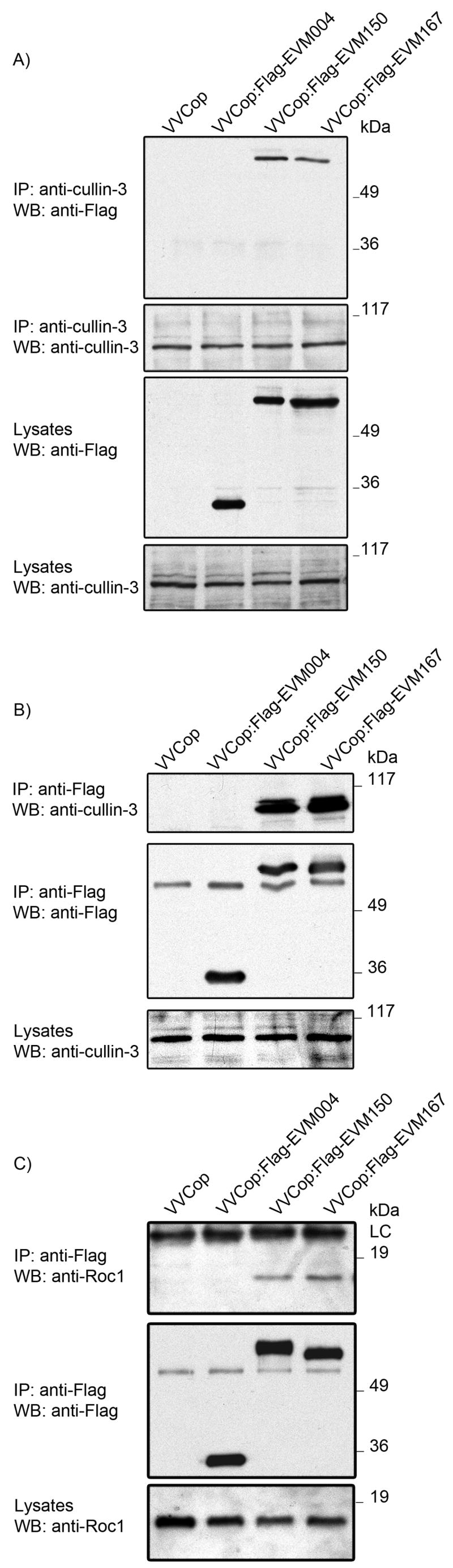
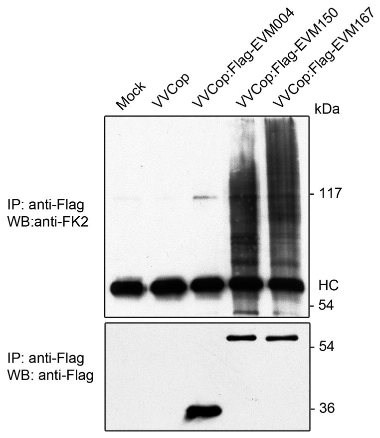
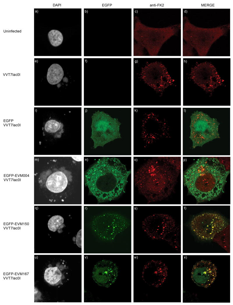
Cellular proteins containing BTB and kelch domains have been shown to function as adapters for the recruitment of substrates to cullin-3-based ubiquitin ligases. Poxviruses are the only family of viruses known to encode multiple BTB/kelch proteins, suggesting that poxviruses may modulate the ubiquitin pathway through interaction with cullin-3. Ectromelia virus encodes four BTB/kelch proteins and one BTB-only protein. Here we demonstrate that two of the ectromelia virus-encoded BTB/kelch proteins, EVM150 and EVM167, interacted with cullin-3. Similar to cellular BTB proteins, the BTB domain of EVM150 and EVM167 was necessary and sufficient for cullin-3 interaction. During infection, EVM150 and EVM167 localized to discrete cytoplasmic regions, which co-localized with cullin-3. Furthermore, EVM150 and EVM167 co-localized and interacted with conjugated ubiquitin, as demonstrated by confocal microscopy and co-immunoprecipitation. Our findings suggest that the ectromelia virus-encoded BTB/kelch proteins, EVM150 and EVM167, interact with cullin-3 potentially functioning to recruit unidentified substrates for ubiquitination.


Figures

Figure 1
Figure 1
Proteins encoded by ectromelia virus contain BTB and kelch domains. (A) Amino acid sequence of BTB domains of EVM004, EVM018, EVM027, EVM150, EVM167 and the BTB domain of Keap1. The ectromelia virus-encoded BTB proteins retain the predicted BTB fold containing a conserved series of alpha helices (grey) and beta sheets (black). Secondary structures were predicted using 3D-PSSM and PYRE. (B) Alignment of the kelch domain of EVM018, EVM027, EVM150 and EVM167 with the kelch domain of Keap1. Alignments were generated using ClustralW. EVM018 and EVM167 are predicted to contain 3 complete kelch repeats while EVM027 and EVM150 are predicted to contain 4 and 5 kelch repeats, respectively.
Figure 2
Figure 2
EVM004, EVM018, EVM027, EVM150 and EVM167 are transcribed early during ectromelia virus infection. (A) Schematic representation of the ectromelia virus BTB/kelch proteins EVM018, EVM027, EVM150, EVM167 and the BTB-only containing protein EVM004. (B) CV-1 cells were infected with ectromelia virus at an MOI 5 in the presence and absence of cytosine arabinoside (AraC) and RNA isolated at the indicated times. RNA was subjected to RT-PCR followed by PCR using gene specific primers to detect transcripts for EVM004, EVM018, EVM027, EVM150, EVM167, EVM058 and cellular β-actin.
Figure 3
Figure 3
EVM150 and EVM167 interact with cullin-3. (A) A schematic representation of cullin-3 based ubiquitin ligase. A single protein containing both BTB and kelch domains serves as the substrate specific adaptor for cullin-3 based ubiquitin ligases. The BTB domain interacts with cullin-3 and the kelch domain recruits substrates for ubiquitination. (B) HEK293T cells were co-transfected with constructs to express EGFP-tagged versions of FPV039BH1, EVM018, EVM027, EVM150, EVM167 and Flag-cullin-3. Lysates were western blotted with anti-Flag to indicate expression of Flag-cullin-3. Immunoprecipitation and western blotting with an antibody specific for EGFP demonstrated expression of FPV039BH1, EVM018, EVM150 and EVM167. Interaction between EVM150 and EVM167 and cullin-3 was demonstrated by western blotting the immunoprecipitations with anti-Flag. (C) Reciprocal immunoprecipitations were performed with anti-Flag to confirm the interaction between EVM150, EVM167 and cullin-3. Western blotting of lysates with anti-EGFP demonstrated expression of FPV039BH1, EVM150 and EVM167. Immunoprecpitation with anti-Flag demonstrated expression of cullin-3 in the co-transfected samples. Interaction between EVM150, EVM167 and cullin-3 was demonstrated by subjecting the immunoprecipitations to western blotting with anti-EGFP.
Figure 4
Figure 4
The BTB domain of EVM150 and EVM167 is required for interaction with cullin-3. (A) HEK293T cells were co-transfected with constructs to express EGFP-tagged BTB-only domains of EVM150 and EVM167 and co-transfected with Flag-cullin-3 and interaction assessed by co-immunoprecipitation. Westen blotting of lysates with anti-Flag indicated expression of Flag-cullin-3. Immunoprecipitation and western blotting with an antibody specific for EGFP demonstrated expression of FPV039BH1, and the BTB-only domains of EVM150 and EVM167. Interaction between the BTB domains of EVM150 and EVM167 and cullin-3 was demonstrated by subjecting the immunoprecipitations to western blotting with anti-Flag. (B) To confirm interaction between the BTB domains of EVM150 and EVM167 and cullin-3 reciprocal immunoprecipitations were performed with anti-Flag. Expression of FPV039BH1, EVM150BTB and EVM167BTB was assessed by western blotting lysates with anti-EGFP. Immunoprecpitation with anti-Flag demonstrated expression of cullin-3 in the co-transfected samples. Interaction between the BTB domains of EVM150 and EVM167 with cullin-3 was demonstrated by subjecting the immunoprecipitations to western blotting with anti-EGFP. (C) The kelch domains of EVM 150 and EVM167 do not interact with cullin-3. HEK293T cells were transfected with constructs to express EGFP-tagged kelch-only domains of EVM150, EVM167 and as a positive control the BTB-only domain of EVM150. Cells were co-transfected with Flag-cullin-3 and interaction assessed by co-immunoprecipitation. Lysates were subjected to western blotting with anti-Flag to indicate expression of Flag-cullin-3. Immunoprecipitation and western blotting with an antibody specific for EGFP demonstrated expression of FPV039BH1, BTB-only domain of EVM150 and the kelch-only domains of EVM150 and EVM167. Lack of interaction between the kelch domains of EVM150 and EVM167 and cullin-3 was demonstrated by western blotting the immunoprecipitations with anti-Flag.
Figure 5
Figure 5
A conserved region in the N-terminus of cullin-3 is necessary for EVM150 and EVM167 interaction with cullin-3. (A) HEK293T cells were co-transfected with constructs to express EGFP-tagged full-length versions of EVM150, EVM167 and co-transfected with Myc-cullin-3 or Myc-cullin-3ΔN41. Interaction was assessed by co-immunoprecipitation with anti-EGFP. Western blotting of lysates with anti-Myc showed expression of Myc-cullin-3 and Myc-cullin-3ΔN41. Immunoprecipitation and western blotting with an antibody specific for EGFP demonstrated expression of FPV039BH1, EVM150 and EVM167. Interaction between EVM150 and EVM167 and cullin-3 but not Myc-cullin-3ΔN41 was determined by western blotting the immunoprecipitations with anti-Myc. (B) Expression of the BTB-only domains of EVM150 and EVM167 retain the ability to interact with Myc-cullin-3 but not Myc-cullin-3ΔN41. (C) EVM150 and EVM167 interact with both cullin-3 and cullin-3ΔRoc1. HEK293T cells were co-transfected with constructs to express EGFP-tagged full-length versions of EVM150, EVM167 and co-transfected with Myc-cullin-3ΔRoc1. As a positive control, EVM150 was co-transfected with Myc-cullin-3. Lysates were subjected to western blotting with anti-Myc to indicate expression of Myc-cullin-3 and Myc-cullin-3ΔRoc1. Immunoprecipitation and western blotting with an antibody specific for EGFP demonstrated expression of FPV039BH1, EVM150 and EVM167. Interaction between EVM150 and EVM167 and cullin-3 and Myc-cullin-3ΔRoc1 was demonstrated by subjecting the immunoprecipitations to western blotting with anti-Myc.
Figure 6
Figure 6
EVM150 and EVM167 localize to cytoplasmic regions during poxvirus infection. HeLa cells were infected with VVCop:Flag-EVM150 (a–c), VVCop:Flag-EVM167 (d–f), VVCop:Flag-EVM004 (g–i) or VV(Cop) (j–l) at an MOI of 5. Twelve hours post infection cells were fixed and stained with DAPI and anti-Flag to visualize the viral factories and EVM150, EVM167 and EVM004.
Figure 7
Figure 7
EVM150 and EVM167 co-localize with Flag-cullin-3 during virus infection. A) HeLa cells were infected with VVT7lacOI (MOI 5) and co-tranfected with pEGFP (a–c), pEGFP-EVM150 (d–f) or pEGFP-EVM167 (g–i) and Flag-cullin-3. Expression was induced by the addition of IPTG. Twelve hours post-infection, cells were fixed and cullin-3 localization visualized by staining with anti-Flag. EGFP fluorescence indicated the localization of EVM150 and EVM167. B) HeLa cells were infected with VVT7lacOI (MOI 5) and co-transfected with pEGFP (a–c), pEGFP-EVM150 (d–f) or pEGFP-EVM167 (g–i) and pGEMT-Flag-cullin-3(200–768).
Figure 8
Figure 8
EVM150 and EVM167 interact with endogenous cullin-3 and Roc1 during infection. HEK293T cells were infected with VV(Cop), VVCop:FlagEVM004, VVCop:FlagEVM150 or VVCop:Flag167 at an MOI 5. Twelve hours post-infection cell lysates were generated and EVM150 and EVM167 interaction with endogenous cullin-3 assessed by co-immunoprecipitation. (A) Endogneous cullin-3 expression was detected by immunoprecipitation and western blotting with a cullin-3 specific antibody. Interaction between EVM150 and EVM167 and cullin-3 was demonstrated by western blotting with anti-Flag. (B) Interaction between cullin 3 and EVM150 and EVM167 was assessed by reciprocal immunoprecipitation using anti-Flag followed by western blotting with anti-cullin 3. (C) Roc1 co-immunoprecipitates with EVM150 and EVM167. Cellular lysates were analyzed by immunoprecipitaiton with anti-Flag followed by western blotting with anti-Roc1.
Figure 9
Figure 9
Conjugated ubiquitin co-immunoprecipitates with EVM150 and EVM167. HeLa cells were infected with VV(Cop), VVCop:FlagEVM004, VVCop:FlagEVM150 or VVCop:FlagEVM167 at an MOI 5. Twelve hours post infection the cells were subjected to immunoprecpitation with anti-Flag. Expression of Flag-EVM150 and Flag-EVM167 was detected by immunoprecipitation with anti-Flag followed by western blotting with anti-Flag-HRP. Co-immunoprecipiation of EVM150 or EVM167 with conjugated ubiquitin was detected by immunoprecipitation with anti-Flag followed by western blotting with the anti-ubiquitin antibody (FK2) which detects only conjugated ubiquitin.
Figure 10
Figure 10
EVM150 and EVM167 co-localize with conjugated ubiquitin. HeLa cells were infected with VVT7lacOI (MOI 5) and transfected with pEGFP, pEGFP-EVM004, pEGFP-EVM150, or pEGFP-EVM167. Expression was induced by treatement with IPTG and twelve hours post-infection cells were fixed and stained with DAPI to visualize nuclei and virus factories and conjugated ubiquitin was detected with an antibody (FK2) that detects conjugated ubiquitin. (A-D) Mock infected HeLa cells display a diffuse staining pattern of conjugated ubiquitin. (E-H) HeLa cells infected with VVT7lacOI demonstrate a punctate localization of conjugated ubiquitin. (I-L) EGFP expression during virus infection does not co-localize with conjugated ubiquitin. (M-P) EGFP-EVM004 does not co-localize with conjugated ubiquitin. (Q-T) EGFP-EVM150 co-localizes with conjugated ubiquitin. (U-X) EGFP-EVM167 co-localizes with conjugated ubiquitin.

Similar articles

Cited by

References

    1. Adams J, Kelso R, Cooley L. The kelch repeat superfamily of proteins: propellers of cell function. Trends Cell Biol. 2000;10(1):17–24. - PubMed
    1. Afonso CL, Tulman ER, Delhon G, Lu Z, Viljoen GJ, Wallace DB, Kutish GF, Rock DL. Genome of crocodilepox virus. J Virol. 2006;80(10):4978–91. - PMC - PubMed
    1. Afonso CL, Tulman ER, Lu Z, Zsak L, Kutish GF, Rock DL. The genome of fowlpox virus. J Virol. 2000;74(8):3815–31. - PMC - PubMed
    1. Ahmad KF, Engel CK, Prive GG. Crystal structure of the BTB domain from PLZF. Proc Natl Acad Sci U S A. 1998;95(21):12123–8. - PMC - PubMed
    1. Ahmad KF, Melnick A, Lax S, Bouchard D, Liu J, Kiang CL, Mayer S, Takahashi S, Licht JD, Prive GG. Mechanism of SMRT corepressor recruitment by the BCL6 BTB domain. Mol Cell. 2003;12(6):1551–64. - PubMed

Publication types

MeSH terms