. 2008 Feb 6;27(3):535-45.
doi: 10.1038/sj.emboj.7601984. Epub 2008 Jan 24.

Estrogen protects bone by inducing Fas ligand in osteoblasts to regulate osteoclast survival

Affiliations

Estrogen protects bone by inducing Fas ligand in osteoblasts to regulate osteoclast survival

Susan A Krum et al. EMBO J. .

Abstract

Estrogen deficiency in menopause is a major cause of osteoporosis in women. Estrogen acts to maintain the appropriate ratio between bone-forming osteoblasts and bone-resorbing osteoclasts in part through the induction of osteoclast apoptosis. Recent studies have suggested a role for Fas ligand (FasL) in estrogen-induced osteoclast apoptosis by an autocrine mechanism involving osteoclasts alone. In contrast, we describe a paracrine mechanism in which estrogen affects osteoclast survival through the upregulation of FasL in osteoblasts (and not osteoclasts) leading to the apoptosis of pre-osteoclasts. We have characterized a cell-type-specific hormone-inducible enhancer located 86 kb downstream of the FasL gene as the target of estrogen receptor-alpha induction of FasL expression in osteoblasts. In addition, tamoxifen and raloxifene, two selective estrogen receptor modulators that have protective effects in bone, induce apoptosis in pre-osteoclasts by the same osteoblast-dependent mechanism. These results demonstrate that estrogen protects bone by inducing a paracrine signal originating in osteoblasts leading to the death of pre-osteoclasts and offer an important new target for the prevention and treatment of osteoporosis.


Figures

Figure 1
Figure 1
Estrogen induces apoptosis in pre-osteoclasts and prevents the formation of mature osteoclasts. (A) Bone marrow was differentiated with M-CSF and RANKL for 8 days in the presence of ethanol (EtOH), 0.1 nM estradiol (E2), 1 nM E2 or 10 nM E2. Cells were then fixed and stained for TRAP. (B) The expression of cathepsin K mRNA or (C) calcitonin receptor mRNA was measured by quantitative real-time PCR from RNA isolated from bone marrow, pre-osteoclast and osteoclast cultures treated with or without 10 nM E2 for the entire course of differentiation. Fully differentiated osteoclasts treated with E2 were never formed and thus there is no value in this figure. The fold induction was normalized to β-actin mRNA. Error bars represent ±1 s.d. (D) Bone marrow was differentiated to the pre-osteoclast or osteoclast state with M-CSF and RANKL and then treated for 7 or 24 h with vehicle (EtOH), 1 nM E2 or 10 nM E2. Cells were fixed and apoptosis was detected by TUNEL. Quantification was performed using a laser scanning cytometer. Data are representative of three independent experiments.
Figure 2
Figure 2
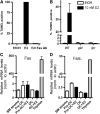
Estrogen-induced apoptosis is mediated by the Fas/FasL pathway. (A) Pre-osteoclasts were differentiated with M-CSF and RANKL for 6 days and then treated for 16 h with vehicle (EtOH) or 10 nM E2, and either a control IgG or 5 pg/μl FasL neutralizing antibody. Cells were fixed and apoptosis was detected by TUNEL. Quantification was performed using a laser scanning cytometer. Data are representative of three independent experiments. (B) Pre-osteoclasts from wild-type, FasL-deficient mice (gld) and Fas-deficient mice (lpr) were differentiated with M-CSF and RANKL for 6 days and then treated for 16 h with vehicle control (EtOH) or 10 nM E2. Cells were fixed and apoptosis was detected by TUNEL. Quantification was performed using a laser scanning cytometer. (C) Fas and (D) FasL mRNA levels were analyzed by quantitative PCR from RNA obtained from bone marrow stroma (BM stroma), pre-osteoclasts (pre-OC) with and without 10 nM E2, osteoclasts with and without 10 nM E2 and thymus.
Figure 3
Figure 3
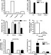
Estrogen increases FasL in osteoblasts. (A) Primary calvarial osteoblasts were differentiated for 3, 5 or 13 days in the presence of 100 μg/ml ascorbic acid and 5 mM β-glycerophosphate. Cells were then treated with 10 nM E2 for 3 h and RNA was obtained. FasL mRNA was analyzed by quantitative PCR. (B) U20S-ERα and MCF-7 cells were deprived of estrogen for 3 days in phenol red-free media containing 5% CDT-FBS. They were then treated with 10 nM E2 for 0, 3, 6 or 12 h and RNA was obtained. FasL mRNA was analyzed by quantitative PCR. (C) U20S-ERα cells and MCF-7 cells were deprived of estrogen for 3 days in phenol red-free media containing 5% CDT-FBS. They were then treated with 10 nM E2 for 45 min. ERα (C) and RNA polymerase II (RNA Pol II) (D) were immunoprecipitated, DNA was isolated and quantitative PCR was performed at the indicated sites of the FasL locus. Each PCR signal was normalized to input. (E) MC3T3 cells were differentiated for 10 days in the presence of 100 μg/ml ascorbic acid. The media were then changed to phenol red-free media containing 5% CDT-FBS for 3 days. The cells were then treated with 10 nM E2 for 3 h and FasL mRNA was analyzed by quantitative PCR. (F) MC3T3 cells were differentiated for 10 days in the presence of 100 μg/ml ascorbic acid. The media were then changed to phenol red-free media containing 5% CDT-FBS for 3 days. The cells were then treated with 10 nM E2 for 45 min. ERα was immunoprecipitated, DNA was isolated and quantitative PCR was performed with primers for the FasL enhancer. Each PCR signal was normalized to input. Error bars in all panels represent ±1 s.d.
Figure 4
Figure 4
Fas is expressed in pre-osteoclasts and FasL is expressed in osteoblasts. (A) Bone marrow cultures treated under osteoclastic conditions for 6 days to the pre-osteoclast stage were fixed and immunostained for Fas. The primary antibody was detected with an anti-rabbit IgG Alexa 488 (green fluorescence). DNA was counterstained with DAPI. The cells were then stained for alkaline phosphatase (dark blue cells). The red arrowhead points to clustering of the Fas receptor proteins next to the osteoblast. Parallel cultures were stained with TRAP to identify pre-osteoclasts. (B) Bone marrow cultures treated under osteoclastic conditions for 6 days to the pre-osteoclast stage were fixed and immunostained for FasL and RUNX2. DNA was counterstained with DAPI. (C) A laser scanning cytometer was used to separate cells with low and high RUNX2. The amount of FasL in the presence or absence of 10 nM E2 was quantified in each of these populations.
Figure 5
Figure 5
FasL is induced by E2 in osteoblasts in vivo. (A) Ovariectomized mice were treated with either vehicle (veh.) or 50 μg/kg E2 for 24 h. Paraffin-imbedded femurs were immunostained with an antibody to FasL (blue) and the sections were counterstained with hematoxylin (pink). BM=bone marrow, CB=cortical bone. (B) The growth plate and (C) diaphysis of E2-treated femurs were immunostained with FasL (blue) and osteoclasts were identified with TRAP (pink). HC=hypertrophic chondrocytes. The cells were counterstained with a nuclear green dye (methyl green). (D) Femurs treated with vehicle alone (veh.) or (E) E2 were immunostained for FasL (red) and RUNX2 (green) and counterstained with DAPI (blue).
Figure 6
Figure 6
Apoptosis of pre-osteoclasts is mediated by ERα. (A) Pre-osteoclasts were differentiated with M-CSF and RANKL for 6 days and then treated for 16 h with vehicle control (ethanol, EtOH), 1 nM E2 and/or 10 nM ICI 182,780 (ICI). Cells were fixed and apoptosis was detected by TUNEL. Quantification was performed with a laser scanning cytometer (LSC). (B) Bone marrow cells from wild-type (WT), ERαKO or ERβKO mice were differentiated with M-CSF and RANKL in the presence or absence of 10 nM E2 to the pre-osteoclast stage. Cells were then fixed and apoptosis was detected by TUNEL. Quantification was performed with a laser scanning cytometer (LSC). (C) Bone marrow cells from wild-type (WT), ERαKO or ERβKO mice were differentiated with M-CSF and RANKL in the presence or absence of 10 nM E2 for 8 days. Cells were then fixed and stained for TRAP. Multinucleated cells, defined as cells that are TRAP positive and containing three or more nuclei, were counted in each well. Three wells were counted for each genotype. (D) Bone marrow cells from wild-type and ERαKO mice were cultured in mesenchymal stem cell media and then differentiated under osteoblastic conditions for 9 days. mRNA for FasL was measured by quantitative PCR. (E) U20S-ERβ cells were deprived of estrogen for 3 days in phenol red-free media containing 5% CDT-FBS. They were then treated with 10 nM E2 for 3 h and RNA was obtained. FasL and Rbbp1 mRNA levels were analyzed by quantitative PCR. (F) U20S-ERβ cells were deprived of estrogen for 3 days in phenol red-free media containing 5% CDT-FBS. They were then treated with 10 nM E2 for 45 min. An anti-Flag antibody was used to immunoprecipitate ERβ and quantitative PCR was performed to detect the Rbbp1 enhancer or the FasL enhancer (+86 kb site). Each PCR was normalized to input.
Figure 7
Figure 7
Osteoblasts are required for estrogen-mediated apoptosis. (A) Human monocytes were differentiated with M-CSF and RANKL to osteoclasts, without or (B) with osteoblasts (MC3T3 cells), and (left) without or (right) with 10 nM E2 for 8 days. The cells were then fixed and stained for TRAP. (C) The number of multinucleated cells per field from (A) and (B) was counted and expressed as a percentage of the vehicle control-treated cells. Each treatment was counted in triplicate and error bars represent ±1 s.d. (D) Bone marrow cells from wild-type or ERαKO mice were co-cultured with or without MC3T3 cells and without or with 10 nM E2 under osteoclastic conditions for 8 days. The cells were then fixed and stained for TRAP. The number of multinucleated cells per field was counted and expressed as a percentage of the vehicle control-treated cells. (E) Cells were cultured as in part D, except that 10 nM E2 was added at day 6 for 16 h. The cells were then fixed and apoptosis was detected by TUNEL. Quantification was performed with a laser scanning cytometer.
Figure 8
Figure 8
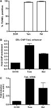
SERMs regulate FasL to induce apoptosis. (A) Pre-osteoclasts were differentiated with M-CSF and RANKL and then treated for 16 h with vehicle (EtOH), 1 μM tam or 10 nM ral. Cells were fixed and apoptosis was detected by TUNEL. Data are representative of three independent experiments. (B) U20S-ERα cells were deprived of estrogen for 3 days in phenol red-free media containing 5% CDT-FBS. Cells were then treated for 45 min with 1 μM tam or 10 nM ral. ChIP was performed with an antibody to ERα and quantitative PCR was performed to detect the FasL enhancer (+86 kb site). Each PCR was normalized to input. (C) Cells were deprived of estrogen as in part B. They were then treated with 1 μM tam or 10 nM ral for 3 h and RNA was obtained. FasL mRNA was analyzed by quantitative PCR. Error bars represent ±1 s.d.

Similar articles

Cited by

References

    1. Boyle WJ, Simonet WS, Lacey DL (2003) Osteoclast differentiation and activation. Nature 423: 337–342 - PubMed
    1. Carroll JS, Liu XS, Brodsky AS, Li W, Meyer CA, Szary AJ, Eeckhoute J, Shao W, Hestermann EV, Geistlinger TR, Fox EA, Silver PA, Brown M (2005) Chromosome-wide mapping of estrogen receptor binding reveals long-range regulation requiring the forkhead protein FoxA1. Cell 122: 33–43 - PubMed
    1. Carroll JS, Meyer CA, Song J, Li W, Geistlinger TR, Eeckhoute J, Brodsky AS, Keeton EK, Fertuck KC, Hall GF, Wang Q, Bekiranov S, Sementchenko V, Fox EA, Silver PA, Gingeras TR, Liu XS, Brown M (2006) Genome-wide analysis of estrogen receptor binding sites. Nat Genet 38: 1289–1297 - PubMed
    1. Clowes JA, Riggs BL, Khosla S (2005) The role of the immune system in the pathophysiology of osteoporosis. Immunol Rev 208: 207–227 - PubMed
    1. Cohen PL, Eisenberg RA (1991) Lpr and gld: single gene models of systemic autoimmunity and lymphoproliferative disease. Annu Rev Immunol 9: 243–269 - PubMed

Publication types

MeSH terms