. 2008 Feb 1;180(3):1854-65.
doi: 10.4049/jimmunol.180.3.1854.

Activation of peroxisome proliferator-activated receptor gamma (PPARgamma) suppresses Rho GTPases in human brain microvascular endothelial cells and inhibits adhesion and transendothelial migration of HIV-1 infected monocytes

Affiliations

Activation of peroxisome proliferator-activated receptor gamma (PPARgamma) suppresses Rho GTPases in human brain microvascular endothelial cells and inhibits adhesion and transendothelial migration of HIV-1 infected monocytes

Servio H Ramirez et al. J Immunol. .

Abstract

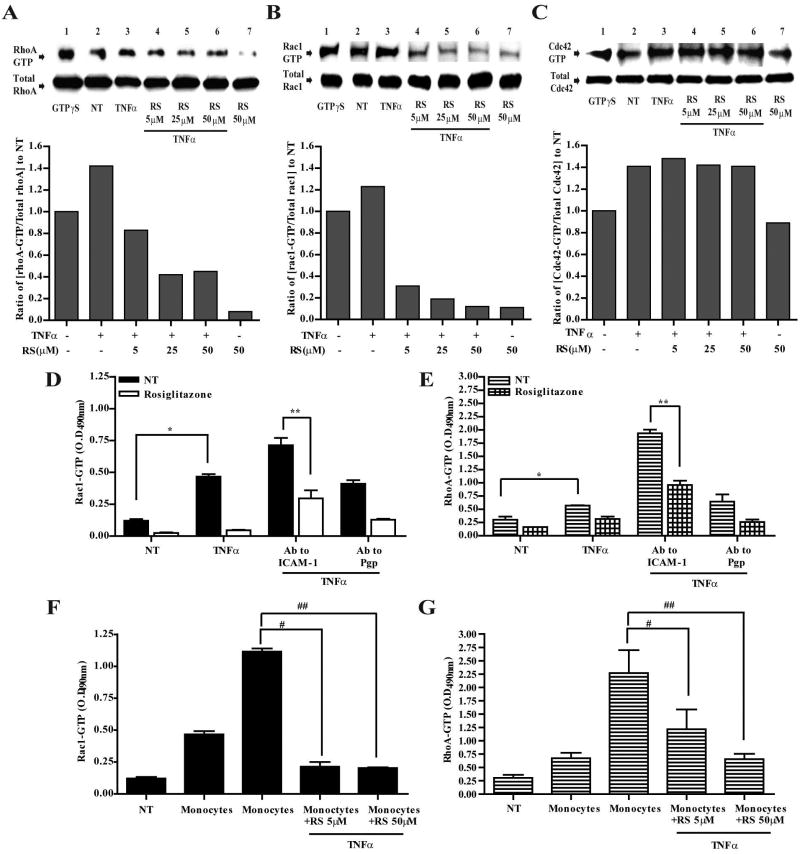
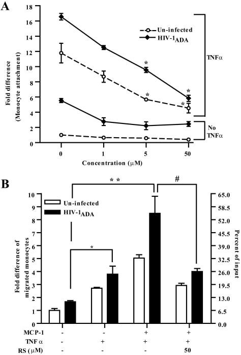
Under inflammatory conditions (including HIV-1 encephalitis and multiple sclerosis), activated brain endothelium enhances the adhesion and transmigration of monocytes across the blood-brain barrier (BBB). Synthetic ligands that activate the peroxisome proliferator-activated receptors (PPARs) have anti-inflammatory properties, and PPAR stimulation prevents the interaction of leukocytes with cytokine stimulated-endothelium. However, the mechanism underlying these effects of PPAR ligands and their ability to intervene with leukocyte adhesion and migration across brain endothelial cells has yet to be explored. For the first time, using primary human brain endothelial cells (BMVEC), we demonstrated that monocyte adhesion and transendothelial migration across inflamed endothelium were markedly reduced by PPARgamma activation. In contrast to non-brain-derived endothelial cells, PPARalpha activation in the BMVEC had no significant effect on monocyte-endothelial interaction. Previously, our work indicated a critical role of Rho GTPases (like RhoA) in BMVEC to control migration of HIV-1 infected monocytes across BBB. In this study, we show that in the BMVEC PPARgamma stimulation prevented activation of two GTPases, Rac1 and RhoA, which correlated with decreased monocyte adhesion to and migration across brain endothelium. Relevant to HIV-1 neuropathogenesis, enhanced adhesion and migration of HIV-1 infected monocytes across the BBB were significantly reduced when BMVEC were treated with PPARgamma agonist. These findings indicate that Rac1 and RhoA inhibition by PPARgamma agonists could be a new approach for treatment of neuroinflammation by preventing monocyte migration across the BBB.


Conflict of interest statement

Disclosures

The authors have no financial conflict of interest.

Figures

Figure 1
Figure 1
PPARγ agonist, rosiglitazone, inhibited monocyte adhesion and migration across human BMVEC in a dose dependent manner. (A) PPAR protein expression profile derived from lysates of primary cultured brain microvascular endothelial cells (BMVEC) and isolated human brain microvessels. Passage 2 BMVEC monolayers and microvessels were lysed and analyzed by western blot using specific antibodies for the three known PPAR isotypes, PPARα (top panel), PPARβ/δ (middle panel), and PPARγ (lower panel). (B and C) BMVEC were treated with the indicated PPAR agonist (fibrate or TZD) in increasing concentrations, after 4 h nuclear extractions were collected and introduced to oligo binding ELISA for PPARα and PPARγ as described in the materials and methods. The values represent the mean ± SEM. (D) Adhesion assays of BMVEC monolayers pretreated for 30 min with increasing concentrations of rosiglitatozone, pioglitazone, and fenofibrate with or without subsequent co-incubation with TNFα (20ng/ml) for additional 4 h. All treatments were removed and calcein-AM labeled monocytes were introduced to the BMVEC, as described in the material methods. Monocytes were allowed to adhere for 15 min to the BMVEC, unattached cells were rinsed and the fluorescence measured. The data is represented as mean ± SEM fold difference, which is the adhesion value from treated BMVEC over the basal adhesion value from untreated cells. Where indicated, the addition of PPARγ antagonist GW9662 (50μM) was used as control. (E) Transendothelial monocyte migration across pre-treated BMVEC monolayers towards CCL2/MCP-1 (30ng/ml) was evaluated after 2 hours as described in the methods section. Data is represented as mean ± SEM fold difference of migration, which is the value from migration of treated cells over the “spontaneous” migration of untreated cells without chemoattractant. All data collected are from at least three independent experiments performed in triplicate. Asterisk denotes statistically significant differences (p<0.01) between untreated and rosiglitazone treated and cytokine stimulated BMVEC.
Figure 2
Figure 2
shRNA mediated PPARγ knockdown in BMVEC eliminates the effects of rosiglitazone on monocyte adhesion and migration. (A) Western blot analysis showing knockdown of endogenous PPARγ expression in BMVEC (72 hours post transfection) with vectors coding shRNA sequences directed to PPARγ. Three candidate sequences were evaluated denoted as shPPARγ-1, shPPARγ-2 and shPPARγ-3. The vector with shPPARγ-2-SC, lane 5, represents the scrambled control for shPPARγ-2, lane 3. (B) Adhesion assays of BMVEC expressing shPPARγ-2, control scramble shPPARγ-2-SC, and mock transfected (NT). After 5 days post-transfection, the cells were treated with increasing concentration of rosiglitazone, and adhesion assays were performed. (C) Migration assays of transfected BMVEC with the knockdown sequence and control. After 5 days, cells were pretreated with rosiglitazone along with or without cytokine stimulation for 4 h; migration was then performed for 2 h and values collected. The data for the adhesion and migration is represented as mean ± SEM fold difference.
Figure 3
Figure 3
PPARγ activation did not alter ICAM-1 and VCAM-1 surface expression of stimulated BMVEC or induce changes to barrier properties. (A) Flow cytometry of BMVEC ICAM-1/CD54 and VCAM-1/106 surface staining following addition of rosiglitazone (RS, 50μM) for 30 min and simultaneous incubation with TNFα (20ng/ml) for 4 h. (B), BMVEC grown to confluence on ECIS electrode chamber slides were used to analyze for differences in TEER. The measurements shown represent recordings acquired at 5 min intervals at the parameters described in the materials and methods. The single arrow designates the pre-treatment with 50μM of rosiglitazone following co-incubation with TNFα as shown by the double arrow. Addition of lindsidomide at 100μM was used as a control for lowering TEER. The data is presented as normalized resistance, which is the resistance measured post treatment over the resistance acquired before treatment introduction (typically ~2600 Ω/cm2). (C) Western blot analysis of TJ proteins ZO-1, occludin-1, and claudin-5 from the cytosolic (Cyto) and membranous (Mem) fractions of lysed BMVEC are shown; treatments are indicated in the figure.
Figure 4
Figure 4
PPARγ regulates activation of RhoA and Rac1 GTPases. GTPases from whole cell extracts were affinity immunoprecipitated from BMVEC after the indicated treatments. The top panels show western blots of the active GTP bound RhoA (A), Rac1 (B), and Cdc42 (C) following affinity precipitation (top panel). Together with the active form of the GTPase are immunoblots showing the total GTPase (lower panels) content of inactive plus the active form from whole cell lysates. The included densitometry analysis depicts the ratios from the normalized values of the active GTPases from all treatments and compared to the normalized value from untreated cells. (D–G), ELISA based GTPase activation assays of Rac1 (D) and RhoA (E) were performed after BMVEC crosslinking of ICAM-1 surface molecules with specific antibodies. Monocyte mediated activation of Rac1 (F) and RhoA (G) was performed by using BMVEC lysates collected after monocyte adhesion, monocytes were removed as previously described [Persidsky, 2006 #26]. The data is presented as the average ± SEM of three independent experiments each performed in triplicate. Symbols in the graph represents statistical significance of (p<0.01). (*) compares values from untreated cells and TNFα treated; (**) indicates values from untreated cells compared with those from TNFα, rosiglitazone and crosslinked with ICAM-1; (#) compares values from TNFα and monocytes with TNFα, monocytes and rosiglitazone (5μM); (##) compares values from TNFα and monocytes with TNFα, monocytes and rosiglitazone (50μM).
Figure 5
Figure 5
Stimulation of PPARγ in activated BMVEC prevents Rac-1 mediated monocyte adhesion. BMVEC were transfected with expression vectors for wild type (WT), constitutively active (CA) and dominant negative (DN) Rac1 and RhoA. Five days post-transfection the cells were treated with two increasing concentrations of rosiglitazone (2μM and 5μM). Figure inserts in A and B show the BMVEC expression of the mutant GTPase by antibodies that detected the hemagglutin epitope (HA) fused to the expressing GTPase (top row), along with the proteins detected by RhoA or Rac1 antibodies (middle row), and actin (bottom row). After transfection and treatments, the BMVEC were used in adhesion assays. The data is presented as the mean ± SEM adhesion fold difference of transfectants with the Rac1 (A) and RhoA (B) mutants. Asterisk indicates statistical significance (p<0.01) when compared with the value from stimulated adherent cells without PPARγ agonist.
Figure 6
Figure 6
Activation of PPARγ in BMVEC inhibits RhoA mediated transendothelial migration of monocytes. BMVEC were transfected with GTPase mutants and allowed to express the transgene for 5 days followed by treatment with or without PPARγ agonist, rosiglitazone, at 5μM and 50μM with or without TNFα co-incubation. The figure shows CCL2/MCP-1 driven monocyte migration across BMVEC expressing RhoA (A) and Rac1 (B) mutants. The migration assay was conducted for 2 h and the relative fluorescence of labeled monocytes was measured. Asterisk indicates statistical significance (p<0.01) when compared with the value from stimulated cells in the presence of CCL2/MCP-1 but without rosiglitazone. Data represented as the migration fold difference mean ± SEM from at least three independent experiments performed in triplicate.
Figure 7
Figure 7
PPARγ activation in BMVEC inhibited adhesion and transendothelial migration of HIV-1 infected monocytes. (A) Adhesion and (B) migration assays using uninfected or HIV-1ADA (MOI 0.01) infected monocytes were performed. For adhesion assays the endothelial cells were exposed to increasing concentrations of rosiglitazone, 1μM, 5μM and 50μM and where indicated cells were also simultaneously incubated with TNFα (20ng/ml) for 4 h. Data is represented as adhesion fold differences as described earlier. For migration assays BMVEC monolayers were pre-incubated with rosiglitazone (50μM) with or without TNFα (20ng/ml) following the same treatments times as before. Data is represented as the migration fold difference mean ± SEM. (*) indicates statistical significance (p<0.01) when compared with the value from migrated cells towards CCL2/MCP-1 but without cytokine stimulation. (**) represents statistical significance (p<0.01) when compared with values from uninfected cells migrating towards CCL2/MCP-1 and monolayers treated with cytokine. (#) indicates statistical significance (p<0.01) of values from infected cells migrated towards CCL2/MCP-1 on monolayers treated with TNFα and rosiglitazone compared with infected cells migrating towards CCL2/MCP-1 but without rosiglitazone treatment.

Similar articles

Cited by

References

    1. Engelhardt B, Ransohoff RM. The ins and outs of T-lymphocyte trafficking to the CNS: anatomical sites and molecular mechanisms. Trends Immunol. 2005;26:485–495. - PubMed
    1. Kanwar JR. Anti-inflammatory immunotherapy for multiple sclerosis/experimental autoimmune encephalomyelitis (EAE) disease. Curr Med Chem. 2005;12:2947–2962. - PubMed
    1. Persidsky Y, Gendelman HE. Mononuclear phagocyte immunity and the neuropathogenesis of HIV-1 infection. J Leukoc Biol. 2003;74:691–701. - PubMed
    1. Stalder AK, Ermini F, Bondolfi L, Krenger W, Burbach GJ, Deller T, Coomaraswamy J, Staufenbiel M, Landmann R, Jucker M. Invasion of hematopoietic cells into the brain of amyloid precursor protein transgenic mice. J Neurosci. 2005;25:11125–11132. - PMC - PubMed
    1. Eugenin EA, Osiecki K, Lopez L, Goldstein H, Calderon TM, Berman JW. CCL2/monocyte chemoattractant protein-1 mediates enhanced transmigration of human immunodeficiency virus (HIV)-infected leukocytes across the blood-brain barrier: a potential mechanism of HIV-CNS invasion and NeuroAIDS. J Neurosci. 2006;26:1098–1106. - PMC - PubMed

Publication types

MeSH terms