. 2008 Jan 22;18(2):129-35.
doi: 10.1016/j.cub.2007.12.057.

Morphine-induced receptor endocytosis in a novel knockin mouse reduces tolerance and dependence

Affiliations

Morphine-induced receptor endocytosis in a novel knockin mouse reduces tolerance and dependence

Joseph A Kim et al. Curr Biol. .

Abstract

Opioid drugs, such as morphine, are among the most effective analgesics available. However, their utility for the treatment of chronic pain is limited by side effects including tolerance and dependence. Morphine acts primarily through the mu-opioid receptor (MOP-R) , which is also a target of endogenous opioids. However, unlike endogenous ligands, morphine fails to promote substantial receptor endocytosis both in vitro, and in vivo. Receptor endocytosis serves at least two important functions in signal transduction. First, desensitization and endocytosis act as an "off" switch by uncoupling receptors from G protein. Second, endocytosis functions as an "on" switch, resensitizing receptors by recycling them to the plasma membrane. Thus, both the off and on function of the MOP-R are altered in response to morphine compared to endogenous ligands. To examine whether the low degree of endocytosis induced by morphine contributes to tolerance and dependence, we generated a knockin mouse that expresses a mutant MOP-R that undergoes morphine-induced endocytosis. Morphine remains an excellent antinociceptive agent in these mice. Importantly, these mice display substantially reduced antinociceptive tolerance and physical dependence. These data suggest that opioid drugs with a pharmacological profile similar to morphine but the ability to promote endocytosis could provide analgesia while having a reduced liability for promoting tolerance and dependence.


Figures

Figure 1
Figure 1. Generation of rMOP-R knock-in mice
a. Schematic of targeting strategy A Sal1-Sac1 genomic fragment containing the MOP-R sequence, including Exons 2 and 3 was modified to contain the rMOP-R sequence (inset). A cassette containing resistance to G418 (Fx-Neo) and flanked by Lox P sites was inserted in the intron downstream of exon 3 for selection of ES clones. b. Detection of homologous recombinants. Genomic DNA was digested with BamHI and subjected to DNA hybridization with a ~1.1kb BglII fragment (see a). Targeted loci were confirmed by the presence of a band at ~8kb. The intact locus gave a band at ~6kb. c. Quantification of endocytosis. MetaMorph software was used to quantify the intensity of receptor signal at the plasma membrane versus the cytosol for each treatment condition and each genotype (MOP-R in black, rMOP-R in white). Data is plotted as the ratio of signal located within 0.3 μm of the surface (peripheral) versus the amount in the cytosol (central). See supplemental Fig. 1 for representative neurons and schematic.
Figure 2
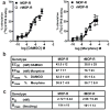
Figure 2. Pharmacological characterization of wild-type MOP-R and rMOP-R knock-in mice
a, b. Receptor-G protein coupling. Agonist mediated GTPγS binding was measured in brain membranes of wild-type MOP-R (squares) and rMOP-R knock-in mice (circles) with increasing concentrations of DAMGO or morphine. Data were analyzed by nonlinear regression using GraphPad Prism software and are presented as means ± SEM of at least three experiments performed in triplicate. There were no significant genotypic differences in either EC50 or Emax. c. Ligand affinity and receptor number. [3H] Naloxone binding in whole brain membranes from MOP-R and rMOP-R mice. Saturation binding assays were performed on membranes (50-100 μg per well) with increasing concentrations of [3H] naloxone (0 to 15 nM, 55.9Ci/mmol). Nonspecific binding was measured in the presence of 10 μM naloxone. Binding parameters were determined by Scatchard analysis of specific binding. Data are means ± SEM of three experiments performed in duplicate. There were no statistically significant differences between the genotypes (One way ANOVA with Tukey’s post-hoc test). Bmax, maximum binding capacity; KD, dissociation constant.
Figure 3
Figure 3. Antinociception in wild-type MOP-R and rMOP-R knock-in mice
a. Enhanced and prolonged morphine-induced antinociception in rMOP-R knock-in mice. Antinociceptive responses were measured with the hot-plate response latency test (56°C) after morphine treatment (10 mg/kg, sc). A response endpoint was defined as latency to either lick the fore- or hindpaws or flick the hindpaws. To avoid tissue damage, mice were exposed to the hot-plate for a maximum of 20s. Data are reported as the mean ± SEM of percent maximum possible effect (MPE) using the following formula: 100% × [(drug response time – basal response time)/(20 s – basal response time)]. A two-way analysis of variance revealed that the MPE curve for rMOP-R mice (n=17) mice was significantly greater and prolonged relative to the MOP-R mice (n=17) as indicated by a significant genotype [p<.001, F(1,7)=28.05] and genotype X time interaction effect [p<.001, F(1,7)=4.97]. b. Dose-dependent morphine-antinociception. Antinociceptive responses were determined with the hot-plate test and data are reported as mean ± SEM of MPE (see a). Separate groups of mice for both genotypes (n=7–9) were injected with the doses of morphine indicated and assessed for antinociception 30 min later. To test whether the antinociceptive responses were mediated by opioid receptors, a final grouping was injected with morphine (10 mg/kg) followed by naloxone (2 mg/kg). rMOP-R knock-in mice showed enhanced antinociception at 3 and 10 mg/kg doses (rMOP-R vs. MOP-R scores for MPE at respective morphine doses, student’s t-test, *p<.03) with the latter dose inducing the maximum possible response (100%) in the mutant mice. At the highest dose tested (50 mg/kg) both genotypes exhibited the maximum possible response (100%). For both genotypes, antinociception induced by 10 mg/kg of morphine was reversed by treatment with 2 mg/kg of the opioid antagonist naloxone (morphine 10 mg/kg with and without naloxone 2 mg/kg treatment for each genotype respectively, student’s t-test +++p<.001). c. MOP-R desensitization in the brainstem following acute morphine treatment. Agonist-mediated [35S]GTPγS binding was measured in brainstem membranes of MOP-R and rMOP-R mice with increasing concentrations of DAMGO. Left Panel. Binding in MOP-R mice was significantly reduced (p<0.01) following acute morphine-treatment (10 mg/kg s.c. 30 min; EC50 = 428 ± 141 μM; open squares ) compared to vehicle-treated mice (EC50 = 2.35 ± 0.9 μM; closed squares). Right Panel. Binding in rMOP-R mice was not significantly changed (p>0.05) following acute morphine-treatment (EC50 = 3.29 ± 1.4 μM; open circles) compared to vehicle-treated mice (EC50 = 1.16 ± 0.6 μM; closed circles). Data were analyzed by nonlinear regression using GraphPad Prism software and are presented as means ± SEM of at least three experiments performed in triplicate. d. Enhanced antinociception in rMOP-R knock-in mice is morphine-specific. Separate groups of mice for both genotypes (n=8-10) were injected with the doses of methadone indicated (1-10 mg/kg,) and assessed for antinociception. Methadone induced a dose-dependent increase in antinociceptive response with no genotypic differences. For both genotypes, antinociception induced by 4 mg/kg of methadone was reversed by treatment with 2 mg/kg of the opioid antagonist naloxone (methadone 4 mg/kg with and without naloxone 2 mg/kg treatment for each genotype respectively, student’s t-test +++p<.001). Thus, enhanced opioid-induced antinociception observed in the rMOP-R knock-in mice is agonist-specific, and naloxone-reversible.
Figure 4
Figure 4. Opioid tolerance and dependence in MOP-R wild-type and rMOP-R knock-in mice. a. Acute morphine tolerance
Mice (n=9) were initially treated with either saline (gray bars) or a high dose of morphine (100 mg/kg, sc, black bars). 24 h later, mice were challenged with an acute equi-antinociceptive dose of morphine (3 mg/kg for rMOP-R and 10 mg/kg for MOP-R, see Fig. 4B). Data are presented as mean ± SEM of MPE. Wild-type MOP-R mice exhibited significant acute tolerance, showing a 43% reduction in MPE when pretreated with 100 mg/kg of morphine compared to saline 24 h before (MOP-R comparing saline vs. morphine pretreatment, student’s t-test, ***p<.001). In contrast, rMOP-R knock-in mice showed no evidence of tolerance and maintained the same level of morphine-induced antinociception whether they were pretreated with 100 mg/kg of morphine or saline 24 h before. b. Chronic morphine tolerance. Mice were treated twice daily with morphine (10 mg/kg, sc) for 5 days and antinociception was assessed following the first injection of morphine each day. Mean ± SEM of MPE across days are presented. A two-way ANOVA revealed that mice treated chronically with morphine (n=17) behaved differently corresponding to genotype as indicated by a significant group effect [F(2,42)=27.95, p<.001] and group X treatment days effect [F(4,84)=12.09, p<.001]. Post-hoc comparisons (Tukey’s) revealed the source of the interaction. rMOP-R knock-in mice treated with 10 mg/kg of morphine had significantly longer response latencies across days compared to wild-type mice treated with the same dose of morphine (10 mg/kg) and rMOP-R knock-in mice chronically treated with a lower (equi-antinociceptive) dose of morphine (3 mg/kg), [rMOP-R 10 significantly different from MOP-R 10 and rMOP-R 3, **p<.01]. Additionally, rMOP-R knock-in mice chronically treated with an equi-antinociceptive dose of morphine (3 mg/kg) showed significantly greater antinociception than did wild-type mice chronically treated with a higher dose of morphine (10 mg/kg) across the tolerance development days (rMOP-R 3 vs. MOP-R 10, ++p<.01). Only wild-type mice chronically treated with morphine (10 mg/kg) showed a significant decrease in antinociception from Day 1 to Day 5 (MOP-R 10 Day 1 vs. Day 5, ###p<.001). Thus, the development of antinociceptive tolerance to morphine was evident in wild-type mice but attenuated in the knock in mice, whether they were chronically treated with the same (10 mg/kg) or equi-antinociceptive (3 mg/kg) dose of morphine. c. Methadone tolerance. Mice were similarly treated to the protocol in experiment in Fig. 6b but injected twice daily with methadone (4 mg/kg, sc) for 5 days. For both genotypes, there was no evidence of tolerance development with both groups expressing comparable levels of methadone-antinociception following the first and last injection. Methadone antinociception was equivalent in both genotypes across all days. d. Naloxone precipitated withdrawal. Groups of mice were chronically treated with 10 mg/kg of morphine (black bars, n=10-12), 4 mg/kg of methadone (grey bars, n=9) or saline (white bars, n=6) at the same intervals described for Fig 4b and c. Mice were challenged with naloxone (2 mg/kg, sc) 30 min following the final treatment injection. Notably, the chronic dose of morphine used (10 mg/kg) corresponded to a functionally higher dose in the rMOP-R knock-in mice relative to wild-type mice (see Fig 3a, b). Standard withdrawal behaviors including jumping, wet-dog shakes, paw licks and paw tremors were scored by an observer blind to genotype. Total withdrawal scores (the sum of all individual withdrawal behaviors) ± SEM are presented and group differences were analyzed with the LSD test. Compared to saline-treated mice, MOP-R wild-type mice displayed a significantly higher incidence of withdrawal when chronically treated with morphine or methadone (MOP-R MOR vs. MOP-R SAL, ***p<.001; MOP-R METH vs. MOP-R SAL, *p<.05), with a higher degree of withdrawal associated with chronic morphine treatment (MOP-R MOR vs. MOP-R METH, ++p<.01). In contrast, rMOP-R knock-in mice displayed similar levels of withdrawal regardless of pretreatment drug. For rMOP-R knock-in mice, both morphine and methadone pretreatment resulted in similar levels of moderate withdrawal, comparable to methadone pretreatment in MOP-R wild-type mice.

Similar articles

Cited by

References

    1. Matthes HW, Maldonado R, Simonin F, Valverde O, Slowe S, Kitchen I, Befort K, Dierich A, Le Meur M, Dolle P, et al. Loss of morphine-induced analgesia, reward effect and withdrawal symptoms in mice lacking the mu-opioid-receptor gene. Nature. 1996;383:819–823. - PubMed
    1. Arden JR, Segredo V, Wang Z, Lameh J, Sadee W. Phosphorylation and agonist-specific intracellular trafficking of an epitope-tagged mu-opioid receptor expressed in HEK 293 cells. J Neurochem. 1995;65:1636–1645. - PubMed
    1. Keith DE, Murray SR, Zaki PA, Chu PC, Lissin DV, Kang L, Evans CJ, von ZM. Morphine activates opioid receptors without causing their rapid internalization. J Biol Chem. 1996;271:19021–19024. - PubMed
    1. Koch T, Schulz S, Pfeiffer M, Klutzny M, Schroder H, Kahl E, Hollt V. C-terminal splice variants of the mouse mu-opioid receptor differ in morphine-induced internalization and receptor resensitization. J Biol Chem. 2001;276:31408–31414. - PubMed
    1. Yu Y, Zhang L, Yin X, Sun H, Uhl GR, Wang JB. Mu opioid receptor phosphorylation, desensitization, and ligand efficacy. J Biol Chem. 1997;272:28869–28874. - PubMed

Publication types